Hitological Changes on the Wound Healing Process of Alkali Burned Mouse Cornea
Ji-Young Lee and Koon-Ja Lee*
Department of Ophthalmic Optics, Daegu Polytechnic College University, Daegu 706-711, Korea
*Department of Optometry, Eulji University, Sungnam 461-713, Korea
(Received October 31, 2008: Revised November 15, 2008: Accepted December 9, 2008)
···
Purpose: To better understand the corneal regeneration after alkali burn regarding the initial clinical progression and the therapy, we investigated the changes of the multi factors following chemical injury in cornea. Methods:
This study was performed to observation on the healing process of alkali burned cornea in aspect of immuno- histochemistry by immunofluorescence or H-E staining and TUNEL assay. Results: The results showed that although a healing process occurred after alkali burn, apoptosis of epithelial, stromal and endothelial cells in the cornea was continuously observed. Neovascularization and expression of α-smooth muscle actin (α-SMA) from limbus and from injured cornea, respectively, were observed after 3 days of alkali burn. Formation of collagen III in corneal stroma and increased expression of chondroitin sulfate are coincident with expression of α-SMA and transforming growth factor-β (TGF-β). Conclusions: These data suggest that medical treatment within 3 days of alkali burn will be effective to inhibit neovascularization and formation of collagen III and chondroitin sulfate.
This study extends our immumohistochemical understanding of healing process in alkali burned cornea, and the results get in this study will be cornerstones in the development of therapeutic agent for accelerating renewal of chemical damaged cornea.
Key words: alkali burn, cornea, neovascularization, immunohistochemistry
···
Introduction
Ocular chemical injuries represent 12~19% of all ocular accidents1, of which corneal alkali burns are the most dangerous because alkali penetrates the surface of the eye very quickly. Alkali burns on the ocular surface can dam- age the corneal and conjunctival epithelium. Eventually, progression of this damage leads to recurrent corneal ero- sion, ulceration, and perforation decreasing visual acuity that primarily depends on the optical clarity of the cor- nea.
The cornea is covered with stratified epithelium that functions to protect the front of the eye from physical and chemical agents. To perform these functions, the cor- neal epithelium must be able to maintain its integrity by cell proliferation and repair any damage due to physical abrasion and chemical wound. Ocular trauma in the form
of an alkali burn to the cornea is a serious clinical prob- lem, which may cause severe and permanent visual impairment2. Even though wound healing process occurs in cornea after alkali burn, the cornea's optical properties are severely modified because of the neovascular vessel formation and rearrangement of collagen fibers.
Penetrated alkaline stimuli induces a strong keratocyte apoptosis and inflammatory reaction characterized by cell infilteration, production of proteolytic enzyme, and cyto- kines3-6. Inflammation and activation of corneal kerato- cytes and epithelial cells are both involved in the patho- genesis of post-alkali injured tissue and can lead to persistent epithelial defects7. Moreover, degradation of the basement membrane by matrix metalloproteinase (MMP) secreted by these cells contributes to the pathogenic ulceration and perforation of the stroma leading to ocular dysfunction8.
Corresponding Author Address: Koon-Ja Lee, Department of Optometry, Eulji University, Sungnam 461-713, Korea TEL: +82-31-740-7182, FAX: +82-31-740-7195, E-mail: kjl@eulji.ac.kr
Keratocytes are responsible for repairing the corneal stromal matrix after injury or infection. After corneal wounding, quiescent keratocytes become activated, mig- rate to the wound area, and begin to secrete a host of substances required for wound repair. During corneal stromal wound healing, keratocytes are activated to become corneal fibroblasts9-11. Activated fibroblasts are recruited to wound sites where their cellular proliferation, matrix deposition, remodeling, and contraction aim to achieve tectonic repair but could result in vision-threa- tening corneal scarring9,11-13. The precise mechanisms responsible for keratocyte activation and migration have been examined to date through studies of substances capable of activating keratocytes. These activating sub- stances include epithelial growth factor14, fibroblast growth factor15, interleukin-116, and transforming growth factor-β (TGF-β)17.
Neovascularization on the ocular surface is a common pathologic condition that can be caused by inflammation, infection, thermal and chemical burns18. Abnormal growth of new blood vessels on the cornea is a major cause of blindness19 and the key reason for corneal graft failure20,21. Scarring and neovascularization of the corneal stroma, which occurs during the healing process, impair a vision.
Opacification resulted from corneal neovascularization remain the most frequent causes of blindness after severe alkali burn trauma22. Despite the routine use of topical therapeutic agents23,24, the inflammatory response can lead to edema, lipid deposition and corneal scarring that may not only significantly alter visual acuity, but also worsen the prognosis of subsequent penetrating keratoplasty. In this study, to better understand the corneal regeneration after alkali burns according to the initial clinical severity and the therapy, we investigate the histopathologic evolu- tions of the multi factors, following chemical burns in cornea.
Materials and Methods
Experimental animal
Five to seven weeks old male C57BL/6 mice were pur- chased from DaeHan biolink (Korea) and 20 g of mice were used in this experiment. All animals were main- tained and treated in accordance with the Association for Research and Vision and Ophthalmology Statement for
the use of Animals in Ophthalmic and Vision Research.
Alkali burned animal model and observation of corneal haze
Mice were anesthetized with ketamin hydrochloride (70 mg/kg) and xylazine hydrochloride (30 mg/kg) by intrap- eritoneal injection. An alkali burn was performed by touching an applicator stick soaked in 1 N NaOH on the central cornea for 10 seconds. The cornea surface was then carefully rinsed with 10 ml of physiological saline for 5 minutes. All mice were anesthetized, corneas were observed by biomicroscope at 0, 1, 3, 7, 14, and 21 day after alkali burn treatment. Corneal haze was scored on scale of 0~4, where 0=no opacity, completely clear cor- nea; 1=mild opacity, iris and lens visible; 2=moderate opacity; 3=severe opacity (iris and lens invisible, obscur- ing iris detail); 4=dense opacity (whole cornea extensive opacity).
Tissue preparation
The eyeballs were enucleated at 0, 1, 3, 7, 14, and 21 days after alkali burn treatment and embedded in liquid OCT compound (Sakura FineTek, Torrance, CA, USA) within a mould. Cornea specimens were centered within the mould that block could be sectioned transversely from the center of the cornea. The mould and tissue were rap- idly frozen and stored at −80oC until sectioning was per- formed. The Corneal sections were cut into 4 mm thickness with a cryostat microtome (Shandon cryotome FE, Thermo, USA) and placed on microscope slides and stored at −80oC until staining was preformed. The sec- tions were evaluated with hematoxylin and eosin stain and for TUNEL, α-smooth muscle actin (α-SMA), TGF- β2, collagen III and chondroitin sulfate assay.
TUNEL assay
To detect fragmentation of DNA associated with apop- tosis in the cornea, the TUNEL (TdT-mediated dUTD nick-end labeling) method was preformed according to the manufacturer's protocol (In situ cell death detection kit, Fluorescein, Roche Diagnostics, Germany). Sections were air-dried and fixed with ice cold methanol (−20oC) for 1 hour. The sections were incubated with permeabili- sation solution (0.2% Triton X-100 in phosphate buffered saline) for 20 minutes at room temperature and stained
with TUNEL reaction mixture at 37oC for 1 hour.
Immunofluorescence staining for CD31, TGF-β2, α- SMA, collagen III and chondroitin sulfate
Air dried sections were fixed with ice cold methanol (−20oC) for 1 hour, and rinsed in phosphate buffered saline and preformed single and double immunofluores- cence staining. For single immunofluorescence labeling, the sections were stained with rat anti-CD31 (1:50, BD phamingen, USA) for overnight at 4oC and stained again with secondary antibody Alexa 594 donkey anti-rat IgG (1:400, invitrogen, USA) at room temperature for 1 hour.
And then sections were counterstained with DAPI (sigma, USA) for 10 minutes at room temperature. For immunof- luorescence labelling of TGF-β2, α-SMA, collagen III and chondroitin sulfate, the sections were stained with rabbit polyclonal antibody for TGF-β2 (1:50, Santa cruz, USA) or with goat polyclonal antibody for collagen III (1:50, Santa cruz, USA) for overnight at 4oC. The sec- ondary antibody, Alexa 488 donkey anti-rabbit IgG (1:400, invitrogen, USA) or donkey anti-goat IgG (1:400, invitrogen, USA) was applied for 1 hour at room temper- ature. The sections were washed, blocked and stained secondly with mouse monoclonal antibody for α-SMA (1:800, Sigma, USA) or with mouse monoclonal antibody for chondroitin sulfate (1:200, Sigma, USA) at room tem- perature for 2 hours then washed and stained with sec- ondary antibody Alexa 594 donkey anti-mouse IgG (1:500, invitrogen, USA) at room temperature for 1 hour.
The sections were mounted by aqueous mounting medium (DAKO, USA).
Results and Discussion
Corneal haze and neovascular vessels evaluated by biomicroscopy
Corneal haze was developed after alkali burn and per- sisted throughout the observation period and increased to an average grade of 4 at day 21 (Fig. 1g). Biomicro- scopic observation of the burned corneal surface shows opaque cornea at day 3 (Fig. 1d). Neovascular vessels were developed from limbus at day 3 (Fig. 1d) and invaded into the central cornea (Fig. 1g).
Histological findings after alkali burn
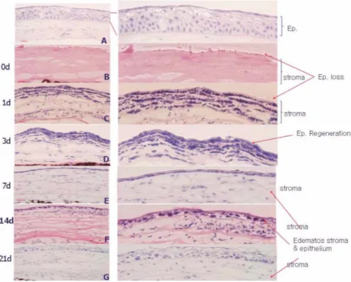
After alkali burn, the corneal epithelium was com- pletely destroyed (Fig. 2, see the arrow). The inflamma- tory cell infiltration was observed at day 1 and 3 and gradually reduced to day 7 in the stroma. Invasion of monocytes and macrophage may play an important role in tissue damage following injury, including alkali burn in the cornea6. Central cornea was covered with one or two layers of epithelial cells (Fig. 2d) at day 3. Epithelial tis- sue was gradually recovered and showed normal thick- ness at day 21. But regenerated epithelial tissue had many empty spaces and lost its regularity (Fig. 2g). The basement membrane changed to a wavy shape at day 0 and 3 but it was turned straight during healing process.
The peroxisome proliferator-activated receptor-γ (PPARγ) could be involved in re-epitheliazation with basement membrane reconstruction in the healing process25.
Data showed that the stroma was moderately edematous and collagen lamella structure was disorganized after alkali burn (Fig. 2). Alkali burns result in a dramatic change of the corneal mineral content26. To regulate the hydration state of the corneal stroma, the endothelial cells play an important role in maintaining corneal transpar- ency. Lack of this regulation may cause swelling, which in turn alters collagen fibril spacing and makes the stroma opaque27.
Keratocyte, inflammatory cell, endothelial cell, and regenerated epithelial cell apoptosis
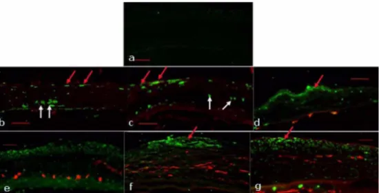
Because the central role of keratocyte apoptosis is acti- vation of the wound healing cascade, we investigated the change of apoptotic keratocyte. After alkali burn, TUNEL positive keratocytes were detected in the corneal stroma at day 0 and 1 (Fig. 3b and c, see the white arrow), and TUNEL positive inflammatory cells began to be detected in the anterior stroma at day 3 (Fig. 3d, see the red arrow).
Increases of incomplete destruction of the entire kerato- cyte population, as shown in Fig. 3, the repaired fibro- blasts probably derive from the surviving keratocytes at the peripheral cornea. Earlier study noted the shortage of repaired fibroblasts in the ulcerated stroma and consid- ered the lack of healthy fibroblasts as one of the major causes of corneal ulceration after severe alkali burn5. We observed TUNEL positive inflammatory cells (Fig. 3d) and densely stained cells in the endothelium (Fig. 3e) and regenerated epithelial tissue (Fig. 3f and g). Apoptosis of
endothelial cells could be the cause of corneal edema and apoptosis of regenerated epithelial cells might be the cause of edematous epithelium shown in this experiment during healing process (Fig. 2g). And we also found TUNEL positive stromal cells during healing process (Fig. 3f and g, see the white arrow).
Although the mechanisms responsible for this phenom- enon remain unknown, there are extensive studies indicat- ing that apoptosis plays an important role in regulating cellularity during corneal wound healing28,29. Moreover, previous studies shown that platelet-activation factor (PAF)
is a strong inducer of apoptosis in corneal keratocytes and myofibroblasts30,31. Therefore, in the wound healing process, PAF might, alone or in combination with other cytokines, induce apoptosis in myofibroblasts.
Formation of the neovascular vessels by alkali burn in the cornea
Immunohistochemical staining of corneal tissue showed significant level of CD31 (red) in the stroma from day 1 after alkali burn (Fig. 3c) and maximal level at day 14 (Fig. 3f) and gradually decreased at day 21 (Fig. 3g). Fig.
Fig. 2. Histological sections (haematoxylin and eosin staining) show differences in the normal and the alkali burn treated cornea (magnification, ×100). a: untreated normal control. The central corneas were observed after alkali burn treatment at 0(b), 1(c), 3(d), 7(e), 14(f), and 21(g) days after alkali burn (bar 100 mm, original magnification ×100).
Fig. 1. Photomicrographs demonstrating corneal clarity or haze in control and experiment cornea after alkali burn treatment. a:
untreated normal control. Corneal haze was monitored by biomicroscopy at 0(b), 1(c), 3(d), 7(e), 14(f), and 21(g) days after alkali burn. The corneal haze was observed through the observation period and neovascular vessels were observed clearly at 7 days, 14 day, and 21 day after alkali burn.
4 shows newly formed vessels were growing from limbus to central part of the cornea. Larger neovessels were detected at the limbal area at day 7 and they grow into
the central cornea (Fig. 4Bd).
A wound healing reaction is orchestrated by a variety of signals derived from endogenous soluble factors. Pre- Fig. 3. TUNEL assay and CD31 stain in alkali burned mouse cornea merged images, indicating co-localization of TUNEL (green) with anti-CD31 (red) antibody staining in the central cornea, staining for corneal neovassels and confirming that vascular endothelial cells undergo apoptosis after alkali burn treatment. a: untreated normal control. The central corneas were observed after alkali burn treatment at 0(b), 1(c), 3(d), 7(e), 14(f), and 21(g) days after alkali burn (bar 100 mm, original magnification, ×100).
Fig. 4. CD31 stain in alkali burned mouse limbus: Immunostaining of the normal and alkali burned limbus. (A) Histological sections (haematoxylin and eosin staining) show differences in the normal and the alkali burn treated limbus, magnification, ×100.
(B) Stain for CD31, show striking differences in the density of newly formed vessels in alkali burn treated mouse limbus, magnification, ×100 (C) Merged image of CD31 and DAPI. a: untreated normal control. The central corneas were observed after alkali burn treatment at 0 day (b), 1 day (c), 3 day (d), 7 day (e), 14 day (f), and 21 day (g). The nuclei of all the cells were counterstained with DAPI, original magnification, ×100.
vious work has demonstrated that neovascularization is initiated soon after trauma32. Neovascularization involves the sprouting of essential new vessels from capillaries and venules from the pericorneal plexus33. One of the hallmarks of corneal stromal scarring is the development of neovascularization. Such neovascularization of the cor- neal stroma likely contributes to stromal opacification and is associated with inflammation. In this study, examina- tion of stromal neovascularization using an antibody to CD31 showed marked staining in alkali burned corneas (Fig. 3). This effect might be attributed to the induction of expression of TGF-β2, as shown in Fig. 5B. It is widely accepted that TGF-β stimulates also neovascular- ization and epithelialization in healing wounds34.
Expression of TGF-β2 in the alkali burned cornea Since various cytokines and matrix-degrading enzymes induced following injury are known to be expressed by activated macrophages, we examined the expression of TGF-β implicated in the tissue destruction in alkali
burned cornea. After alkali burn TGF-β2 was expressed in the regenerated epithelial and stromal tissue (Fig. 5B).
The expression of TGF-β2 at day 3 in the epithelial tis- sue (Fig. 5Bd) and the level of stromal expression of TGF-β2 being particularly intense at day 21 (Fig. 5Bg).
A number of growth factors and cytokines are believed to be involved in the tissue destruction and late scarring that occur in the cornea after alkali burn. Reduction of TGF- β2 levels seems to contribute to the reduction of mac- rophage invasion at the early stage of alkali burn35.
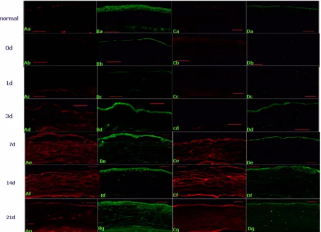
Increase of α-SMA positive cells by alkali burn Another hallmark of scar tissue formation and opacifi- cation of the corneal stroma is the well established trans- differentiation of keratocytes into α-SMA-positive myo- fibroblasts13,36,37. Examination of the expression pattern of SMA in injured corneas showed that, at day 3, many epi- thelial and stromal cells in injured corneas expressed α- SMA indicating the presence of myofibroblasts (Fig.
5Ad). At day 7, the anterior stroma was infiltrated with
Fig. 5. Immunohistochemical localization of α-SMA, TGF-β2, Chondroitin sulfate, and collagen III in alkali burned mouse cornea.
a: untreated; normal control. The central corneas were observed after alkali burn treatment at 0 day (b), 1 day (c), 3 day (d), 7 day (e), 14 day (f) and 21 day (g), original magnification.
more myofibroblasts in the alkali burned cornea (Fig.
5Ae) and higher expression of α-SMA cell was observed at day 14 after alkali burn (Fig. 5Af). Stromal repopula- tion after alkali injury by repair fibroblasts, also called myofibroblasts, is a necessary event because these cells have the unique capacity to remodel the damaged stroma by producing collagen and extracellular matrix muco- polysaccharides.
Induction of chondroitin sulfate and collagen III TGF-β is known to increase the synthesis of Type I, II, III, and VII collagen38,39. TGF-β inhibits the transcription of metalloproteinases that degrade collagen, thereby fur- ther increasing accumulated collagen protein. Evidence suggests that Type I collagen synthesis is stimulated by TGF-β through both transcriptional40,41 and posttranscrip- tional mechanisms42. Shah et al.43 showed that neutraliza- tion of TGF-β1 and -β2 leads to decreased scarring in adult rodents. We measure the level of TGF-β2 and col- lagen III resulting in scar formation. In this experiment the expression of TGF-β2 was increased in the epithelial and stromal tissue after alkali burn (Fig. 5Bg) and the collagen III (green) was observed at the regenerated epi- thelial cells (Fig. 5D) and stromal tissue at day 14 and 21 (Fig. 5Df, Dg). And chondroitin sulfate (red) expression was increased (Fig. 5C) and more pronounced at the 7 and 14 day (Fig. 5Ce, Cf).
Conclusion
Taken together, in this study, although a healing process occurred after alkali burn, development of apoptosis on the epithelial tissue was observed. Edema was also found on healed epithelial tissue. Apotosis of epithelial, stromal and endothelial cells was continuously developed after alkali burn. Formation of neovascular vessel from limbus was observed after three days of alkali burn. After 3 days from alkali burn, expression of α-SMA from injured cor- neas were observed, which it is possible due to the for- mation of myofibroblasts. Formation of collagen III at stroma and increased expression of chondroitin sulfate are coincident with expression of α-SMA and TGF-β. The results suggest that medical treatment within 3 days of alkali burn will be effective to inhibit neovascularization and formation of collagen III and chondroitin sulfate.
This study extends our immumohistochemical understand- ing of healing process in alkali burned cornea, and the results get in this study will be cornerstones in the devel- opment of therapeutic agent for accelerating renewal of chemical damaged cornea.
References
1. Reim M., Redbrake C., and Schrage N., “Chemical and thermal injuries of the eyes. Surgical and medical treat- ment based on clinical and pathophysiological findings”, Arch. Soc. Exp. Ophthalmol., 76(2):103-124(2001).
2. Araki-Sasaki K., Ohashi Y., Sasabe T., Hayashi K., Watanabe H., Tano Y., and Handa H., “An SV40-immor- talized human corneal epithelial cell line and its character- ization”, Invest. Ophthalmol. Vis. Sci., 36(3):614-621(1995).
3. Berman M., Dolman C. H., and Davison P. F., “Character- ization of collagenolytic activity in the ulcerating cornea”, Exp. Eye Res., 11(2):255-257(1971).
4. Kenyon K. R., Berman M., Rose J., and Gage J., “Preven- tion of stromal ulceration in the alkali-burned rabbit cornea by glued-on contact lens: evidence for the role of polymor- phonuclear leukocytes in collagen degradation”, Invest.
Ophthalmol. Vis. Sci., 18(6):570-587(1979).
5. Pfister R. R. and Burstein N., “The alkali burned cornea, I:
epithelial and stromal repair”, Exp. Eye Res., 23(5):519- 535(1976).
6. Sotozono C., He J., and Kinoshita S., “Cytokine expression in the alkali-burned cornea”, Curr. Eye Res., 16(7):670- 676(1997).
7. Bazan H. E., Reddy S. T., and Lin N., “Platelet-activating factor (PAF) accumulation correlates with injury in the cor- nea”, Exp. Eye Res., 52(2):481-491(1991).
8. Daniels J. T., Geerling G., Alexander R. A., Murphy G., Khaw P. T., and Saarialho-Kere U., “Temporal and spatial expression of matrix metalloproteinases during wound healing of human corneal tissue”, Exp. Eye Res., 77(6):
653-664(2003).
9. Gan L. and Fagerholm P., “Leucocytes in the early events of corneal neovascularization”, Cornea, 20(1):96-99(2001).
10. Nakamura K., “Interaction between injured corneal epithe- lial cells and stromal cells”, Cornea, 22:S35-S47(2003).
11. Wilson S. E., Liu J. J., and Mohan R. R., “Stromal-epithe- lial interactions in the cornea”, Prog. Retin. Eye Res., 18(3):293-309(1999).
12. Daniels J. T., Schultz G. S., Blalock T. D., Garrett Q., Gro- tendorst G. R., and Dean M., “Mediation of transforming growth factor-beta(1)-stimulated matrix connective tissue growth factor in contractile scarring”, Am. J. Pathol., 163(5):2043-2052(2003).
13. Ten Dijke P., Goumans M. J., Itoh F., and Itoh S., “Regula- tion of cell proliferation by Smad proteins”, J. Cell Phys-
iol., 191(1):1-16(2002).
14. Weimar V. L., “Activation of initial wound healing responses in rat corneas in organ culture by mesodermal growth factor”, Invest, Ophthalmol. Vis. Sci., 18(5):532- 535(1979).
15. Hecquet C., Morisset S., Lorans G., and Adolphe M.,
“Effects of acidic and basic fibroblast growth factors on the proliferation of rabbit corneal cells”, Curr. Eye Res., 9(5):429-433(1990).
16. Girard M. T., Matsubara M., and Fini E., “Transforming growth factor-beta and interleukin-1 modulatemetallopro- teinase expression by corneal stromal cells”, Invest. Oph- thalmol. Vis. Sci., 32(9):2441-2445(1991).
17. Woost P. G., Brightwell J., Eiferman R. A., and Schultz G.
S., “Effect of growth factors with dexamethasone on heal- ing of rabbit corneal stromal incisions”, Exp. Eye Res., 40(1):47-60(1985).
18. Lee P., Wang C. C., and Adamis A. P., “Ocular neovascu- larization: an epidemiologic”, Surv. Ophthalmol., 43(3):
245-269(1998).
19. Whitcher J. P., Srinivasan M., and Upadhyay M. P., “Cor- neal blindness: a global perspective”, Bull. WHO, 79(3):
214-221(2001).
20. Price M. O., Thompson R. W. Jr,, and Price F. W. Jr., “Risk factors for various causes of failure in initial corneal grafts”, Arch. Ophthalmol., 121(8):1087-1092(2003).
21. Weisbrod D. J., Sit M., and Naor J., “Outcomes of repeat penetrating keratoplasty and risk factors for graft failure”, Cornea, 22(5):429-434(2003).
22. Chang J. H., Gabison E. E., Kato T., and Azar D. T., “Cor- neal neovascularization”, Curr. Opin. Ophthalmol., 12(4):
242-249(2001).
23. Den S., Sotozono C., Kinoshita S., and Ikeda T., “Efficacy of early systemic betamethasone and cyclosporine A after corneal injury via inflammatory cytokine reduction”, Acta Ophthalmol. Scand., 82(2):195-199(2004).
24. Kuckelkorn R., Schrage N., Keller G., and Rebrake C.,
“Emergency treatment of chemical and thermal eye burns”, Acta Ophthalmol. Scand., 80(1):4-10(2002).
25. Saika S., Yamanaka O., Okada Y., Miyamoto T., Kitano A., Flanders K. C., Ohnishi Y., Nakajima Y., Kao W. W., and Ikeda K., “Effect of overexpression of PPAR gamma on the healing process of corneal alkali burn in mice”, Am.
J. Physiol., Cell. Physiol., 293(1):C75-C86(2007).
26. von Fischern T., Lorenz U., Burchard W. G., Reim M., and Schrage N. F., “Changes in mineral composition of rabbit corneas after alkali burn”, Graefes Arch. Clin. Exp. Oph- thalmol., 236(7):553-558(1998).
27. Gipson I. K., “Anatomy of the conjuntiva, cornea, and lim- bus”, In The Cornea Little Brown. MA. USA, pp. 3-24 (1994).
28. Wilson S. E., “Analysis of the keratocyte apoptosis, kerato- cyte proliferation, and myofibroblast transformation responses after photorefractive keratectomy and laser in situ keratom-
ileusis”, Trans. Am. Ophthalmol. Soc., 100:411-433(2002).
29. Wilson S. E. and Kim W. J., “Keratocyte apoptosis: Impli- cations on corneal wound healing, tissue organization, and disease”, Invest. Ophthalmol. Vis. Sci., 39:220-226(1998).
30. He J. and Bazan H. E., “Synergistic effect of platelet-acti- vating factor and tumor necrosis factor-alpha on corneal myofibroblast apoptosis”, Invest. Ophthalmol. Vis. Sci., 47(3):883-891(2006).
31. Ottino P., He J., Axelrad T. W., and Bazan H. E., “PAF- induced furin and MT1-MMP expression is independent of MMP-2 activation in corneal myofibroblasts”, Invest. Oph- thalmol. Vis. Sci., 46(2):487-496(2002).
32. Daniels J. T. and Khaw P. T., “Temporal stimulation of corneal fibroblast wound-healing activity by differentiating epithelium in vitro”, Invest. Ophthalmol. Vis. Sci., 41(12):
3754-3762(2000).
33. Yaylali V., Ohta T., and Kaufmann S. C., “In vivo confocal imaging of corneal neovascularization”, Cornea, 17(6):646 -653(1998).
34. Eskild-Jensen A., Koff J. D., Kjolseth L. H., Andersen T., Christensen M., Baandrup U., and Hjortdal V. E., “Endoge- nous TGF-beta 1 and TGF-beta 2 are not essential for epi- thelialization and neovascularization in the hairless mouse ear wound model”, Ann. Chir. Gynaecol., 86(3):248-254 (1997).
35. Andresen J. L. and Ehlers N., “Chemotaxis of human kera- tocytes is increased by platelet-derived growth factor-BB, epidermal growth factor, transforming growth factor-alpha, acidic fibroblast growth factor, insulin-like growth factor-I, and transforming growth factor-beta”, Curr. Eye Res., 17(1):79-87(1998).
36. Tesch G. H., Schwarting A., Kinoshita K., Lan H. Y., Roll- ins B. J., and Kelley V. R., “Monocyte chemoattractant protein-1 promotes macrophage-mediated tubular injury, but not glomerular injury, in nephrotoxic serum nephritis”, J. Clin. Invest., 103(1):73-80(1999).
37. Tomasek J. J., Gabbiani G., Hinz B., Chaponnier C., and Brown R. A., “Myofibroblasts and mechano-regulation of connective tissue remodelling”, Nat. Rev. Mol. Cell Biol., 3(5):349-363(2002).
38. Penttinen R. P., Kobayashi S., and Bornstein P., “Trans- forming growth factor beta increases mRNA for matrix proteins both in the presence and in the absence of changes in mRNA stability”, Proc. Natl. Acad. Sci., 85(4):1105- 1108(1988).
39. Ryynanen J., Sollberg S., and Olsen D. R., “Transforming growth factor-beta up-regulates type VII collagen gene expression in normal and transformed epidermal kerati- nocytes in culture”, Biochem. Biophys. Res. Commun., 180(2):673-680(1991).
40. Ritzenthaler J. D., Goldstein R. H., and Fine A., “Regula- tion of the alpha 1(I) collagen promoter via a transforming growth factor beta activation element”, J. Biol. Chem., 268(18):13625-13631(1993).
41. Rossi P., Karsenty G., and Roberts A. B., “A nuclear factor 1 binding site mediates the transcriptional activation of a type I collagen promoter by transforming growth factor- beta”, Cell, 52(3):405-414(1988).
42. Raghow R., Postlethwaite A. E., and Keski-Oja J., “Trans- forming growth factor-beta increases steady state levels of
type I procollagen and fibronectin messenger RNAs post- transcriptionally in cultured human dermal fibroblasts”, J.
Clin. Invest., 79(4):1285-1288(1987).
43. Shah M., Foreman D. M., and Ferguson M. W., “Neutralis- ing antibody to TGF-beta 1, 2 reduces cutaneous scarring in adult rodents”, J. Cell Sci., 107:1137-1157(1994).
알칼리 화상을 입은 마우스 각막에서 상처 치유과정 중 관찰된 조직학적 변화
이지영·이군자*
대구산업정보대학 안경광학과, *을지대학교 안경광학과
투고일(2008년 10월 31일), 수정일(2008년 11월 15일), 게재확정일(2008년 12월 9일)
목적: 알칼리 화상 후 초기 임상적 손상반응의 진행과 치료를 위한 각막 재생의 이해를 높이기 위하여, 화학적 손 상 후 동반하는 다양한 인자에 대한 면역조직화학적 변화를 조사하였다. 방법: 알칼리 화상을 입은 각막의 자가치 유과정을 면역형광염색법과 H-E 염색, 그리고 TUNEL assay를 통해 면역조직화학적 측면에서 관찰하였다. 결과: 화 상 후 각막의 치유는 진행되었지만 각막기질(stroma)과 내피세포의 세포사는 지속적으로 관찰되었다. 각막가장자리 의 혈관신생과 손상된 각막의 α-SMA의 발현은 알칼리 화상 3일 후부터 나타났으며, 각막기질에서의 콜라젠 III(collagen III)의 형성과 콘드로이친황산(chondroitin sulfate)의 발현은 α-smooth muscle actin(α-SMA)와 transforming growth factor-β(TGF-β)의 발현증가와 일치하는 결과를 얻었다. 결론: 각막혼탁을 막기 위해서는 알칼 리 화상 후 3일 이내에 혈관신생, 콜라젠 및 콘드로이친황산의 형성을 억제하는데 주력하는 치료가 효과적일 것이 라 사료된다. 이 연구는 알칼리 화상을 입은 각막의 치유과정에 있어서의 면역조직화학적 지식을 제공함으로써, 각 막의 재생을 촉진하는 치료제의 개발과 이용에 초석이 되리라 사료된다.
주제어:알칼리 화상, 각막, 각막혈관신생, 면역조직화학