• 검색 결과가 없습니다.

Endovascular treatment for pseudoaneurysm after carotid blowout syndrome

N/A
N/A
Protected

Academic year: 2021

Share "Endovascular treatment for pseudoaneurysm after carotid blowout syndrome"

Copied!
4
0
0

로드 중.... (전체 텍스트 보기)

전체 글

(1)

INTRODUCTION

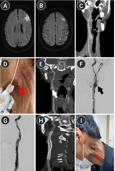
Carotid artery (CA) aneurysms are rare and account for approxi- mately 0.4%–4% of all peripheral artery aneurysms [1]. Pseudo- aneurysms are the most common type of CA aneurysms [2].

Trauma is the most common cause of CA pseudoaneurysm, fol- lowed by surgery and/or radiotherapy for head and neck cancer [3]. Pseudoaneurysms are not covered by healthy, well-vascular- ized tissue and can therefore rupture easily [4]. Carotid blowout

Endovascular treatment for

pseudoaneurysm after carotid blowout syndrome

Chong Hyuk Chung, MD 1 ; Young-Nam Roh, MD, PhD 2 ; Seo Hyeon Lee, MD 3 ; Yeong Seok Jeong, MD 3 ; Jeong-Ho Hong, MD, PhD 3 ; Sung-Il Sohn, MD, PhD 3 ; Hyungjong Park, MD 3

1

Division of Rheumatology, Department of Internal Medicine, Wonkwang University College of Medicine, Iksan, Republic of Korea

2

Department of Surgery, Keimyung University School of Medicine, Daegu, Republic of Korea

3

Department of Neurology, Keimyung University School of Medicine, Daegu, Republic of Korea

CASE REPORT

Received: September 24, 2020 Revised: November 9, 2020 Accepted: November 26, 2020 Corresponding Author:

Hyungjong Park, MD

Department of Neurology, Keimyung University School of Medicine, 1035 Dalgubeol-daero, Dalseo-gu, Daegu 42601, Korea

Tel: +82-53-258-7747 Fax: +82-53-258-4380

E-mail: [email protected]

Background: Pseudoaneurysms of the carotid artery are rare and can be both a cause and consequence of carotid blowout syndrome (CBS) in patients with head and neck cancer. Surgical or endovascular treatment approaches may be useful for managing this condi- tion.

Case Report: A 55-year-old man presented with a pulsatile mass surrounding the carotid bifurcation in the neck. He reported a histo- ry of CBS that occurred as a surgical complication while removing a benign neck cyst. Additionally, he reported a history of laryngeal cancer that underwent complete remission after surgery and radiotherapy. The mass was diagnosed as a pseudoaneurysm. Surgical treatment proved challenging owing to the patient’s history of neck surgery and radiotherapy; therefore, stent-graft deployment was performed and the absence of pulsations was checked through the neck mass.

Conclusion: Endovascular treatment may be a useful therapeutic strategy in cases of post-CBS pseudoaneurysm in patients undergo- ing surgery and/or radiotherapy for head and neck cancer.

Keywords: Aneurysm, False; Carotid artery injuries; Stents J Neurocrit Care 2020;13(2):115-118

https://doi.org/10.18700/jnc.200026

syndrome (CBS) is the rupture of the CA and its branches, which can cause life-threatening complications due to massive blood loss, as well as neurological complications such as cerebral infarc- tion [5]. A pseudoaneurysm of the CA, while being one of the main known causes of CBS, can also occur as a surgical complica- tion for CBS [1,6].

Pseudoaneurysms are treated using open surgery or with endo- vascular treatments, such as parent vessel sacrifice, using detach- able balloons or coil embolization and stent-graft placement [7,8].

eISSN 2508-1349

115 www.e-jnc.org

© 2020 The Korean Neurocritical Care Society

This is an Open Access article distributed under the terms of the Creative Commons Attribution Non-Commercial License (http://creativecommons.org/licenses/by-nc/4.0/)

which permits unrestricted noncommercial use, distribution, and reproduction in any medium, provided the original work is properly cited.

(2)

Currently, endovascular treatment is regarded as a better alterna- tive to open surgery in selected patients [7]. We report a case of post-CBS pseudoaneurysm that was treated successfully using en- dovascular therapy in a patient with a history of surgery and radio- therapy for laryngeal cancer.

CASE REPORT

A 55-year-old man was diagnosed with laryngeal cancer 23 years prior to presentation; complete tumor resolution was observed after partial laryngectomy and radiotherapy. Approximately 1 year before his visit, a palpable mass surrounding the left carotid bifur- cation was observed. Ultrasonography revealed a subcutaneous, solid, and well-demarcated ovoid mass that distorted the left inter- nal jugular vein thus necessitating excision. The left internal CA (ICA) was injured intraoperatively during neck dissection which resulted in active bleeding that was managed by an emergency pri- mary suture at the site of injury. The following day, the patient was observed to be drowsy, with an altered mental status, and with se- vere dysarthria. Diffusion-weighted magnetic resonance imaging revealed border-zone and cortical infarctions in the left cerebral hemisphere (Fig. 1A and B ). Computed tomography angiography (CTA) showed multifocal long-segment stenosis of the left com- mon CA (CCA) (Fig. 1C). Volume replacement was performed in addition to blood pressure control to maintain cerebral perfu- sion pressure. The patient showed complete recovery a month lat- er and was discharged with aspirin (100 mg), clopidogrel (75 mg), and atorvastatin (80 mg) for multifocal stenosis extending along the CCA to the ICA. Histopathologic findings of the neck mass revealed benign soft tissue without any evidence of inflam- mation or tumor cells. No bleeding or abnormal vessel formation was observed at the site of CBS; the patient was therefore instruct- ed to follow up with the outpatient department.

During follow-up, a pulsating mass was detected in the vicinity of the previous site of injury (Fig. 1D). This mass gradually in- creased in size and was clearly identified a year after the develop- ment of the CBS. CTA revealed a new pseudoaneurysm immedi- ately beneath the skin in addition to multifocal long-segment ste- nosis of the left CCA (Fig. 1E). Because of the patient’s history of partial laryngectomy and radiotherapy, we expected that perform- ing surgical ligation would be technically challenging; therefore, a self-expandable stent graft was used to treat the post-CBS pseudo- aneurysm. We inserted a 10-F sheath through the right femoral artery and an 8-F guiding catheter (Shuttle; Cook, Bloomington, IN, USA) was into the proximal portion of the left CCA. Left CCA angiography revealed a pseudoaneurysm at the carotid bulb with multifocal long-segment stenosis of the left CCA (Fig. 1F).

A microcatheter (Headway 17; Microvention, Tokyo, Japan) was placed at the orifice of the external CA (ECA) and coil emboliza- tion (Tornado, Cook) of the left ECA was performed to prevent a type II endoleak after stent placement. A 0.035-inch guidewire was placed at the petrous portion of the left ICA after confirming that coil embolization had completely blocked blood flow through the ECA. A 9× 60-mm self-expandable stent graft (Covera Plus;

Bard Medical, Covington, GA, USA) was selected based on the dimensions of the affected CA (the graft size was at least 1 mm greater than the diameter of the CCA). The self-expandable stent graft was then advanced over the 0.035-inch guidewire and de- ployed to extend between the distal segment of the left CCA and the proximal ICA, ensuring that the pseudoaneurysm was com- pletely covered. Left CCA angiography was performed after stent graft deployment and showed the absence of a pseudoaneurysm.

We subsequently placed a carotid stent into the stenotic segment of the left CCA, which, if left untreated, could have served as a po- tential source of emboli thereby predisposing the patient to isch- emic stroke. A 9× 50 mm carotid stent (Carotid Wallstent; Bos- ton Scientific, Santa Clara, CA, USA) was advanced over a 0.014- inch microwire (Transend; Stryker, Kalamazoo, MI, USA) and deployed from immediately beneath the orifice of the left proxi- mal ICA, partially overlapping the previously deployed self-ex- pandable stent graft. Left CCA angiography revealed no pseudo- aneurysms, in-stent stenosis, or in-stent thrombosis (Fig. 1G).

Carotid duplex ultrasonography was performed the following day and revealed no stenotic flow, in-stent stenosis, or thrombosis.

Moreover, the patient did not show any postoperative neurologi- cal symptoms. A CTA was performed 7 days postoperatively which confirmed the disappearance of the pseudoaneurysm with good stent patency (Fig. 1H). After 3 months, the pulsatile mass disappeared completely (Fig. 1I).

DISCUSSION

A pseudoaneurysm is defined as a loss of integrity of the three lay- ers of the arterial wall that can result in a contained rupture of blood vessels; this differs from a true aneurysm in which the three vascular walls are intact but dilated [9]. Pseudoaneurysms of the ICA are known to present as a growing pulsatile mass in the lateral aspects of the neck [10].

Radiotherapy following head and neck cancer surgery is known as an important cause of CA pseudoaneurysm that can lead to CBS [11]. The CA receives 80% of its blood supply from the vasa vasorum in its adventitia (the outermost layer of the CA) [4]; ra- diotherapy results in injury to the adventitia of the CA and/or its major branches, resulting in the weakening of the arterial wall https://doi.org/10.18700/jnc.200026 116

Chong Hyuk Chung, et al. • Endovascular treatment for pseudoaneurysm

(3)

[5,12]. Thus, radiotherapy itself can be the main cause of pseudo- aneurysm formation in CA. In addition, weakening of the arterial wall due to radiotherapy after head and neck cancer surgery can increase the risk of pseudoaneurysm formation after surgical re- pair for CBS.

Treatment modalities for pseudoaneurysms can be categorized into surgical repair and endovascular treatment [7,8]. Surgical re-

pair involves surgical ligation of the affected ICA followed by vas- cular bypass or anastomosis; this procedure sacrifices the ICA.

Furthermore, surgical ligation can predispose patients to cerebral ischemia, and bypass or anastomosis cannot be performed when the aneurysms are located near the skull base. Additionally, the open access approach is challenging in patients who previously underwent surgery or radiotherapy for head and neck cancer [8].

Endovascular treatment involves deconstructive methods, such as occlusion of the affected ICA using a balloon or coil, and recon- structive methods, such as stent-graft placement [5,7]. Because stent-graft placement is less invasive and has lower complication rates [13], it may be a useful strategy for managing post-CBS an- eurysms especially in patients with a history of surgery and/or ra- diotherapy for head and neck cancer. Nevertheless, the following factors should be considered prior to stent-graft placement for CBS: (1) it is necessary to assess the patient’s anatomical suitabili- ty for the procedure, as this method is only feasible in patients without excessive vascular tortuosity, to ensure easy access for the stent delivery system, and in those with patent femoral or iliac ar- teries to ensure placement of a large-caliber (10–11F) vascular sheath [14]; (2) stent-grafts are more thrombogenic than bare stents and warrant long-term aggressive antiplatelet therapy which involves a high risk for bleeding complications [15]; and (3) en- doleak is a common complication of stent-graft placement. Spe- cifically, reconstitution of the proximal branches of the ECA for lesions in the vicinity of the carotid bifurcation is associated with a risk for developing a type II endoleak from collateral vessels [16]; however, embolization of the main trunk of the ipsilateral ECA can reduce this risk [14].

In our case, the patient was relatively young and had a favorable vascular anatomy and was therefore considered to be a suitable candidate for endovascular treatment. In addition, severe adhe- sions were detected intraoperatively between the CA and the sur- rounding connective tissue, which occurred at the time of CBS development. Based on these findings, we performed endovascu- lar treatment for a post-CBS pseudoaneurysm. Furthermore, we performed ECA embolization to prevent a type II endoleak.

In conclusion, the anatomical constraints associated with surgi- cal treatment of post-CBS pseudoaneurysm make endovascular treatment an appealing alternative. This case illustrates the useful- ness of stent-graft placement in treating post-CBS pseudoaneu- rysms.

ARTICLE INFORMATION

Ethics statement

This study was reviewed and approved by the Institutional Re-

B

D

G

F

I

A C

E

H

Fig. 1. (A, B) Cortical and border-zone infarctions in the left cerebral hemisphere. (C) Multifocal long-segment stenosis of the left common carotid artery extending to the proximal internal carotid artery on computed tomography angiography.

(D) A pulsating mass in the vicinity of the previous injury site of the carotid artery (red arrow). (E) Pseudoaneurysm immediately beneath the skin (white arrow). (F) Pseudoaneurysm (black arrow) with long-segment stenosis of the left common carotid artery on digital subtraction angiogram. (G) Non-visualization of pseudoaneurysm after deployment of a self-expandable stent graft. (H) The disappearance of the pseudoaneurysm with good stent patency on computed tomography angiogram 7 days after endovascular treatment. (I) The disappearance of the pulsatile mass in the vicinity of the previous injury site of the carotid artery (red arrowhead) after 3 months.

117

https://doi.org/10.18700/jnc.200026

(4)

view Board of Keimyung University Dongsan Hospital (IRB No.

2020-09-068). The requirement to obtain informed consent was waived by the Board.

Conflict of interest

Dr. JH Hong is an editorial board member of the journal but was not involved in the peer reviewer selection, evaluation, or decision process of this article. There are no other potential conflicts of in- terest relevant to this article to declare.

Funding

This work was supported by a National Research Foundation of Korea (NRF) grant funded by the Korean government (MSIT) (No. 2020R1G1A007100).

ORCID

Chong Hyuk Chung https://orcid.org/0000-0001-5806-0733 Young-Nam Roh https://orcid.org/0000-0002-0799-1578 Seo Hyeon Lee https://orcid.org/0000-0001-8357-8956 Yeong Seok Jeong https://orcid.org/0000-0003-4778-7413 Jeong-Ho Hong https://orcid.org/0000-0002-8235-9855 Sung-Il Sohn https://orcid.org/0000-0002-6900-1242 Hyungjong Park https://orcid.org/0000-0002-6112-2939 Author contributions

Conceptualization: HP, Data curation: CHC, YNR, SHL, YSJ, JHH, SIS, Writing–original draft: CHC, HP, Writing–review &

editing: HP.

REFERENCES

1. Faggioli GL, Freyrie A, Stella A, Pedrini L, Gargiulo M, Taranti- ni S, et al. Extracranial internal carotid artery aneurysms: results of a surgical series with long-term follow-up. J Vasc Surg 1996;23:587-94.

2. Bellosta R, Sesana M, Baglini R, Luzzani L, Talarico M, Sarcina A. Endovascular treatment of a symptomatic carotid artery an- eurysm with a stent graft. Vasc Endovascular Surg 2008;42:276- 8.

3. Li Z, Chang G, Yao C, Guo L, Liu Y, Wang M, et al. Endovascu- lar stenting of extracranial carotid artery aneurysm: a systematic review. Eur J Vasc Endovasc Surg 2011;42:419-26.

4. Citardi MJ, Chaloupka JC, Son YH, Ariyan S, Sasaki CT. Man- agement of carotid artery rupture by monitored endovascular therapeutic occlusion (1988-1994). Laryngoscope 1995;105:

1086-92.

5. Chaloupka JC, Putman CM, Citardi MJ, Ross DA, Sasaki CT.

Endovascular therapy for the carotid blowout syndrome in head and neck surgical patients: diagnostic and managerial consider- ations. AJNR Am J Neuroradiol 1996;17:843-52.

6. Lee CW, Yang CY, Chen YF, Huang A, Wang YH, Liu HM. CT angiography findings in carotid blowout syndrome and its role as a predictor of 1-year survival. AJNR Am J Neuroradiol 2014;

35:562-7.

7. Troutman DA, Mohan CR, Samhouri FA, Sohn RL. Endovas- cular repair of carotid artery pseudoaneurysm after carotid end- arterectomy with self-expanding covered stents-a long-term fol- low-up. Ann Vasc Surg 2010;24:954.e13-6.

8. Attigah N, Külkens S, Zausig N, Hansmann J, Ringleb P, Hakimi M, et al. Surgical therapy of extracranial carotid artery aneu- rysms: long-term results over a 24-year period. Eur J Vasc Endo- vasc Surg 2009;37:127-33.

9. Pearson SE, Choi SS. Pseudoaneurysm of the internal carotid artery: a case report and review of the literature. Arch Otolaryn- gol Head Neck Surg 2005;131:454-6.

10. Grossi RJ, Onofrey D, Tvetenstrand C, Blumenthal J. Mycotic carotid aneurysm. J Vasc Surg 1987;6:81-3.

11. Chaloupka JC, Roth TC, Putman CM, Mitra S, Ross DA, Low- licht RA, et al. Recurrent carotid blowout syndrome: diagnostic and therapeutic challenges in a newly recognized subgroup of patients. AJNR Am J Neuroradiol 1999;20:1069-77.

12. Lesley WS, Chaloupka JC, Weigele JB, Mangla S, Dogar MA.

Preliminary experience with endovascular reconstruction for the management of carotid blowout syndrome. AJNR Am J Neuroradiol 2003;24:975-81.

13. Bond KM, Brinjikji W, Murad MH, Cloft HJ, Lanzino G. Endo- vascular treatment of carotid blowout syndrome. J Vasc Surg 2017;65:883-8.

14. Chang FC, Lirng JF, Luo CB, Guo WY, Teng MM, Tai SK, et al.

Carotid blowout syndrome in patients with head-and-neck can- cers: reconstructive management by self-expandable stent- grafts. AJNR Am J Neuroradiol 2007;28:181-8.

15. Tielliu IF, Verhoeven EL, Zeebregts CJ, Prins TR, Span MM, van den Dungen JJ. Endovascular treatment of popliteal artery aneurysms: results of a prospective cohort study. J Vasc Surg 2005;41:561-7.

16. Chang FC, Lirng JF, Luo CB, Wu HM, Guo WY, Teng MM, et al. A self-expandable stent overlapped with a stent-graft as a cause of endoleak in a patient with carotid blowout syndrome.

Clin Radiol 2008;63:948-51.

https://doi.org/10.18700/jnc.200026 118

Chong Hyuk Chung, et al. • Endovascular treatment for pseudoaneurysm

수치

Fig. 1. (A, B) Cortical and border-zone infarctions in the left  cerebral hemisphere. (C) Multifocal long-segment stenosis  of the left common carotid artery extending to the proximal  internal carotid artery on computed tomography angiography

참조

관련 문서

Therefore depending on the size of the fracture in the veneering porcelain, intraoral repair methods using surface treatment and composite resins may

Therefore, head and neck massage is an exercise program that is effective for brain function activation and stress factors in middle-aged women, and it is judged that it can be

treat wide wide wide wide necked necked necked necked aneurysms aneurysms aneurysms aneurysms that that that that selection selection selection selection of of

Serum uric acid levels and risk for vascular disease in patients with metabolic syndrome... Prevalence if the metabolic syndrome in a Turkish

For my study, this being diagnosed with prostate cancer receiving treatment in patients with complementary and alternative therapies for their experience

In this study, two types of treatment methods, ozone treatment and combination of alkaline and ozone treatment, was used for character's variation and

Bismuth-containing quadruple therapy as second-line treatment for Helico- bacter pylori infection: effect of treatment duration and antibiotic resistance on the eradication

Improvement in Gastrointestinal Symptoms After Cognitive Behavior Therapy for Refractory Irritable Bowel Syndrome.. Mindfulness-based therapies in the treatment