137
Analysis of Condition Changing on Dose Variation using Intraoral Radiation Devices
Youngjae Kim*, Yongtak Lee**, Hyeoncheol Song**
Dept. of Radiological Technology Gwangyang Health College*, Dept. of Dental Hygiene Gwangyang Health College**
구내 촬영용 방사선 장치의 촬영조건에 따른 선량변화 분석
김영재*, 이용탁**, 송현철**
광양보건대학교 방사선과*, 광양보건대학교 치위생과**
Abstract
This study is investigated dose change on intra-oral radiography when same conditions under the others unit and same unit under the different exposed conditions.
Three different radiation devices were studied. Exposure to the upper anterior, premolar and molar on the variant time and dose measure was using semiconductor radiation dose meter. Obtained film density value was analyzed to the belong in the range of diagnosis.
Results for dose of each region were less dissimilar between the maximum and minimum. Its value was different 10 times as many as 3 times. In addition, the range of film density was 2.10 ∼ 2.95. These values were exceeded on the allow density of diagnostic value '0.25 ∼ 2.0'.
Even if the same device and the same condition, measured dose was considerable differance and film density
was showed show the inappropriate density range. Those can be caused the patient's re-take and patient's diagnostic errors so patients has affected direct and indirect radiological harm. Therefore, dental radiography devices will be required periodical maintenance and also provided standard on the exposure and processing conditions.
Key Words : intraoral radiography, image quality, dose change
요약
본 연구는 구강 내 방사선 촬영 시 동일조건 하에서 촬영장비의 기종을 다르게 한 경우와 동일 기종에서 노출조건을 일정하게 하였을 경우 획득되는 방사선의 선량변화를 알아보고자 하였다.
장비의 기종에 따른 변화를 알아보기 위해 3대의 각기 다른 촬영 장치를 이용하여 상악 전치부, 소구치부, 대구치부 의 3가지의 촬영조건을 이용하여 선량을 측정하였고 장치의 재현성 검사를 위해서 각 장치에 동일한 조건으로 3회 조 사하여 선량변화를 알아보았다. 선량의 측정은 반도체 측정기를 사용하였으며, 필름의 농도값을 획득하여 진단에 유효 적인 범위에 만족하는지 분석해 보았다.
Corresponding Author: Youngjae Kim E-mail:[email protected], Tel: +82-(0)10-5645-6160 Kwangyang Dept. of Radiological Technology,Health College, Gwangyang-eup, Gwangyang-si, Jeollanam-do, Korea
같은 촬영기를 사용하여 실험한 결과 각 부위별로 선량의 차이는 최대값과 최소값이 적게는 3배 많게는 10배의 선 량 차이를 보였다. 흑화도의 범위 또한 적절한 범위인 0.25∼2.0를 초과한 2.10∼2.95의 허용범위를 보였다.
동일한 촬영기로 특정 부위를 같은 조건에서 촬영하더라도 측정된 선량값은 큰 차이를 보였으며 필름의 농도 분석 의 결과 적합한 농도 분포를 보이지 않았던 것으로 나타났다. 이는 환자의 재촬영을 발생시킬 수 있으며 진단시 오류를 일으킬 수 있어 환자의 피폭에 직·간접적인 피해를 일으킬 수 있다. 따라서 치과 방사선 촬영장치 또한 정기점검이 필 요할 것으로 생각되며 표준화된 촬영조건과 현상조건이 제공되어야 한다고 사료된다.
중심단어: 구내촬영, 피폭선량, 선량변화
Ⅰ. Instruction
Medical radiation generating devices are affected diagnosis and treatment of diseases directly or indirectly.
For this reason, systematic management plan should be carried out. Because to be the best image quality is obtained. in this work is QA (quality assurance).[1]
Through it, we acquired that reducing radiation exposure can obtain high-quality of radiation medical imaging, reducing image reading process and preventing retake about the relative error. Through in this action, we can reduce radiation exposure of patients as well as indirect exposure of workers.
Oral and Maxillofacial Radiology Quality Assurance in Radiation adjustment of the device and related equipment in accordance with several studies are underway.[2-12]
American Academy of Oral and Maxillofacial Radiology(AAOMR) by dentists is proposed the standard of radiation protection methods when image aquired and diagnostic imaging, film processing for radiation equipment. This standard quality management is needed on a constant of all radiation equipments.[13] And in Korea, Medical Law Article[4] 'Diagnostic radiation generator on the device's safety management rule', diagnostic radiation generator unit should be performing during installation, measuring and test on every three years.[14] Inspection criteria for diagnosis of radiation generating devices are stated as following. 'Changes factor in dose should be less than 0.05'. But it isn't during the shooting period about reproducibility of the irradiation dose. Thus, we will find out acquired dose whether constant or uncertain on the different models or even the same model under the same
exposure conditions. If radiation exposure is not adequacy for image, the image was not diagnostic data. It may be raise re-take exposure, and patient have redundancy radiation exposure.
This study was investigated the film density of image on same shooting conditions on the different types of inter-oral radiation generator unit under the same condition and different condition on same unit. and than we provided adequate data and standardized on the appropriate oral dose of radiation during image acquisition.
Ⅱ. Materials and Methods
1. Experiment Material1.1 Intra-Oral Radiation Generating Device Radiation generating devices in G college were investigated on exposure under the same tube voltage and current[Table 1]. The testing equipment was quality control and periodically maintenance was performed on December 15, 2011
X-ray machines No. Tube current Tube voltage(Fix)
MAX-GLS-01 1 10 mA 60 kVp
MAX-GLS-02 1 10 mA 60 kVp
STANDARD
DENTAL X-RAY 1 10 mA 60 kVp
Total 3 10 mA 60 kVp
[Table 1] The surveyed dental x-ray machines
We used the dosimetry matrials with Semiconductor Dosimetry (MiniTrace γ, SAPHYMO , Germany) and GX-770 (Gendex, Des Planies, USA).
The film we used Insight (Kodak Medical, NY, USA) and used prosessor is XP-400 (DONGA, Korea, Auto Film Processor) and used Statistical Analysis was Mann-Whitney test its data was statistic value (p-value) less than 0.01 is be statistically significant. and film densitometer (Model RPSM110905RT6, Newconic Co, USA) we used.
2. Experiment Method
2.1 Dosimetry
The distance from the radiation generating devices' source (focal spot) to radiation dose meter was 42 cm.
Using MAX-GLS-01, measured the dose on upper anterior maxilla, premolar region and molar region under selecting the each of the area. The exposure time of MAX-GLS-02 was 0.4, 0.6 and 0.8 (time) and STANDARD DENTAL X-RAY was selected 0.7, 0.9 and 1.2 (time). These three different exposure conditions were measured five times. and then measured dose used by Semiconductor measurement MiniTrace γ (SAPHYMO , Germany)
2.2 Film Density Measurement
Each of the equipment exposed to the same conditions was measured the maximum, average, minimum dose, reproduced by GX-770 (Gendex, Des Planies, USA)[Table 1]. and Dental film (Insight, Kodak Medical, NY, USA) was exposed to each of the three sheets. Exposured film was developed by using auto processing device XP-400 (DONGA, Korea) then, film density measured by film densitometer (Model RPSM110905RT6, Newconic Co, USA) on the 5 random side places of film. The value was evaluated average of five films density, and than obtained the maximum, average, minimum density of the film, according to the different kind of device type.
2.3 Statistical Analysis
Compared the exposure of mouth on same kind of oral radiation generating devices under the conditions of exposure maximum, average and minimum. and using 'Mann-Whitney test' was compared that exposure conditions on the different oral radiation devices according to the maximum, average, and minimum exposure.
Ⅲ. Result
1. Dose of Devices' Type1.1 MAX-GLS-01
The dose was measured that upper anterior maxilla's value was 1,476 ± 850 μGy, premolar region was 1,957
± 452 μGy, and molar region was 2,095 ± 1,012 μGy these numerical value means the average and standard deviation. Measured maximum dose was upper anterior maxilla 3,025 μGy, premolar region 3,951 μGy, and molar region was 4,685 μGy. The minimum dose upper anterior maxilla was 224 μGy, premolar region was 263 μGy, and molar region was 386 μGy[Table 2]. Even so, between the same device and exposure conditions, exposure dose was up to 12 ~ 15 times by comparison the minimum dose.
Exposure time *
Exposure dose (μGy)
Mean ± SD Minimum Maximum
Upper anterior
maxilla 1,476 ± 850 224 3,025
Upper premolar 1,957 ± 452 263 3,951
Upper molar 2,095 ± 1,012 386 4,685 Table 2. Surface exposure dose of MAX-GLS-01
(* : Manufacture’s guide)
1.2 MAX-GLS-02
The MAX-GLS-02 was exposing to 0.4 (time) condition. The dose were measured 412 ± 105 μGy, 658
± 230 μGy on 0.6 (time), and 875 ± 325 μGy on 0.8 (time) these numerical value means the average and standard deviation. Measured dose were 691 μGy when 0.4, 1,315 μGy when 0.6 and 1,457 μGy when 0.8. The minimum dose were 202 μGy when 0.4, 170 μGy when 0.6 and 178 μGy when 0.8[Table 3]. MAX-GLS-02's exposure condition in the period was constancy, the maximum dose to 3 ~ 8 times by comparison the minimum dose.
Exposure time
Exposure dose (μGy)
Mean ± SD Minimum Maximum
0.4 412 ± 105 202 691
0.6 658 ± 230 170 1,315
0.8 875 ± 325 178 1,457
Table 3. Surface exposure dose of MAX-GLS-02
1.3 STNADARD DENTAL X-RAY
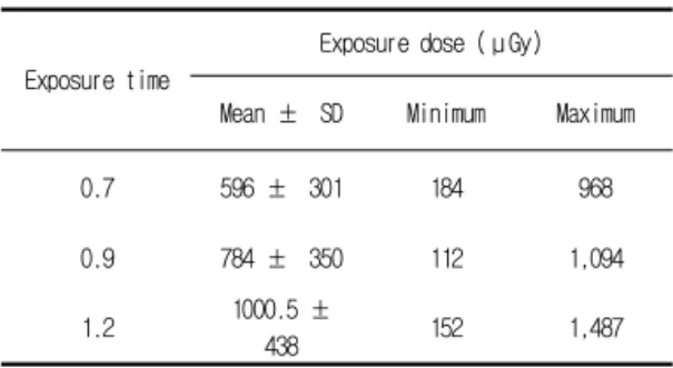
The STANDARD DENTAL X-RAY exposed to 0.7 (time) condition. The dose was measured 596 ± 301 μGy, 784 ± 350 μGy on 0.9 (time), and 1000.5 ± 438 μGy on 1.2 (time) these numerical value means the average and standard deviation. The maximum dose measured 968 μ Gy when 0.7, 1,094 μGy when 0.9 and 1,487 μGy when 1.2. The minimum dose were 184 μGy when 0.7, 112 μ Gy when 0.9 and 152 μGy when 1.2(Table 3).
STANDARD DENTAL X-RAY of X-ray exposure conditions in the period was constancy, the maximum dose to 5 ~ 10 times by comparison the minimum dose.
Exposure time
Exposure dose (μGy)
Mean ± SD Minimum Maximum
0.7 596 ± 301 184 968
0.9 784 ± 350 112 1,094
1.2 1000.5 ±
438 152 1,487
Table 4. Surface exposure dose of STANDARD DENTAL X-RAY
2. Film Density of Devices' Type
The MAX-GLS-01's mean value of maximum, average, minimum film density was 4.12, 3.75 and 1.09.
MAX-GLS-02 was 3.44, 2.84 and 0.97, and STANDARD DENTAL X-RAY was 2.22, 1.39 and 0.02 [Table 5]. Film density was compared maximum and average, average and minimum, maximum and minimum values using Mann-Whitney test (when p<0.01, the data was statistically significant.) it was no statistically significant difference in every model (p <0.01).
Dental x-ray machine
Exposure dose
Radiographic density (Mean ± SD) p-Value
MAX-GLS-01
Maximum 4.12 ± 0.11
* Mean 3.75 ± 0.31
Minimum 1.09 ± 0.02
MAX-GLS-01
Maximum 3.44 ± 0.05
* Mean 2.84 ± 0.01
Minimum 0.97 ± 0.02
STANDARD DENTAL X-RAY
Maximum 2.22 ± 0.03
* Mean 1.39 ± 0.03
Minimum 0.02 ± 0.00
(*: p<0.01, by Mann-Whitney test) Table 5. Film densities
Ⅳ. Discussion
The quality of radiographic images was an important factor to consideration. Because images, obtained from patient, has affect to the diagnosis and future treatment of patient. In addition, unnecessary radiation dose should be incur increasing patient exposed dose when obtaining image and cause unacceptability clinically image quality, as a result it bring re-take to patient. It was result to increase the amount of radiation exposure dose of patient. To overcome those things, required the regular quality control of radiation generating device.[3] However, the dental radiation device's life was different. and Film processing was not consistency. These overcome made patients higher dose more than investigated.[16,17]
The study of Yakoumakis study said that general used 52 radiation device's dose has different each of them by measuring the average surface dose.[4] It was due to random -based their experience- conditions when getting image even if same model device, and exposure conditions such as time, tube voltage, tube current has not been standardized. Additionally, Farman's research of 108 dental clinics said that exposure time was used various, despite of same phantom.[15] It means that workers had not using the recommended exposure time in clinical practice but using of their knowledge and experience when determine the exposure time. Therefore, improving image quality and reducing patient's exposed dose was need to standardize on the shooting conditions and film processing. Several studies show that exposure conditions when acquiring image and prosessing using by their experience.[4,16] Jung's study said that the percent of turnover rate workers -dental hygienist- was 54% in survey of 447 dental hygienists.[18] It means that workers, turnover of the previous dental clinic, were determine the shooting conditions based on the their experience as former place.
Gang claimed that even small amount of radiation dose has incur the probabilistic risk of radiation resided in the critical organs, cancer or genetic effects.[19] It means that
small amount of radiation has incur to the chronic changes in biological tissues, organs furthermore cause abnormalities. Exposure conditions were set differently for each site(upper anterior maxilla, upper premolar, upper molar). so the recording value was different[Table 2].
According to this study by the same dose of exposure measured under constant conditions was various range of value[Table 2, Table 3, Table 4]. Although the dose was 3
~ 10 times difference even if same kind of radiation generating device's exposure conditions such as tube voltage, tube current, exposure time, radiation source - measuring instrument distance were constant. Professor of Oral and Maxillo-facial Radiology Council of Korea recommended that diagnosable appropriate film density was 1.75, the range of density is 0.25 ~ 2.[20] In this study, a range of radiographic density was 2.10 to 2.95. It was exceeded on the acceptable range. It means those images can be caused diagnostic errors when reading.
Ⅴ. Conclusion & Result
Exposure radiation dose can be varied under the circumstances by being used radiation device and a new device, or the same model of radiation exposure. It cause by period the purchase of equipment and equipment's aging. It means that the dental hygienists, turnover working place, are exposure their experience in the past, it might be incur inappropriated diagnosic errors and needed to re-take the patient. Although the obtained dose value was very tiny, radio-biological harms on probabilistic effect - it has no threshold dose-. so, should be avoid the unnecessary exposure. It could be increase the risk from radiation. Re-take means that increase unnecessary radiation exposure dose to patients. Therefore, we aware although exposure conditions and radiation unit were same, exposed patient dose was different. and we should be determine appropriate exposure conditions for reducing exposed patient dose and accept high quality radiography.
To do this, we should be need to the standardized appropriate exposure and processing conditions of device,
before using radiation generating devices.
The Study's limitations were that the testing devices were only on-campus. With reference to this article, in clinical studies should be conducted.
Reference
[1] 대한구강악안면방사선학교수협의회. 방사선 촬영장비 및 관
련기기의 관리. 구강악안면방사선학. 제3판. 서울: 나래출판 사, pp.60-7, 2001.
[2] Geist JR, Katz JO. Radiation dose-reduction techniques in North American dental schools. Oral Surg Oral Med Oral Pathol Oral Radiol Endod Vol. 93, pp.496-505, 2002.
[3] Platin E, Ludlow JB. Knowledge and adoption of radiographic quality assurance guidelines by general dentists in North Carolina. Oral Surg Oral Med Oral Pathol Oral Radiol Endod Vol. 79, pp.122-6, 1994.
[4] Yakoumakis E, Tierris C, Tsalafoutas I, Stefanou E, Panayotakis G, Proukakis C. Quality control in dental radiology in Greece.
RadiatProt Dosim; Vol. 80, pp.89-93, 1998.
[5] Farman AG , Hines VG . Radiation safety and quality assurance in North American dental schools. J Dent Educ Vol 50, pp.304-8, 1986.
[6] Jensen OE , Handelman SL, Iker HP. Use and quality of bitewing films in private dental offices. Oral Surg Oral Med Oral Pathol Vol. 63, pp. 249-53, 1987
[7] Goren AD, Sciubba JJ, Friedman R, Malamud H. Survey of radiologic practices among dental practitioners. Oral Surg Oral Med Oral Pathol Vol. 67, pp.464-8, 1989.
[8] Kantor ML, Hunt RJ, Morris Al. An evaluation of radiographic equipment and procedures in 300 dental offices in the United States. J Am Dent Assoc Vol. 120, pp.547-50, 1990.
[9] Nakfoor CA, Brooks SL. Compliance of Michigan dentists with radiographic safety recommendations. Oral Surg Oral Med Oral Pathol Vol. 73, pp.510-513, 1992.
[10] Bohay RN, Kogon SL, Stephens RG. A survey of radiographic techniques and equipment used by a sample of general dental practitioners. Oral Surg Oral Med Oral Pathol Vol. 78, pp.806-810, 1994.
[11] Platin E, Janhom A, Tyndall D. A quantitative analysis of dental radiography quality assurance practices among North Carolina dentists. Oral Surg Oral Med Oral Pathol Oral Radiol Endod Vol. 86, pp.115-20, 1998.
[12] Tugnait A, Clerehugh V, Hirschmann PN. Radiographic equipment and techniques used in general dental practice: a survey of general dental practitioners in England and Wales. J Dent Vol. 31, pp.197-203, 2003.
[13] Quality Assurance Committee of American Academy of Dental Radiology. Recommendations for quality assurance in dental radiography. Oral Surg Oral Med Oral Pathol Vol. 55, pp.421-423, 1983.
[14] 대한민국 의료법 제32조의 2. 진단용 방사선 발생장치의 안 전 관리에 관한 규칙 및 진단용방사선발생장치의 검사 기준
[15] Yakoumakis EN, Tierris CE, Stefanou EP, Phanourakis IG, Proukakis CC. Image quality assessment and radiation dose in intraoral radiography. Oral Surg Oral Med Oral Pathol Oral Radiol Endod Vol. 91, pp362-368, 2001.
[16] Conney P, Gavin G, Rajan J, Malone JF. Radiation protection problems with dental radiological equipment. Radiat Prot Dosim Vol. 57, pp.339-342, 1995.
[17] Button TM, Moore WC, Goren AD. Causes of excessive bitewing exposure: Results of a survey regarding radiographic equipment in New York. Oral Surg Oral Med Oral Pathol Oral Radiol Endod Vol. 87, pp.513-517, 1999.
[18] 정연화. 치과위생사의 이직 결정에 영향을 미치는 요인에 관 한 연구. 한국치위생교육학회지 Vol. 3, pp. 183-194, 2003.
[19] 강성숙, 조봉혜, 김현자, 두부규격방사선사진 촬영시 주요장 기의 등가선량, 유효선량 및 위험도, 대한구강악안면방사선 학회지, Vol. 25, No. 2, pp.309-318, 1995.
[20] 대한구강악안면방사선학교수협의회. X선 필름, 증감지 및 격 자,구강악구강악안면방사선학. 제3판. 서울: 나래출판사 pp.26-40, 2001.