Physiologic and molecular regulation of amniotic fluid - Intramembranous absorption -
인제대학교 의과대학 일산백병원 산부인과학교실
최 형 민
임신중 양수의 기능은 태아를 보호하는 공급원(reservoir)로서 정상 양수량은 태아의 정상 발달에 필수적이다. 사람에 있어 양수과소증은 태아 폐발육부전증(lung hypoplasia)과 같은 태아 기형이나 자궁내 태아발육지연 등과 연관되어 있 으며, 또한 양수과다증은 폐쇄성 태아 기형, 조기진통 등과 관련되어 있다.1,2 따라서 양수량의 이상은 주산기 이환률 과 사망률 증가와 밀접한 관계가 있다. 그러나 이와 같은 중요성에도 불구하고 임신중 정상 양수량의 유지를 조절하 고 있는 모체태아태반계(maternal-fetal-placental unit)의 기능은 아직까지 잘 이해되지 못하고 있다. 따라서 여기서는 이 와 같은 양수의 양을 조절하고 있는 기전의 최신지견에 대하여 알아보고자 한다.
I. 양수의 생성과 흡수(Dynamics of amniotic fluid)
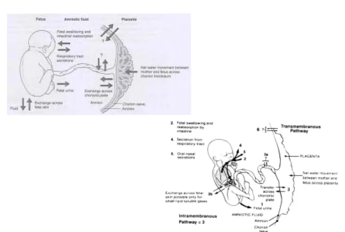
임신초기의 배아(embryo)는 피부가 4세포층(4-cell layers)으로 구성되어 있고 각화가 되어 있지 않아서 태아 피부를 통해 조직액과 용해물질이 양막강으로 전달되어 양수의 성분은 태아의 조직액과 유사하다. 한편 양수가 양막강으로 들어가고 나오는 데에는 여러 가지 경로가 있지만 크게 네 가지로 볼 수 있다(Figure 1). 즉 양수의 생성에는 태아의 소변(fetal urine)과 태아 폐의 지방성 분비물(lung liquid secretion)이 두 가지 중요한 경로이고, 양수의 흡수에는 태아가 양수를 삼키는 것(swallowing)과 태반의 막을 통하여 태아의 혈액 내로 이동하는 것(막내흡수: intramembranous absorption)이 두 가지 중요한 경로이다.3
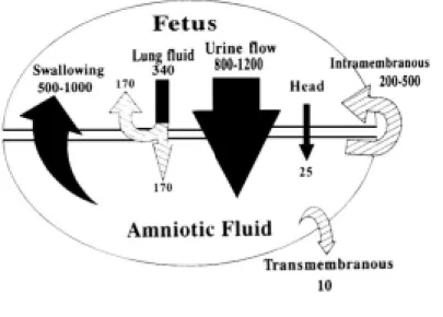
태아의 소변은 오랫동안 임신 후반기의 양수에 중요한 구성요소로 여겨져 왔으며 이는 무신장(agenisis of kidney)이나 요로폐쇄 등의 기형이 있는 태아에서 임신 제 2삼분기 중반 이후에는 양수량이 거의 없는 것에 기인하였다. 약 임신 12주 경부터 태아의 신장에서 소변을 만들어 양막강 내로 배설을 시작하고 이후 점차적으로 양수의 양은 증가하여 임신 36주의 만삭 경에는 약 700-1000 mL에 이른다. 그러나 만삭이 더 가까워지면 오히려 양수의 양은 감소하며 임신 이 분만 예정일을 지나게 되면 그 양은 더욱 적어지게 된다(Figure 2).3 태아의 신장은 독특한 특성을 갖고 있는데 네프 론의 형성(nephrogenesis)과 사구체의 발달(glomerularization)은 임신 후반기에 완성되어 신장 1 gm 혹은 체중 1 kg 당 태 아 사구체 여과율(fetal glomerular filtration rate)은 신생아 혹은 성인의 30% 정도 밖에 안된다. 그러나 하루에 정상 태아 의 소변 생성속도는 임신 후반기의 총 양수양(600-1000 mL)보다 크다(800-1000 mL). 이러한 태아의 소변 생성속도는 여과된 물의 약 20%가 배출된다는 것을 반영하며 신생아나 성인에서 약 1-2% 정도가 배출되는 것과 크게 구별된다.4 이는 근위세뇨관(proximal tubule)의 미성숙으로 인한 재흡수가 감소되어 있기 때문이며, 이와 같은 사구체 여과율과 근 위세뇨관의 재흡수율은 분만 직후부터 즉시 크게 상승하게 된다. 또한 이 두 가지 신장 기능의 성숙은 분만 전에도
glucocorticoid의 투여로 유도될 수 있다.5
Fig. 1. Schematic showing the 6 routes for fluid entry into and exit from the amniotic sac for the late gestation fetus. (Am J Obstet Gynecol 1980;138:575-86)
Fig. 2. Nomogram showing amniotic fluid as a function of gestational age.
(Reproduction with permission from Brace RA, Wolf EJ. Normal amniotic fluid volume changes throughout pregnancy. Am J Obstet Gynecol 1989;161:382-8)
태아의 폐에서도 분비물을 만드는데 이는 태아의 폐 성숙에 중요할 뿐아니라 양수의 생성에도 많은 기여를 한다.
과거에는 태아의 폐가 양수를 흡수하는 것으로 알려져 있었으나 최근의 많은 연구에서 많은 양의 분비물이 폐에서 생성되며 이는 진통과 분만 중에 감소하고,6 오직 태아 저산소증이나 심한 가사가 있을 때에만 arginine vasopression, cathecholamine, cortisol과 같은 호르몬 등의 영향으로 양수를 흡수한다는 사실이 밝혀졌다. 임신 후반기 1/3에서 1/2 동 안 태아 폐액의 분비량은 태아 무게 1 kg 당 100 mL/day 정도이며 이는 태아 폐의 성장에 필요한 양보다 약 100배 많은 양이다. 분비액은 기관을 통하여 폐를 빠져나가며 이는 태아의 호흡운동이 있을 때 더욱 두드러진다.7 분비액의 일부가 양수내로 유입된다는 것은 양수내에 폐의 계면활성제가 존재함으로 입증되었고 이를 임상적으로 태아 폐성숙 의 지표로도 사용할 수 있다. 최근의 양(sheep)을 통한 여러 동물실험연구에 의하여 분비된 폐 분비액의 약 절반 정도 가 양수 내로 유입되며 나머지 반은 다시 태아가 삼키는 것을 알게 되었다.
태아의 위는 임신 약 11주 경에 액체로 팽만되어 있으며 임신 18주 경에 연하운동을 관찰할 수 있고8 그 양은 과거 에는 200-1500 mL/day로 측정되기도 했으나, 최근의 보고에 의하면 하루에 태아 몸무게의 약 20-25%의 양수를 삼키는 것으로 밝혀졌다. 삼키는 양수의 양은 태아의 몸무게에 비례하여 증가하며(100-300 mL/kg/day), 그 양은 정상 성인의 경우(40-60 mL/kg/day)보다 많다. 임신 초기에는 연하운동이 어떻게 조절되는 지는 알 수 없으나, 임신 제 3삼분기에는 태아의 중추신경계와 구갈성 기전(dipsogenic mechanism)이 중요한 역할을 하고 있다. 즉 태아 혈장 삼투압이 증가하면 연하가 증가되고, 반대로 급격한 저혈압이나 저산소증은 태아의 연하(swallowing)를 감소시킨다.9-11 한편 태아에게 angiotensin II를 주입하거나, 모체의 금식, 혹은 모체에 당(glucose)주입 등에 의해서는 태아의 연하운동은 영향을 받지
않았다.12,13 위장관 폐쇄나 신경학적인 이유로 태아의 연하운동이 크게 감소하거나 없어진 경우에는 양수과다증이 유
발될 수 있다.
한편 태아의 식도폐쇄가 있으면서도 양수과다증이 없는 경우에는 종종 기관-식도 누공(tracheoesophageal fistula)이 발견되는데 이를 통하여 양수가 위로 들어갈 수 있다. 그러나 단독적으로 식도폐쇄만 있는 경우에도 약 15%
에서는 양수과다증이 발견되지 않는데 이는 소변과 폐액이 꾸준히 생성되고 있는 점을 감안한다면 양수량의 조절에 있어 막내흡수의 역할을 명백히 반영하고 있는 것을 알 수 있다.14
이밖에 적은 양이지만 태아의 비강에서 분비되는 액체(oro-nasal secretion)와 경막양수이동(transmembranous pathway) 즉 양막, 융모막, 자궁벽을 통한 모체 혈관으로 이어지는 경로도 양수량의 조절에 관여하는 것으로 알려져 있다(Figure 3).3
Fig. 3. Summary of water flows into and out of the amniotic space in late gestation.
(Modified with permission from Gilbert WM, Brace RA. Amniotic fluid volume and normal flows to and from the amniotic cavity. Semin Perinatol 1993;17:150-7)
II. 막내흡수(Intramembranous absorption)
막내흡수는 양수에 노출된 태아의 혈관을 통하여 양수를 구성하는 수분이나 용질(solute)들이 태아 순환계로 직접 이동하는 경로이다. 이는 주로 태반의 혈관과 같은 양수에 직접 노출된 태반의 태아 측 혈관에서 일어나며, 적은 양은 탯줄이나 태아 피부를 통해서도 이동하게 된다. 삼투압과 양수량의 변화로부터 계산을 해보면 하루에 약 240 mL의 양수가 막내흡수를 통하여 이동하며 시간에 따른 삼투압 차이로 교정을 하면 임신 말기에는 약 하루에 400 mL의 양수 가 이 경로로 흡수되는 것을 알 수 있다.3 이 경로는 주로 막내 투과성(intramembranous permeability)을 결정하는 인자와 표면적(surface area)에 의하여 조절된다.15 투과성이 약간만 변해도 막내 투과속도는 많은 영향을 받는데 예를 들면 태 아 신장, 폐, 양막이나 융모막에서 분비되는 prostaglandin이 양수 내로 유입되면 막내 투과성에 변화를 일으켜 직접적 인 양수량의 변화를 가져온다. 또 하나의 영향 인자로 생각되는 것은 양수와 혈장의 삼투압의 차이로서 다양한 동물 실험들에서 태아의 신장이 많은 양의 수분과 용질을 양막강 내로 전달시키는 능력이 있음을 확인하였고, 다른 연구에 서는 수일 동안의 모체의 탈수상태에서는 양수량이 감소됨을 보고하였다.15 그러므로 수액 주입을 통한 태아 혈장 삼 투압의 감소는 막내흡수를 감소시킨다.15
최근의 보고에 의하면 혈관내피성장인자(vascular endothelial growth factor: VEGF)가 막내흡수에 일차적인 조절을 담당 하는 것으로 보고되고 있다.16 다양한 연구에서 태아의 소변 생성, 태아의 연하작용, 폐액의 생성 등에 의하여 양수량 의 조절이 이루어지고 있다는 것을 보여주었으나, 이는 태아의 필요에 의해 유기적으로 조절되는 것이지 양수량의 회복이나 유지를 위한 조절은 아니므로 오직 막내경로를 통하여서만 양수량은 조절될 수 있는 것이다.4
사람에 있어서 이와 같은 막내이동은 태반을 덮고 있는 태아측 표면인 태반막의 부분과 융모판(chorionic plate)의 태 아측 표면으로 분출되고 있는 태아 미세혈관(fetal microvessels)으로 구성되어 있다. 초기 연구에 의하면 태반의 태아측 표면은 수분의 흡수에 중요한 이동경로로 밝혀졌으며 이것이 간접적으로 사람에서 막내이동의 존재를 증명하는 것이 었다.17 다른 연구에 의하면 사람에 있어서 아미노산의 이동은 다른 고분자단백질들(high-molecular-weight-proteins)의 이 동 정도에 비하여 10배를 나타내었는데 이는 두 물질이 다른 경로로 이동한다는 것을 시사한다.18,19 즉 저분자물질들 (low-molecular-weight substances)은 막내이동뿐 아니라 태아의 연하운동에 의하여 이루어지고, 반면에 고분자물질들은 태아의 연하운동에 의하여 주로 이루어지고 있다는 것이다. 또한 최근의 연구에 의하면 사람의 태반에서 양수내 glucose가 양막부위를 통하여 태아측으로 빨리 이동할 수 있는 것이 밝혀졌다.20 즉 이와 같은 관찰은 막내흡수가 양수 내 glucose의 주이동경로가 된다는 것이다. 최근까지 약 20년 동안 원숭이나 양을 이용한 여러 동물실험에서 이와 같은 막내이동의 과정은 많이 알려지기 시작하였으며, 특히 태아의 연하를 통한 양수의 이동과정을 없앤 후의 양을 통한 여러 동물실험에서 막내이동이 양수의 조절에 중요한 이동경로이며 이는 여러 조건들(모체 수분 공급, 모체 탈수, 저 산소증 유발, 인위적 양수주입, 여러 약제들)의 변화에 따라 양수량의 조절에 관여하는 것이 밝혀졌다. 이와 같이 양수 량의 유지에 중요한 막내흡수를 조절하는 중요한 인자는 태아막과 혈관의 혈관성(vascularity)와 투과성(permeability)을 조절하여야 하는데 최근 들어 앞에서 언급하였던 혈관내피성장인자가 이의 중요한 조절을 담당하고 있는 것으로 시 사되고 있다.21
III. 태반과 태아막에서의 VEGF와 VEGF 수용체
(VEGF and VEGF receptors in placenta and fetal membranes)
혈관내피성장인자는 특히 혈관내피세포에 작용하여 분화와 성장 그리고 이동(migration)을 유발하는 혈관성인자 (angiogenic fator)이다.22 또한 혈관내피성장인자는 단백질의 분출과정(extravasation)을 유도하는 강력한 혈관의 투과성 을 증가시키는 인자이다.23,24 혈관내피성장인자의 생물학적 작용은 두 가지 수용체, 즉 kinase insert domain-containing recreptor (KDR)와 fms-like tyrosine kinase (Flt-1)에 의하여 나타난다.25,26 따라서 혈관내피성장인자가 막내흡수에 중요한 인자라면 태아막, 막내혈관 등에서 이와 같은 수용체가 발현되어야 하는데 사람에서 임신 중 융모세포영양막(villous cytotrophoblasts)과 합포영양막(syncytiotrophoblasts)에서 혈관내피성장인자 mRNA와 VEGF 단백질이 모두 표현되었다.27-29 또한 Flt-1수용체 mRNA와 단백질도 혈관내피성장인자가 존재하였던 융모세포영양막에서 같이 존재하는 것이 확인되
었다.30,31 반면 KDR수용체는 주로 태반의 혈관내피세포에서 표현되었다.31,32 사람의 태반막에서 혈관내피세포 mRNA
와 단백질은 양막(amnion)과 영양막(trophoblasts), 그리고 융모막(chorion)의 결체조직에서 발견되나27 양에서는 양막의 상피와 융모세포영양막에서 주로 표현된다.21,33 또한 KDR수용체도 양막의 상피에서 많이 표현되며 막내혈관의 내피 세포에서도 관찰된다.21,34 이와 같은 표현결과는 임신기간이 만삭에 가까울수록 증가하며 더 많이 표현된다. 따라서 혈관내피성장인자의 작용의 주요 부위는 양막임을 시사하고 있다(Figure 4).21
사람의 혈관내피성장인자 구성의 다른 종과의 차이점은 혈관내피성장인자 peptide 6번째에 glycine이 붙어 있어서 아 미노산이 하나 더 있는 것이다.35 사람과 양에서 혈관내피성장인자는 다양한 분자형태로 나타나는데 VEGF165/
VEGF164가 주 형태이다.36,37 기능성 분석에 의하면 사람에 있어서는 여러 isoform중에서 VEGF121, VEGF145, VEGF165, VEGF189, VEGF206가 중요한 기능을 하고 있는 것으로 알려져 있다.38
Fig. 4. Cellular localization of VEGF (A) and KDR (B) in the fetal membranes of an ovine fetus at 142 days gestation. (Bar, 100 µm; Am, amniotic epithelium; Ch, chorionic cytotrophoblast; ECM, extracellular matrix; fbv, fetal intramembranous blood vessel; sms, vascular smooth muscle around intramembranous blood vessel.)
혈관내피성장인자가 막내흡수를 조절하는 기전은 아직까지 명확하게 밝혀지지는 않았다. 그러나 지금까지의 연구 에 의하면 혈관내피성장인자가 태아의 연하를 제거하였던 동물실험에서 양수과다증이 발현되지 않는 점16과 이는 혈 관내피성장인자에 의한 막내흡수가 증가되어서라고 생각되며, 이 때에 태아막에서 혈관내피성장인자의 유전자 표현 이 증가되어 있는 것(up-regulation)으로 알 수 있다(Figure 5).21 이 과정에서 태아막에서와 달리 태반에서의 혈관내피성 장인자수용체의 mRNA와 KDR은 증가되지 않는 것으로 보아 그 기전은 태반에서의 수용체의 증가에 의한 것이 아니 고 혈관내피성장인자에 의한 막내 혈관들의 혈관내피세포의 성장과 분화가 증가되었기 때문으로 추측된다.21,39,40
Fig. 5. Northern blot showing VEGF up- regulation of its own expression in primary cultures of ovine amnion cells.
자궁동맥의 혈류를 지속적으로 감소시켜서 태아에 저산소증(nonanemic hypoxia)을 유발하는 동물실험41에서 태아의 연하가 감소하고 소변의 배출이 증가하므로 양수의 양은 증가하여야 하나 저산소증의 초기 시기를 지나면 나중에는 결국 양수량은 변화가 없음을 관찰하였다. 이는 결국 다른 경로 즉 막내흡수가 증가되어 나타나는 현상으로 생각할 수 있는데 이와 같은 저산소증은 잘 알려진 혈관내피성장인자 표현의 중요한 자극조건이므로 이와 같은 저산소증이 태아나 모체에 일어날 경우 혈관내피성장인자의 유전자 표현이 증가되어서 막내흡수를 통한 양수의 흡수가 일어나서 생각한 만큼 양수량의 변화는 초래되지 않는다.21 저산소증이 초래되었을 때 양막과 융모막에서의 혈관내피성장인자 mRNA는 보통 때보다 약 5배 정도의 증가가 관찰되었다.39 또 다른 혈관내피성장인자 표현의 자극 인자로는 혈관내의 용적팽창(intravascular volume expansion)을 들 수 있는데 정맥주사로 생리적 식염수를 주입하였던 동물실험에서 예상보 다 적은 양수량의 증가(12.8% of expected volume)가 관찰되었는데 이는 역시 혈관내피성장인자의 표현의 증가로 막내 흡수를 통한 양수량의 조절로 생각된다. 이 때에도 태아막에서 혈관내피성장인자 mRNA가 보통 때보다 약 2-4배 정도 높게 관찰되었다.15,42,43
IV. 태반과 태아막에서의 VEGF의 유도물질
(Mediators of VEGF expression in placenta and fetal membranes)
태반과 태아막에서 혈관내피성장인자의 유전자 표현을 증가시키는 인자들로는 prostaglandins (PG)과 다른 성장인자 들, 즉 epidermal growth factor (EGF), transforming growth factor (TGF), platelet-derived growth factor (PDGF), platelet-activating factor (PAF) 등을 들 수 있다. 이들 인자들은 모두 양막, 태아의 소변, 양수 등에 존재하고 있으며 또한 혈관내피 성장인자의 표현 증가를 유도할 수 있으므로 현재 막내흡수의 조절 인자들로 고려되고 있는 것이다. 지금까지 의 여러 연구에 의하면 이들 인자들은 독립적으로 혹은 상호작용에 의하여 혈관내피성장인자의 유전자 표현을 증가시키는 것으로 알려지고 있다.21 예를 들어 EGF는 사람의 양막에서 PG합성과정의 가장 중요한 효소인 cyclooxygenase-2 (COX-2)를 활성화시키고 PGE2의 합성을 증가시킨다. 또한 PAF는 사람의 태아막과 혈관내피세포에서 COX-2 mRNA를 증가시킬 뿐만 아니라 PGE2의 합성을 증가시킨다.44,45 한편 이와 같은 여러 인자들의 출처로는 현재 태아의 폐나 신장이 고려되고 있으며 이는 양수가 이들 기관에서 만들어지고 있기 때문이다.46 저산소증은 혈관내피 성장인자 유전자 표현의 중요한 자극으로 앞에서 언급하였는데 사람의 제대정맥의 혈관내피세포에서 저산소증 시에 COX-2 유전자 표현의 증가(COS-2 mRNA와 단백질의 증가)가 관찰되었고,47,48 쥐를 대상으로 혈관내피성장인자의 표현 을 유도할 수 있는 저산소증 유발 실험에서도 쥐의 뇌와 폐에서 COX-2 유전자의 표현이 증가되어 있었다.49 따라서 지금까지의 여러 동물실험을 통한 연구를 종합하여 보면 혈관내피성장인자의 유전자 표현을 증가시키는 여러 다양한 조건들에서 혈관내피세포의 직접적인 활성화, 혹은 혈관내피성장인자의 표현을 유도할 수 있는 여러 인자들에 의한 상호작용 등에 의하여 양수량의 조절을 위한 막내흡수가 조절되고 있다고 할 수 있다.21
V. 막내흡수의 조절기전(Mechanism of intramembranous absorption)
막내이동에 관여하는 주요 구성요소는 앞에서 언급하였듯이 태아막과 태반의 태아 측과 연결되어 있는 미세혈관들 이다. 따라서 이들의 구성요소에 따른 이동과정이 막내흡수를 조절하고 있는 것이다. 이들 구성요소에서 혈관내피성 장인자 유전자 표현이 어떻게 활성화되는 지는 현재까지 4가지 정도의 가설이 제기되고 있다(Figure 6).21
Fig. 6. A proposed diagram of the intramem- branous pathway showing possible mechanisms mediating amniotic fluid absorption and the involvement of VEGF and caveolin-1 in the regulation of caveolae transport of fluid through the amnion and chorion.
1) 태아막과 태반의 태아측 표면에서 혈관성이 증가에 의한 막내흡수가 일어날 수 있는 표면적을 증가시킨다 (membrane surface area and angiogenesis) (Figure 7, Figure 8).
Fig. 7. Mitogenic effect of VEGF on ovine amnion cells in primary culture. Proliferation of amnion cells was significantly enhanced by VEGF in a dose-dependent manner from 105 ng/mL over 11-day period of VEGF treatment.
Fig. 8. Growth curves of primary cultures of ovine umbilical vein endothelial cells (OUVEC) treated with VEGF at 5 ng/mL maximally stimulated the proliferation of OUVEC above the control (without VEGF treatment) over a 12-day period.
2) 막내 혈관을 통한 혈류량을 증가시킨다.
3) 수동적 막투과성(passive membrane permeability)을 증가시킨다.
4) 태반막을 통한 소포성 이동(vesicular transfer)의 형태로 bulk fluid transfer를 유도한다.
일반적으로 막내흡수 과정은 양수의 삼투압이나 농도가 태아혈액의 것보다 낮으므로 이 차이로 인한 양막과 융모 막을 통한 수동적인 이동(passive transport)과정으로 주로 생각할 수 있다.50 즉 수분이나 sodium, chloride와 같은 용질(solute)들은 이와 같은 이동과정에 의해 수분은 주로 양수 내에서 태아 측으로, 용질은 반대로 이동하게 된
다.50,51 또한 이 이동과정의 다른 인자로서는 정역학적인 차이(hydrostatic gradient)를 들 수 있는데 여러 논문에서
보면 urea나 chloride같은 용질들이 삼투압의 차이와 반대로 이동하는 것을 관찰할 수 있으며 이는 양막이나 혈관 내피세포의 정역학적인 압력 차이로 인하여 이동된다고 할 수 있다(Figure 9, Figure 10).50-52 이 확산과정(diffusive mechanism)에 혈관내피성장인자가 관여하며 태아막을 통하여 bulk flow 형태의 이동이 있게 된다. 사람을 비롯한 여러 동물들에서 혈관내피세포뿐아니라 태반을 포함한 태아막에서 많은 수의 막성 소포(vesicle) 혹은 caveolae가 발견되는데53 이는 모세혈관 내피세포에 존재하는 60-80 mn 정도의 membrane-bound vesicle이다. 이 caveolae를 통한 단백질이나 수액의 이동기전이 막성이동의 주 기전으로 생각되고 있는데, 혈관내피성장인자의 양수량의 조절과정이 태아막을 통한 소포성이동(vesicular transport)의 유도에 의하여 이루어진다고 한다.21,54 즉 혈관내피세포의 배양실험에 서 보면 혈관내피성장인자와 caveolae의 형성은 양성관계(positive relationship)가 있으며,55 혈관내피성장인자가 모세혈관 내피세포의 창(fenestrae)을 형성하는 것을 배양실험을 통하여 관찰하였다.56 또한 사람의 제대정맥내피세포의 in vitro 실 험에서도 혈관내피성장인자와 caveolae의 상호작용을 확인하였다.52,57
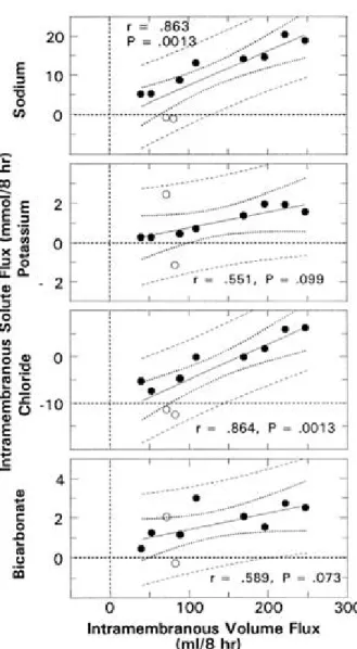
Fig. 9. Bivariate regression relationships between intramembranous flux of sodium, potassium, chloride, and bicarbonate and volume fluxes (n=10). A positive flux corresponds to the exiting of the AF and vice versa.
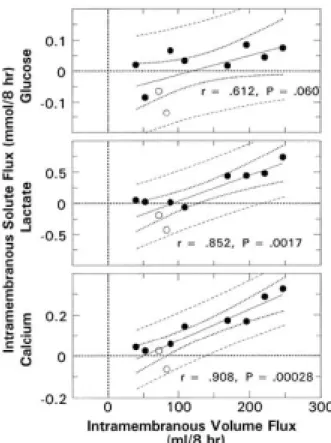
Fig. 10. Bivariate regression relationships between intramembranous flux of glucose, lactate, calcium, and volume fluxes (n=10). A positive flux corresponded to the exiting of the AF and vice versa.
VII. 결론(Conclusions)
최근의 여러 동물실험을 통한 연구들로 인하여 양수량의 조절에 관한 많은 이해가 이루어지고 있다. 또한 양수량의 조절과정 중 양수의 흡수에 관여하는 경로인 막내이동에 관한 개념도 확립되고 있다. 즉 막내흡수과정에서 양막, 융 모막, 그리고 태반의 태아측 표면에서 혈관내피성장인자의 표현이 중요한 조절과정(key regulator)이다. 이 혈관내피성 장인자의 막내흡수 조절의 가능한 기전은 여러 가지가 제시되고 있으며 그 중 소포에 의한 transcytosis의 유도가 중요 한 기전이라고 생각된다.21
참고문헌
1. Knoduri GG, Fewell JE. Oligohydramnios. In: Brace RA, Ross MG, Robilliard JE, eds. Fetal and neonatal body fluids: The scientific basis for clinical practice. Perinatology Press, 1989:157-74.
2. Healy DL. Polyhydramnios: An alternative hypothesis. In: Brace RA, Ross MG, Robilliard JE, eds. Fetal and neonatal body fluids: The scientific basis for clinical practice. Perinatology Press, 1989:175-85.
3. Brace RA. Physiology of amniotic fluid volume regulation. Clin Obstet Gynecol 1997;40:280-9.
4. Ross MG, Brace RA, and the NIH Workshop Participants. National Institute of Child Health Development Conference Summary: Amniotic fluid biology - basis and clinical aspects. The J Maternal-Fetal Med 2001;10:2-19.
5. Ervin MG, Berry LM, Ikegami M, Jobe AH, Padbury JF, Plok DH. Single dose fetal betamethasone administration stabilizes postnatal glomerular filtration rate and alters endocrine function in premature lambs. Pediatr Res 1996;40:645-51.
6. Brace RA, Wlodek ME, Cock ML, Harding R. Swallowing of lung liquid and amniotic fluid by the ovine fetus under normoxic and hypoxic conditions. Am J Obstet Gynecol 1994;171:764-70.
7. Hooper SB, Harding R. Fetal lung liquid: A major determinant of the growth and functional development of the fetal lung.
Clin Exp Pharmacol Physiol 1995;22:235-47.
8. Abramovich DR. Fetal factors influencing the volume and composition of liquor amnion. J Obstet Gynaecol Br Commonw 1970;77:865-77.
9. Ross MG, Sherman DJ, Ervin MG, Day L, Humme J. Stimuli for fetal swallowing: Systemic factors. Am J Obstet Gynecol 1989;161:1559-65.
10 Sherman DJ, Ross MG, Day L, Humme J, Ervin MG. Fetal swallowing: Response to graded maternal hypoxemia. J Appl Physiol 1991;71:1856-61.
11. Ross MG, Sherman DJ, Ervin MG, Day L, Humme J. Fetal swallowing: Response to systemic hypotension. Am J Physiol 1990;258:130-4.
12. Ross MG, Idah R. Correlation of maternal plasma volume and composition with amniotic fluid index in normal human pregnancy. J Maternal-Fetal & Neonatal Med 2004;15:104-8.
13. Ross MG, Kullama LK, Ogundipe OA, Chan K, Ervin MG. Ovine fetal swallowing response to intracerebroventricular hypertonic saline. J Apple Physiol 1995;78:2267-71.
14. Fanaroff AA, Martin RJ, Miller MJ. Identification and management of high-risk problems in the neonate. In Creasy RK, Resnik R. Maternal-Fetal Medicine: Principles and Practices. 3rd ed. Philadelphia Saunders, 1999:1135-72.
15. Daneshmand SS, Cheung CY, Brace RA. Regulation of amniotic fluid volume by intramembranous absorption in sheep: Role of passive permeability and vascular endothelial growth factor. Am J Obstet Gynecol 2003;188:786-93.
16. Matsumoto LC, Cheung CY, Brace RA. Effect of esophageal ligation on amniotic fluid volume and urinary flow rate in fetal sheep. Am J Obstet Gynecol 2000;182:699-705.
17. Abramovich DR, Page KR. Pathways of water exchange in the fetoplacental unit at mid-pregnancy. J Obstet Gynaecol Br Commonw 1972;79:1099-102.
18. Gitlin D, Kumate J, Morales C, Noriega L, Arevalo N. The turnover of amniotic fluid protein in the human conceptus. Am J Obstet Gynecol 1972;113:632-45.
19. Heller L. Intrauterine amino acid feeding of the fetus. In: Bode H, Warshaw J, eds. Parenteral nutrition in infancy and childhood. Plenum Press, 1974:206-13.
20. Schrder HJ, Dehne K, Andreas TH, Rago S, Rybakowshki CH. Diffusive transfer of water and glucose across the chorionic plate of the isolated human term placenta. Placenta 1999;20:59-63.
21. Cheung CY. Vascular endothelial growth factor activation of intramembranous absorption: A critical pathway for amniotic fluid volume regulation. J Soc Gynecol Investig 2004;11:63-74.
22. Leung DW, Cachianes G, Kuang WJ, Goeddel DV, Ferrara N. Vascular endothelial growth factor is a secreted angiogenic mitogen. Science 1989;246:1306-9.
23. Conolly DT. Vascular permeability factor: A unique regulator of blood vessel function. J Cell Biochem 1991;47:219-23.
24. Senger DR, Perruzzi CA, Feder J, Dvorak HF. A highly conserved vascular permeability factor secreted by a variety of human and rodent tumor cell lines. J Cancer Res 1986;46:5629-32.
25. DeVries C, Escobedo JA, Ueno H, Houck K, Ferrera N, Williams LT. The fms-like tyrosine kinase, a receptor for vascular endothelial growth factor. Science 1992;255:989-91.
26. Ferrara N, Davis-Symth T. The biology of vascular endothelial growth factor. Endocr Rev 1997;18:4-25.
27. Sharkey AM, Charnock-Jones DS, Boocock CA, Brown KD, Smith SK. Expression of mRNA for vascular endothelial growth factor in human placenta. J Reprod Fertil 1993;99:609-15.
28. Jackson MR, Carney EW, Lye SJ, Ritchie JW. Localization of two angiogenic growth factors (PDECGF and VEGF) in human placentae throughout gestation. Placenta 1994;15:341-53.
29. Shiraishi S, Nakagawa K, Kinukawa N, Nakano H, Sueishi K. Immunochemical localization of vascular endothelial growth factor in the human placenta. Placenta 1996;17:111-21.
30. Cooper JC, Sharkey AM, McLaren J, Charnock-Jones DS, Smith SK. Localization of vascular endothelial growth factor and its receptor, flt, in human placenta and deciduas by immunohistochemistry. J Reprod Fertil 1995;105:205-13.
31. Clark DE, Smith SK, Sharley AM, Charnock-Jones DS. Localization of VEGF and expression of its receptors flt and KDR in human placenta throughout pregnancy. Hum Reprod 1996;11:1090-8.
32. Vuckovic M, Ponting J, Terman BI, Niketic V, Seif MW, Kuman S. Expression of the vascular endothelial growth factor receptor KDR, in human placenta. J Anat 1996;188:361-6.
33. Bogic LV, Brace RA, Cheung CY. Cellular localization of vascular endothelial growth factor in ovine placenta and fetal membranes. Placenta 2000;21:203-9.
34. Bogic LV, Brace RA, Cheung CY. Developmental expression of vascular endothelial growth factor (VEGF) receptors and VEGF binding in ovine placenta and fetal membranes. Placenta 2001;22:265-75.
35. Cheung CY, Brace RA. Ovine vascular endothelial growth factor: Nucleotide sequence and expression in fetal tissues.
Growth factors 1998;16:11-22.
36. Anthony FW, Wheeler T, Elcock CL, Pickett M, Thomas EJ. Short report: Identification of a specific pattern of vascular endothelial growth factor mRNA expression in human placenta and cultured placental fibroblasts. Placenta 1994;15:557-61.
37. Cheung CT, Singh M, Ebaugh MJ, Brace RA. Vascular endothelial growth factor gene expression in ovine placenta and fetal membranes. Am J Obstet Gynecol 1995;173:753-9.
38. Ferrara N, Houck K, Jakeman L, Leung DW. Molecular and biological properties of the vascular endothelial growth factor family of proteins. Endocr Rev 1992;13:18-32.
39. Matsumoto LC, Bogic LV, Brace RA, Cheung CY. Prolonged hypoxia up-regulates vascular endothelial growth factor messenger RNA expression in ovine fetal membranes and placenta. Am J Obstet Gynecol 2002;186:303-10.
40. Matsumoto LC, Bogic LV, Brace RA, Cheung CY. Fetal esophageal ligation induces VEGF expression in fetal membranes.
Am J Obstet Gynecol 2001;184:175-84.
41. Cock ML, McCrabb GJ, Wlodek ME, Harding R. Effects of prolonged hypoxemia on fetal renal function and amniotic fluid volume in sheep. Am J Obstet Gynecol 1997;165:707-13.
42. Sawdy RJ, Slater DM, Dennes WJ, Sullivan MH, Bennett PR. The roles of the cyclooxygenases types one and two in prostaglandin synthesis in human fetal membranes at term. Placenta 2000;21:54-7.
43. Brace RA, Cheung CY. Amniotic fluid volume response to amnio-infusion of amniotic fluid versus Lactated Ringer’s solution in fetal sheep. J Soc Gynecol Investig 2004;11:363-8.
44. Alvi SA, Brown NL, Bennett PR, Elder MG, Sullivan MH. Corticotrophin-releasing hormone and platelet- activating factor
induce transcription of the type-2 cyclooxygenase gene in human fetal membranes. Mol Hum Reprod 1999;476-80.
45. Erchov AV, Bazan NG. Induction of cyclooxygenase-2 gene expression in retinal pigment epithelium cells by photoreceptor rod outer segment phagocytosis and growth factors. J Neurosci Res 1999;58:254-61.
46. Faber JJ, Anderson DF. Regulatory response of intramembranous absorption of amniotic fluid to infusion of exogenous fluid in sheep. Am J Physiol 1999;299:R236-42.
47. Ji YS, Xu Q, Schmedtje JF Jr. Hypoxia induces high-mobility-group protein I(Y) and transcription of the cyclooxygenase-gene 2 in human vascular endothelium. Circ Res 1998;83:295-304.
48. Schmedtje JF Jr, Ji YS, Liu WL, Dubois RN, Runge MS. Hypoxia induces cyclooxygenase-2 via the NF-kappa B transcription factor in human vascular endothelial cells. J Biol Chem 1997;272:601-8.
49. Cheung CY, Brace RA. Amniotic fluid volume and composition in mouse pregnancy. J Soc Gynecol Investig 2005;12:558-62.
50. Brace RA, Vermin ML, Huijssoon E. Regulation of amniotic fluid volume: Intramembranous solute and volume fluxes in late gestation fetal sheep. Am J Obstet Gynecol 2004;191:837-46.
51. Mann SE, Ricke EA, Torres EA, Taylor RN. A novel model of polyhydramnios: Amniotic fluid volume is increased in aquaporin 1 knockout mice. Am J Obstet Gynecol 2005;192:2041-6.
52. Adams EA, Choi HM, Cheung CY, Brace RA. Comparison of amniotic and intramembranous unidirectional permeabilities in late-gestation sheep. Am J Obstet Gynecol 2005;193:247-55.
53. Lyden TW, Anderson CL, Robinson JM. The endothelium but not the syncytiotrophoblast of human placenta expresses caveolae. Placenta 2002;23:640-52.
54. Michel CC, Curry FE. Microvascular permeability. Physiol Rev 1999;79:703-61.
55. Vasile E, Hong Q, Dvorak HF, Dvorak AM. Caveolae and vesiculo/vacuolar organelles in bovine capillary endothelial cells cultured with VPF/VEGF on floating matrigel/collagen gels. J Histochem Cytochem 1999;47: 159-67.
56. Feng Y, Venema VJ, Venema RC, Tsai N, Behzadian MA, Caldwell RB. VEGF-induced permeability increase is mediated by caveolae. Invest Ophthalmol Vis Sci 1999;40:157-67.
57. Liu J, Razani B, Tang S, Terman BI, Ware JA. Lisanti MP. Angiogenesis activators and inhibitors differentially regulate caveolin-1 expression and caveolae formation in vascular endothelial cells. J Biol Chem 1999;274:15781-5.
약 력 최 형 민
교수1982년 연세대학교 의과대학 입학
1988년 연세대학교 의과대학 졸업
연세대학교 의과대학 석사, 박사 (전공: 산부인과) 1988-1992년 연세대학교 의과대학 신촌세브란스병원 인턴, 레지던트 1996-1997년 연세대학교 의과대학 산부인과학교실 연구강사 (전공: 모체태아 의학)
1998-2000년 연세대학교 의과대학 산부인과학교실 전임강사 2000-현재 인제대학교 일산백병원 산부인과 교수 (부교수) 2003년 7월-2005년 2월 미국 연수
University of California San Diego (UCSD), Department of Reproductive Medicine,
Perinatology Lab에서 Research Professor (visiting scholar) 연수 및 연구 내용: Physiologic and Molecular regulation
of Amniotic fluid