294
책임저자: 하종원, 서울시 종로구 연건동 28번지
110-744, 서울대학교병원 외과 Tel: 02-2072-2991, Fax: 02-766-3975 E-mail: [email protected]
접수일:2010년 5월 26일, 게재승인일:2010년 7월 2일 본 연구는 교육과학기술부의 지원으로 이루어졌음(과제번호:
800-20080766).
쥐의 허혈성 하지 모델에서 VEGF를 함유한 Nanoparticle의 투여 효과
서울대학교 의과대학 외과학교실
안상현ㆍ민상일ㆍ김성엽ㆍ민승기ㆍ양한광ㆍ김상준ㆍ하종원
Effect of Nanoparticle with VEGF in Mouse Ischemic Hindlimb Model
Sang Hyun Ahn, M.D., Sang-Il Min, M.D., Seong Yup Kim, M.D., Seung-Kee Min, M.D., Han-Kwang Yang, M.D., Sang Joon Kim, M.D., Jongwon Ha, M.D.
Department of Surgery, Seoul National University College of Medicine, Seoul, Korea
Purpose: Vascular endothelial growth factor (VEGF) is one of the factors regulating angiogenesis. For angiogenesis, the local concentration of VEGF has to be maintained. Because of its short half-life, VEGF has been conjugated with nanoparticles. Some nanoparticles, such as poly (lactic-co-glycolic acid (PLGA)) or polyethylenimine (PEI) are commonly used in this field, but have weak points such as faster release than expected and cell toxicity.
We investigated the effect of core/shell nanoparticles including lecithin lipid cores in the ischemic hindlimb model.
Methods: Mice were anesthetized and a region of the common femoral artery and vein was ligated and excised.
Hindlimb ischemic mice (n=28) were divided randomly into four groups: Control group (normal saline, n=7), mouse VEGF group (mVEGF, n=7), nanoparticle including mVEGF group (N-mVEGF, n=7), and nanoparticle/
hydrogel mouse VEGF group (NH-mVEGF, n=7). The drug was injected postoperatively into the thigh muscle of the ischemic limb. Perfusion, capillary number and H&E stain were assessed 28 d after treatment.
Results: The capillary number increased in N-mVEGF and mVEGF group (P=0.026). Improvements of ischemic limb perfusion were inferior in N-mVEGF, NH-mVEGF groups (P=0.006) compared to other groups. Mice received N-mVEGF, NH-mVEGF treatment showed significant inflammation in the H&E staining.
Conclusion: Sustained VEGF delivery via core/shell nanoparticle with lecithin core did not show improved perfusion rate despite an increase in capillary number. Furthermore, vacuolization and induction of inflammation requiring a different composition of nanoparticle should be tested. (J Korean Surg Soc 2010;79:294-299) Key Words: Hindlimb ischemia, Vascular endothelial growth factor (VEGF), Nanoparticle
중심 단어: 허혈성 하지, 혈관내피성장인자, 나노입자
서 론
말초 동맥 질환의 유병률은 3∼10%이고 70세 이상에선
15∼20%로 현재처럼 고령 인구가 증가할수록 환자수는 더 욱 늘어날 전망이다.(1) 이들 중 병변의 위치에 따라 수술적 치료가 도움이 될 수도 있지만 혈관의 수술적 재건을 위해 필요한 자가 혈관의 한계로 제한되는 경우가 있으며,(2) 이 경우 혈관 신생은 증상 개선을 위한 또 하나의 치료 방법이 될 수 있다.
혈관 신생은 지난 30년 동안 심근경색, 상처 치유 지연, 신경병증, 망막병증, 이식 실패 등에서 병리학적으로 호전 시킬 수 있는 치료 방법으로 고려되고 있다.(3-5) 혈관을 신 생시키는 방법 중 vascular endothelial growth factor (VEGF)
는 in vivo 실험에서 여러 차례 혈관 신생 효과를 입증했지 만 정맥 투여 시 몇 가지 부작용을 보였다.(6) 더욱이 인체 를 대상으로 시행된 VIVA 연구에서는 recombinant human VEGF의 정맥 투여는 위약 대조군과 비교 했을 때 통계학 적으로 차이가 없었다.(7) 이 실험의 참여자 중 일부에서는 정맥을 통한 전신 투여로 비특이적인 혈관 신생과 고혈압, 부종 등의 부작용을 보였다.(8-10)
그러므로 VEGF의 최적의 치료 효과를 얻기 위해서는 정 맥을 통한 전신 투여 보다는 국소적이면서 지속적인 분비 가 필요할 것이며 이를 위해 새로운 형태의 약물전달체계 가 요구 된다.(11) 나노입자(nanoparticle)에 VEGF를 결합하 면 약물이 서방형으로 수일에서 수주간 분비되어 지속적으 로 근위부위에 혈관 신생을 자극할 것이다.(12,13) 나노입자 는 미세분자(micromolecule)보다 더 효과적으로 세포에 의 해 섭취되며 손상 부위에 직접 투여가 가능하다.(14) 현재 다양한 종류의 나노입자가 연구되고 있으며 이 중 lecithin lipid core를 가진 core/shell 나노입자는 in vitro 실험에서 VEGF와 안정적으로 결합하여 지속적으로 VEGF를 방출하 였다.(11)
이 연구는 VEGF를 함유하고 있는 core/shell nanoparticle 를 이용하여 쥐의 허혈성 사지(ischemic limb)에서 치료 효 과의 가능성을 보고자 하였다.
방 법
1) 실험 동물
실험에 사용된 쥐는 7주 된 BALB/c 종으로 중앙실험동물 에서 구입 후 본 연구기관에서 적응을 위해 7주 동안 식이 제한 없이 사육되었다. 수컷으로 무게는 24∼26 g이었다.
본 실험은 서울대학교병원 동물실험윤리위원회(SNUH-IACUC) 의 승인 후 진행되었다.
2) 나노입자(nanoparticle)
지질핵(lipid core)을 가지고 있는 core/shell 나노입자(신소 재공학과, 한남대학교, 대전광역시, 대한민국)로 VEGF를 지속적으로 방출하도록 고안되었다. 지질핵은 protein-loaded lecithin으로 구성되어 있고 polymeric shell은 Pluronics [poly(ethylene oxide)-poly(propylene oxide)-poly(ethylene ox- ide) triblock copolymer, F-127]으로 구성되어 있다. Core/shell 나노입자와 VEGF는 정전기 상호작용으로 결합되어 있어 polymeric shell의 물리적 붕괴에 의해 방출된다.(11)
3) 수술적 처치
쥐의 복강 내에 케타민(ketamine HCI) 60 mg/kg을 투여하 여 전신 마취를 하였다. 왼쪽 하지의 피부를 약 2 cm가량 절개하여 혈관을 확인한 후 아래배벽동맥(inferior epigastric artery) 아래 부분부터 표재대퇴동맥(superficial femoral ar- tery)의 중간 지점까지 결찰 후 분리하였다. 수술 후 무작위로 4개의 군으로 할당하여 생리식염수(n=7), mVEGF (mouseVEGF, PeproTech Inc., USA)(n=7), N-mVEGF (nanoparticle contained mVEGF)(n=7), NH-mVEGF (Nanoparticle/hydrogel-mVEGF) (n=7)을 수술 후 당일 날 VEGF 20μg을 대퇴부 근육 투여하 거나 동일양의 VEGF가 함유된 나노입자 40μl를 여러 차례 나누어 투여하였다.
4) 레이져 도플러 관류 영상(laser Doppler perfusion imaging)
수술 전, 수술 당일, 수술 후 3일, 1주, 2주, 3주, 4주째에 실험동물을 케타민을 사용하여 마취하고 털을 제거한 후 양측 하지에서 레이저 도플러(Moor Instruments, Devon, UK) 로 혈류량을 측정하였다. 수술한 하지의 혈류량을 수술하 지 않은 반대쪽 다리(nonischemic limb)의 혈류량과의 비율 로 표시하였다.
5) 알카리성 인산분해효소 염색(alkaline phosphatase staining) 및 판독
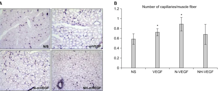
BALB/c 쥐를 4주째 안락사 시키고 장딴지근과 경골근을 채취하였다. 근육의 중간 부위의 냉동 절편을 만든 후 슬라 이드 조직에 아세톤으로 5분간 고정시키고 공기 중에서 건 조하였다. BCIP/NBT buffered substrate tablet을 용해하여 슬 라이드에 분주한 후 37oC에서 약 30분간 방치하고, 증류수 로 세척한 후 표본을 제작하였다. 모세혈관의 수는 광학현 미경의 200배의 배율에서 무작위로 6곳을 한 명의 연구원 이 관찰한 후 평균값을 구하였다.
6) H&E 염색
수술 후 4주째에 수술 한 하지에서 근육을 채취하여 파라 핀 묻힘(paraffin embedding)한 후 절삭한 슬라이드 조직을 hematoxylin과 eosin 염색하여 현미경으로 염증 정도와 괴사 등을 관찰하였다. Image J프로그램(NIH, Bathesda, MD, USA)을 이용하여 전체 세포 중 공포의 비율을 나타내었다.
Fig. 1. H&E staining of the muscles on week 4 (A: ×400), and percent of vacuolar degeneration (B). N/S = normal saline; mVEGF
= mouse vascular endothelial growth factor; N-mVEGF = nanoparticle mouse vascular endothelial growth factor; NH-mVEGF = nanoparticle/hydrogel mouse vascular endothelial growth factor.
Fig. 2. Laser Doppler blood perfusion (A) and blood perfusion (B) in ischemic hind limbs is measured before, just after, on day 3, and at weeks 1, 2, 3 and 4 after right femoral artery ligation.
7) 통계분석
실험 결과 값은 각 그룹별로 평균과 표준편차로 표기하 였다. 변수들간의 유의성은 Kruskal-Wallis test법과 Mann- Whitney법을 이용하여 분석하였다. 유의수준 0.05 이하인 경우 통계적 유의성을 인정하였고 모든 통계 분석은 SPSS 17.0 프로그램을 이용하였다.
결 과
1) H&E 염색
생리식염수를 투여한 실험군에서 지방으로 대체된 공포 형성 비율은 10.556%인 반면 mVEGF 투여 그룹에서는 5.564%로 적었다. mVEGF 투여는 근육이 허혈에 의해 지방
으로 대치 되는 것을 억제하는 소견을 보였다. 반면 N-mVEGF는 공포 형성 비율이 34.955%, NH-mVEG는 28.352%로 생리식염수 군보다 비율이 높았고 염증 세포침 윤이 많은 부분 주위의 근육세포가 파괴된 염증반응 소견 을 보였다(Fig. 1).
2) 레이저 도플러 관류 영상(laser Doppler perfusion imaging)
혈류량 비율은 수술 직후 모든 그룹에서 20% 정도까지 감소한 후 2주까지는 완만하게 상승하였다(Fig. 2). 생리식 염수를 투여한 군에서 4주째 혈류량 비율의 평균 값은 64.69%로 가장 높았고. mVEGF을 투여한 군은 혈류량 비율 평균은 55.25%로 낮았지만 두 군간의 통계학적 의미는 없 었다(P=0.209). N-mVEGF투여 군에서는 가장 낮은 수치인
Fig. 3. (A) Cryosections of muscle obtained from the mice at day 28 were stained for alkaline phosphatase. Representative microscopic photographs of capillaries identified by staining of alkaline phosphatase. (B) Quantification of capillary density on the tissue sections.
The ratio of the number of capillaries to the number of muscle fiber was measured. N/S = normal saline; mVEGF = mouse vascular endothelial growth factor; N-mVEGF = nanoparticle mouse vascular endothelial growth factor; NH-mVEGF = nanoparticle/hydrogel mouse vascular endothelial growth factor.
39.7%를 나타냈고 NH-mVEGF 투여군은 44.13%로 생리식 염수를 투여한 군에 비해 통계학적 의미가 있게 감소하였 다(P=0.006). 생리식염수를 투여한 군과 mVEGF를 투여한 군에서는 4주째 실험 종료 시까지 계속 혈류량 비율이 증가 하는 양상인 반면 N-mVEGF, NH-mVEGF를 투여한 군은 2 주 때부터 혈류량 증가 비율이 감소하였다.
3) 알카리성 인산분해효소 염색
생리식염수를 투여한 군에서 capillary/muscle fiber를 계수 하였을 때 평균값은 0.59 (SD=0.11)이었고 mVEGF을 투여 한 군에서는 평균값이 0.72 (SD=0.07), N-mVEGF을 투여한 군의 평균값은 0.89 (SD=0.14)로 통계적으로 의미 있게 높 았다(P=0.026). NH-mVEGF군은 평균값이 0.68 (SD=0.20)로 생리식염수를 투여한 군과 비교하였을 때 통계적으로 의미 가 없었다(P=0.383)(Fig. 3). VEGF 나노입자를 투여한 군은 비록 도플러 관류 영상에서 혈류량을 증가시키지는 못했으 나 모세혈관의 수는 의미 있게 증가하는 소견을 보였다.
고 찰
나노입자를 이용한 약물 전달 체계(delivery system) 연구 에서 나노입자의 성분은 주요한 관심 분야이다. 오래 전부 터 in vitro나 in vivo 실험에 polyethylenimine (PEI)는 유전자
전달물(gene delivery)로 사용되어 왔다. PEI는 넓은 범위의 pH에서 proton-sponge effect로 인해 높은 유전자핵내 주입 효율(high transfection efficiency)을 가지고 있다.(15-17) 하지 만 PEI 자체의 높은 세포 독성으로 임상적으로 사용하기엔 한계가 있다.(17)
Polymeric nanosphere로 poly (lactic-co-glycolic acid) (PLGA) 는 PEI의 대안으로 약물과 유전자 전달에 널리 이용되고 있 는 물질이다.(18-20) PLGA nanosphere는 안정성이 뛰어나고 세포내이입(endocytosis)에 의해 세포 안으로 쉽게 섭취되며 표면에 리간드를 부착하여 특정 기관에 도달할 수 있는 장 점이 있다. 하지만 이런 많은 장점에도 불구하고 PLGA는 대부분의 단백질 약물을 보호하는 캡슐에 비해 소수성 (hydrophobic)이다. 이런 성질은 단백질 약물의 저장과 in vivo 방출 시 안정성에 문제가 된다.(21) Bioerosion phase 동 안에 단백질 약물의 방출을 위해 PLGA의 수화(hydration)와 분해(degradation)가 필수조건인데 이 과정에서 젖산(lactic acid)과 글리콜산(glycolic acid)이 형성되어 미세환경을 산 성화시킨다. 이런 환경에선 단백질 약물은 가수분해(hydrolytic degradation)와 응집(aggregation)이 되어 변성된다.(22) Lipid core를 가진 lecithin은 새로운 약물 전달 물질로 수 용성이고 분해 과정 중 단백질이 변성되는 것을 억제하는 것으로 알려져 있다.(23) 본 실험을 위해 lecithin에 VEGF를 부하(load)시켜 만든 core/shell nanoparticle은 in vitro 실험에
서 9일에 걸쳐 85%가 서서히 방출되도록 제작하였다. 본 나노입자는 lecithin의 조성 비율에 따라 방출기간을 조절할 수 있다.(24) 이 실험은 lecithin core/shell nanoparticle을 근육 내 투여한 첫 in vivo 결과로 나노입자를 투여한 군에서 주 변 조직의 심한 염증 반응을 유발하였다. 나노입자가 붕괴 되는 과정에서 발생한 염증으로 수술부위가 제대로 아물지 않고 많은 양의 VEGF를 한번에 투여하는데 어려움이 있었 다. 4주째 생검 후 염색된 조직에서 염증 세포의 침윤이 관 찰 되었고 이는 나노입자가 전부 흡수되지 않고 여전히 남 아서 만성 염증 반응을 일으키는 것으로 해석된다. 근육 조 직에 만성 염증 반응으로 인해 생리식염수를 투여한 군보 다 4주째 혈류량이 낮았다. 알카리성 인산분해효소 염색에 서 보이는 capillary/muscle fiber증가는 VEGF의 지속적인 분 비에 의한 모세혈관의 신생인지 혹은 약물 주입 후 일어난 급성 염증 반응에 의한 결과인지 추가적인 실험이 필요하 겠다. 다만 본 기관에서 core/shell 나노입자만을 쥐의 외이 에 주입한 실험에서도 혈관이 신생되는 소견을 보여 급성 염증에 의한 혈관 신생을 배제할 수 없다(결과 미보고). 발 표된 다른 논문에서도 lecithin을 함유한 core/shell 나노입자 가 쥐의 각막의 신생 혈관에서 의미 있게 증가하였다는 보 고가 있을 뿐 혈류의 증가와 관련된 보고는 없다.(25) VEGF의 다른 기능으로 헴-옥시게나제-1 (heme-oxygen- ase-1)를 조절하여 항산화, 세포자멸억제(antiapoptotic), 항염 증 효과가 있다.(26) VEGF만 단독 투여한 군의 H&E 염색 소견에서 공포형성이 적은 것은 세포자멸억제 효과의 결과 로 생각할 수 있다. 혈관신생과 달리 하지의 허혈 직후부터 급성 염증이 진행되는 수시간 동안에 VEGF의 1회 투여 만 으로도 효과가 있는 것으로 생각되며 나노입자가 포함된 군에서는 나노입자에 의한 지속적 염증 반응으로 그 효과 가 상쇄되어 공포 형성이 증가한 것으로 생각된다.
함수나노입자(hydrogel nanoparticle)은 최근 약물 전달 관 련 연구에 주목 받는 물질이다. 수화성의 hydrogel은 생체조 직과 유사성이 높아 계면장력(interfacial tension)이 매우 낮 다.(27) VEGF를 함유한 함수나노입자는 현재 개발 단계에 있는 물질로 in vitro 실험이 진행 중이다. 이 함수나노입자 는 나노입자에 propylene glycol monocaprylate를 첨가한 것 으로 상온에서는 액체 상태를 유지하다가 인체 주입 후에 는 고체로 되는 성질이 있다. 따라서 나노입자 단독 주입보 다 장시간 국소 분비가 될 것으로 기대하였으나 실험 결과 lecithin lipid core/shell 나노입자와 비슷하였다. 따라서 lec- ithin lipid core/shell 나노입자와 함께 함수나노입자 자체의
염증 유발 정도에 대한 추가 실험이 필요할 것이다.
결 론
본 실험에 사용된 나노입자는 in vitro 실험에서 VEGF 같 은 단백질 약물의 안정성은 높였지만 생체 내에서 염증반 응을 유발하는 문제점이 있었다. 나노입자 자체에 의한 염 증반응이 없고 VEGF의 함유량이 높은 약물전달 시스템의 개발은 혈류량 개선을 기대할 수 있을 것이나, 본 나노입자 는 넣을 수 있는 VEGF의 양의 제한과 생체에서 염증 반응 의 유발로 인해 다른 물질을 이용한 나노입자의 개발이 고 려해야 할 것이다.
REFERENCES
1) Hirsch AT, Haskal ZJ, Hertzer NR, Bakal CW, Creager MA, Halperin JL, et al. ACC/AHA 2005 Practice Guidelines for the management of patients with peripheral arterial disease (lower extremity, renal, mesenteric, and abdominal aortic): a collabo- rative report from the American Association for Vascular Surgery/Society for Vascular Surgery, Society for Cardiovascular Angiography and Interventions, Society for Vascular Medicine and Biology, Society of Interventional Radiology, and the ACC/AHA Task Force on Practice Guidelines (Writing Committee to Develop Guidelines for the Management of Patients With Peripheral Arterial Disease): endorsed by the American Association of Cardiovascular and Pulmonary Rehabilitation; National Heart, Lung, and Blood Institute;
Society for Vascular Nursing; TransAtlantic Inter-Society Consensus; and Vascular Disease Foundation. Circulation 2006;
113:e463-654.
2) Hall H. Modified fibrin hydrogel matrices: both, 3D-scaffolds and local and controlled release systems to stimulate angio- genesis. Curr Pharm Des 2007;13:3597-607.
3) Ferrara N, Alitalo K. Clinical applications of angiogenic growth factors and their inhibitors. Nat Med 1999;5:1359-64.
4) Folkman J. Diagnostic and therapeutic applications of angio- genesis research. C R Acad Sci III 1993;316:909-18.
5) Ware JA, Simons M. Angiogenesis in ischemic heart disease.
Nat Med 1997;3:158-64.
6) Simons M, Ware JA. Therapeutic angiogenesis in cardio- vascular disease. Nat Rev Drug Discov 2003;2:863-71.
7) Henry TD, Annex BH, McKendall GR, Azrin MA, Lopez JJ, Giordano FJ, et al. The VIVA trial: vascular endothelial growth factor in Ischemia for vascular angiogenesis. Circula- tion 2003;107:1359-65.
8) Qaum T, Xu Q, Joussen AM, Clemens MW, Qin W, Miyamoto K, et al. VEGF-initiated blood-retinal barrier breakdown in
early diabetes. Invest Ophthalmol Vis Sci 2001;42:2408-13.
9) Martin A, Komada MR, Sane DC. Abnormal angiogenesis in diabetes mellitus. Med Res Rev 2003;23:117-45.
10) Yang R, Ogasawara AK, Zioncheck TF, Ren Z, He GW, DeGuzman GG, et al. Exaggerated hypotensive effect of vas- cular endothelial growth factor in spontaneously hypertensive rats. Hypertension 2002;39:815-20.
11) Oh KS, Han SK, Lee HS, Koo HM, Kim RS, Lee KE, et al.
Core/Shell nanoparticles with lecithin lipid cores for protein delivery. Biomacromolecules 2006;7:2362-7.
12) Murphy WL, Peters MC, Kohn DH, Mooney DJ. Sustained release of vascular endothelial growth factor from mineralized poly(lactide-co-glycolide) scaffolds for tissue engineering.
Biomaterials 2000;21:2521-7.
13) Zisch AH, Lutolf MP, Hubbell JA. Biopolymeric delivery ma- trices for angiogenic growth factors. Cardiovasc Pathol 2003;
12:295-310.
14) Suri SS, Fenniri H, Singh B. Nanotechnology-based drug de- livery systems. J Occup Med Toxicol 2007;2:16.
15) Abdallah B, Hassan A, Benoist C, Goula D, Behr JP, Demeneix BA. A powerful nonviral vector for in vivo gene transfer into the adult mammalian brain: polyethylenimine. Hum Gene Ther 1996;7:1947-54.
16) Godbey WT, Wu KK, Mikos AG. Size matters: molecular weight affects the efficiency of poly(ethylenimine) as a gene delivery vehicle. J Biomed Mater Res 1999;45:268-75.
17) Benns JM, Maheshwari A, Furgeson DY, Mahato RI, Kim SW. Folate-PEG-folate-graft-polyethylenimine-based gene de- livery. J Drug Target 2001;9:123-39.
18) Panyam J, Zhou WZ, Prabha S, Sahoo SK, Labhasetwar V.
Rapid endo-lysosomal escape of poly(DL-lactide-co-glycolide) nanoparticles: implications for drug and gene delivery. FASEB J 2002;16:1217-26.
19) Stern M, Ulrich K, Geddes DM, Alton EW. Poly (D, L-lac- tide-co-glycolide)/DNA microspheres to facilitate prolonged transgene expression in airway epithelium in vitro, ex vivo and in vivo. Gene Ther 2003;10:1282-8.
20) Jeon O, Kang SW, Lim HW, Chung JH, Kim BS. Long-term and zero-order release of basic fibroblast growth factor from heparin-conjugated poly(L-lactide-co-glycolide) nanospheres and fibrin gel. Biomaterials 2006;27:1598-607.
21) Dai C, Wang B, Zhao H. Microencapsulation peptide and pro- tein drugs delivery system. Colloids Surf B Biointerfaces 2005;41:117-20.
22) Sinha VR, Trehan A. Biodegradable microspheres for protein delivery. J Control Release 2003;90:261-80.
23) Robertson D, Hellweg T, Tiersch B, Koetz J. Polymer-induced structural changes in lecithin/sodium dodecyl sulfate-based multilamellar vesicles. J Colloid Interface Sci 2004;270:
187-94.
24) Yang Z, von Ballmoos MW, Diehm N, Baumgartner I, Kalka C, Di Santo S. Call for a reference model of chronic hind limb ischemia to investigate therapeutic angiogenesis. Vascul Pharmacol 2009;51:268-74.
25) Choi WI, Yoon KC, Im SK, Kim YH, Yuk SH, Tae G.
Remarkably enhanced stability and function of core/shell nanoparticles composed of a lecithin core and a pluronic shell layer by photo-crosslinking the shell layer: in vitro and in vivo study. Acta Biomater 2010;6:2666-73.
26) Bussolati B, Mason JC. Dual role of VEGF-induced heme- oxygenase-1 in angiogenesis. Antioxid Redox Signal 2006;8:
1153-63.
27) Ratner BD, Hoffman AS. Synthetic hydrogels for biomedical applications. In: Andrade JD, editor. Hydrogels for Medical and Related Applications. Washington: American Chemical Society; 1976. p.1-36. (ACS symposium series; vol 31).