자궁내막증의 병인: 초기 병변 형성을 중심으로
부산대학교 의과대학 산부인과학교실 나 용 진
Pathogenesis of Endometriosis: Early Lesion Formation Yong Jin Na
Department of Obstetrics and Gynecology, College of Medicine, Pusan National University, Busan, Korea [Korean. J. Reprod. Med. 2007; 34(2): 57-66.]
서
론"자궁강 (uterine cavity) 이외의 장소에 기능성 자 궁내막 선 (gland)과 기질 (stroma) 조직이 존재하 는 상태"로 정의되는 자궁내막증은 월경통, 성교 통 (dyspareunia), 만성 골반통 및 수태능력의 저하 (subfertility)와 같은 특징적인 증상 및 징후들을 나 타낼 수 있으나 아직도 발병기전은 명확히 밝혀지 지 않았다.
본 종설에서는 자궁내막증의 발병과 관련된 여 러 가지 가설들과, 특히 복막 자궁내막증 발생의 핵심적인 과정으로 생각되는 초기 자궁내막증 병 변 형성의 여러 단계들을 관련된 문헌들과 함께 정리하여 살펴봄으로써 자궁내막증의 발병기전과 관련된 현재까지의 연구들에 대한 이해를 돕고, 나 아가 앞으로의 연구방향을 모색해 보고자 하였다.
자궁내막증의 조직기원 (Histogenesis of endometriosis) 1. 이식설 (Implantation theory)
1921년 Sampson에 의하여 최초로 제시되었으며
월경으로 탈락된 자궁내막 세포들이 난관을 경유, 복강으로 역류하여 복막과 난소에 착상하여 성장 함으로써 자궁내막증이 발생한다는 이론이다.
1월 경혈의 역류는 난관이 막히지 않은 가임기 여성에 서 흔히 관찰되는 소견으로, Halme 등은 월경 주위 기에 복강경 수술을 시행하였던 난관이 개통된 여 성의 90%에서 혈액이 섞인 복막액을 확인하였다.
2어떤 하나의 이론만으로 자궁내막증의 모든 경우 들을 설명할 수는 없지만 이식설이 자궁내막증 발 생의 유력한 기전임을 시사하는 다음과 같은 많은 증거들이 있다. ① 복막 자궁내막증 병변은 골반 의 dependent portion에 주로 발생하며 난소, 전방 및 후방 cul-de-sac, 자궁천골 인대 등의 순으로 관 찰된다.
3② 월경으로 탈락된 자궁내막은 세포배양 에서 생존 가능하다.
4③ 자궁 또는 질의 기형으로 월경혈의 유출경로가 막힌 경우 자궁내막증의 발 생이 증가한다.
5④ 동물실험에서 인위적인 utero- pelvic fistula를 만들어 월경혈의 역류를 유도하면 자궁내막증이 유발된다.
6⑤ 초경이 빨리 시작되거 나 월경주기가 짧은 경우, 또는 월경과다가 있는 경우 자궁내막증의 발생이 증가한다.
7주관책임자: 나용진, 우) 602-739 부산광역시 서구 아미동 1가 10번지, 부산대학교 의과대학 산부인과학교실
Tel: (051) 240-7282, Fax: (051) 248-2384 e-mail: [email protected]
2. 체강상피 화생설 (coelomic metaplasia theory) 및 유도설 (induction theory)
Sampson의 이식설을 지지하는 설득력 있는 많 은 증거들이 있지만 자궁내막증의 발생을 설명할 수 있는 또 하나의 중요한 가설인 체강상피 화생 설은 복막과 늑막에 존재하는 체강상피에서 유래 한 중피세포 (mesothelial cell)의 자연발생적 화생 (metaplasia)에 의해 자궁내막증이 발생할 것이라는 이론이다. 체강상피 화생설로부터 변형된 유도설 (induction theory)은 복강으로 유입된 월경기 자궁내 막의 변성 (degeneration)에 의해 유리된 endogenous factor들이 난소의 germinal epithelium과 복막 중피 세포의 화생을 유도하여 자궁내막 조직으로 변화 시킨다는 가설이다.
8적어도 일부 경우들에서는 자 궁내막증이 자연발생적 또는 유도된 체강상피의 화생에 의해 발생할 것임을 시사하는 다음과 같은 증거들이 있다.
9① 자궁내막증은 초경이 시작되지 않은 소녀와 월경을 경험해 보지 않은 여성에서도 보고된 바 있다. ② 자궁내막 세포는 해부학적 결 함이 없는 한, 직접 흉부로 유입될 수 없기 때문에 착상설은 늑막 및 폐의 자궁내막증을 설명할 수 없다. 변성이 일어난 자궁내막 세포들로부터 복막 액 (우측 hemi-diaphragm을 통해 흉강과 연결)으로 유리된 스테로이드 호르몬 또는 화학물질에 의한 자극으로 늑막의 화생이 유발된다는 설이 유력하 다. ③ 자궁내 (eutopic)와 이소성 (ectopic) 자궁내 막이 형태 및 기능적 측면에서 서로 많은 차이를 보이는 것은 자궁내막증의 병변이 정상 자궁내막 조직의 자가이식물 (autotransplants)이라는 견해와 일치되지 않는다.
체강상피 화생설은 자궁내막 세포와 복막세포가 모두 체강상피로부터 기원한다는 사실에 기초하여 제기된 이론이지만 다음과 같은 문제점들도 지적 되고 있다. ① 복막세포가 metaplastic transformation 이 가능하다면 이러한 현상은 남성에게도 나타나 야 한다. ② 체강상피는 복막강 뿐만 아니라 흉강 도 구성하고 있지만 자궁내막증은 거의 대부분 복
막강 내에서 발생한다. ③ Metaplastic process는 연 령의 증가와 관련된 과정이지만 자궁내막증은 젊 은 가임기 여성에서만 발생한다.
체강상피 화생설로 복막강, 흉강, 요로계 및 소 화기계, 배꼽 등에 생긴 자궁내막증을 설명할 수 있지만 자궁내막 세포의 혈행성 또는 림프성 전파 에 의해서도 자궁내막증이 발생할 수 있음을 시사 하는 보고들도 있다.
10,11한편, 수술흉터나 회음부에 서 발견되는 자궁내막증은 제왕절개술, 기타 골반 강내 수술 또는 외음절개술의 봉합을 시행할 때 의인성 (iatrogenic)으로 자궁내막 조직이 직접 이식 될 수 있다는 가설로 가장 잘 설명될 수 있다.
3. 복막, 난소, 심부 직장-질 (deep rectova- ginal) 자궁내막증의 발병기전은 동일한가?
월경으로 난관을 통해 역류한 자궁내막 조직이 생존, 부착, 증식, 침습 및 혈관생성의 과정을 거쳐 자궁내막증의 복막내 병변을 형성한다는 어느 정 도의 견해의 일치는 있으나 난소의 자궁내막증과 특정한 형태의 심부 자궁내막증의 발병기전에 대 해서는 아직도 논란의 여지가 많은 실정이다.
12~13발생위치, 병변의 모양과 호르몬에 대한 반응성이 서로 다르기 때문에 복막의 자궁내막증, 난소의 자 궁내막증 및 직장-질 중격의 adenomyotic nodule은 서로 다른 발병기전을 가진 각각의 독립적인 질환 이라는 의견이 제시된 바 있으나
14심부 자궁내 막증은 직장-질 중격으로부터가 아니라 pouch of Douglas에 착상한 자궁내막증의 표재성 병변으로 부터 발생한다는 견해도 있다.
15자궁내막증의 초기 병변 형성 (Early endometriosis lesion formation)
1. 복강내 방어기전 회피 (evading defense mechanism in the peritoneal cavity)
세포자멸사 (apoptosis)로 흔히 불리우는 program-
med cell death는 다세포 생물의 항상성에 관여하는
필수 과정으로 자궁내막에서는 분비기 말 및 월경
기에 노후된 세포들을 기능층으로부터 제거함으로 써 항상성을 유지시킨다고 한다.
16,17Gebel 등은 정 상 여성에 비해 자궁내막증 환자의 자궁내 자궁내 막에서 세포자멸사의 유의한 감소를 관찰함으로써 세포자멸사에 대한 저항성이 복강으로 유입된 자 궁내막 세포의 생존 가능성을 증가시켜 자궁내막 증의 발병에 관여할 수 있다고 하였다.
17세포자멸 사와 관련된 anti-apoptotic gene인 B-cell lymphoma/
leukemia-2 (Bcl-2)의 발현은 정상 자궁내막에서는 월경주기에 따른 차이를 보이며, 증식기에 가장 현 저하다. Meresman 등은 정상 여성에 비해 자궁내 막증 환자의 증식기 자궁내 자궁내막에서 Bcl-2의 발현이 유의하게 증가함을 관찰하였고,
18Goumenou 등은 자궁내막증의 병변 조직에서 월경주기에 따 른 차이를 보이지 않는 지속적인 Bcl-2의 발현을 보고한 바 있다.
19월경혈의 역류에 의해 복강으로 유입된 자궁내 막 세포들이 대부분의 여성들에서 제거되는 기전 은 아직 명확히 밝혀지지 않았으나 서로 다른 여 러 종류의 세포들이 관여하는 복막내 면역감시체 계가 그 역할을 수행할 것으로 추정되고 있다.
20Haney 등은 복강으로 유입된 월경혈이 염증반응을 유발한다는 사실을 최초로 보고하였다.
21물론 이 러한 염증반응의 생리적인 역할은 복강내에 존재 하는 이소성 세포와 조직을 제거하는 것이지만 그 리 효율적인 시스템은 아니기 때문에 Koninckx 등 은 월경이 있으면서 난관이 막히지 않은 모든 여 성에서는 microscopic endometriosis가 간헐적으로 존재할 것이라고 하였다.
22자궁내막증에서 관찰되 는 세포매개성 및 체액성 면역의 변화들은 면역반 응의 결함이 역류된 월경혈에 포함된 조직 부스러 기들 (debris)의 효율적인 제거를 방해하여 자궁내 막증의 발병에 관여할 수 있음을 시사하고 있다.
이러한 면역학적 이상이 자궁내막증의 원인인지 또는 결과인지의 여부는 아직 명확히 밝혀지지 않 았지만 자궁내막증의 발병에 중요한 역할을 할 것 으로 생각되고 있다.
23또한 자궁내막증 환자의 자 궁내막 세포에서는 면역감시체계를 피할 수 있는
세포들에서 볼 수 있는 특징적인 소견들이 관찰되 는데
13면역 인식에 중요한 major histocompatibility complex (MHC) class I 항원의 발현을 변화시켜 자 연세포독성세포 (natural killer, NK cell)에 대한 저 항성을 나타내며,
24Soluble HLA 또는 intercellular adhesion molecule (sICAM)-1과 같은 혈액을 순환하 는 항원을 생성하여 면역 인식을 방해하며,
25Fas 매개 반응에 의해 면역세포들의 세포자멸사를 유 도한다고 한다.
26선천적 면역의 장애는 자궁내막증의 발병과정에
서 면역학적 요인의 중요성을 뒷받침할 수 있는
소견으로 관심의 대상이 되어 왔다.
13대식세포
(macrophage)는 선천적 면역반응의 중요한 요소로
침범한 미생물의 인식, 탐식 및 파괴로 숙주의 방
어기전에 관여하며, 세포자멸사에 의한 세포들과
세포의 부스러기들을 제거하는 청소부 (scavenger)
로서의 역할도 담당한다.
23대식세포들은 복막액에
정상적으로 존재하지만 자궁내막증 환자의 경우
는 그 수와 활성이 많이 증가되어 있으며,
22,27활
성이 증가된 복막액내 대식세포들과 혈중 단핵구
(monocyte)들은 성장인자들과 cytokine들을 분비하
여 이소성 자궁내막 세포들의 성장을 촉진시키면
서 이들을 제거하는 청소부로서의 역할은 억제시
켜 자궁내막증의 발병을 조장할 수 있다고 한다.
28NK 세포는 선천성 면역반응에 관여하는 또 하나
의 중요한 요소로 해로운 세포들을 인식하여 제거
할 수 있다.
29자궁내막증 환자의 복막액내 NK 세
포들의 수에 관한 여러 연구들은 서로 상이한 결
과를 보이지만
27,30,31기능에 대한 연구들은 NK 세
포들의 세포독성 (cytotoxicity)이 저하되어 있으며,
이는 advanced stage에서 더욱 현저하다는 일관성
있는 결과를 나타내고 있다.
31,32자궁내막증 환자
의 말초혈액 및 복막액내 NK 세포들에서 관찰되
는 killer inhibitory receptor (KIR)의 과발현 (over-
expression)은 이와 관련된 가능성 있는 기전들 중
한 가지로 제시되고 있다.
33,342. 복막표면 부착 (adhesion to the peritoneal surface)
Groothuis 등과 Koks 등은 제 방향으로 떨어져 나온 (antegradely shed) 월경기 자궁내막 조각들 뿐 만 아니라 생검으로 얻은 증식기와 분비기 자궁 내막 조각들도 복막 중피층 (mesothelial layer)의 결 손으로 기저막 및/또는 세포외바탕질 (extracellular matrix)이 노출된 부위에만 부착될 수 있으며,
35,36이러한 결과로 미루어 결손이 없는 복막 중피층은 월경기 자궁내막이 복막에 부착되는 것을 방해하 여 자궁내막증 발생의 장애물 (barrier)로 작용할 것 이라고 하였다.
37복막 중피세포의 약한 표면은 종 양세포, 월경기 자궁내막 또는 동반되는 염증세포 에 의해 손상되기 쉬운데,
29Koks 등과 Demir 등은 월경혈로부터 분리한 세포들이 종양세포와 유사하 게 복막 중피세포의 형태학적 변화를 유발할 수 있 음을 관찰한 결과 월경기 자궁내막은 복막 중피층 에 해로운 영향을 미쳐 스스로 부착할 수 있는 결 손 부위를 만들 수 있을 것이라고 하였다.
38,39그러 나 이들의 연구결과와는 대조적으로 Witz 등은 증 식기 및 분비기 자궁내막에서 분리 배양한 각각의 자궁내막 상피세포 및 기질세포들이 결손이 없는 복막 중피층에도 1시간 이내에 부착되며, 18~24시 간 이내에는 복막 중피층을 침범하는 것을 관찰하 였다.
40이후, 이들은 월경기 자궁내막에서 분리 배 양한 자궁내막 상피세포 및 기질 세포들 역시 결손 이 없는 복막 중피층에 1시간 이내에 부착됨을 확 인한 바 있다.
41세포부착물질들 (cell adhesion molecules), 특히 integrin과 cadherin은 세포와 세포 및 세포와 세포 외바탕질의 부착에 관여한다.
42자궁내막증 환자의 자궁내 자궁내막에서 관찰되는 세포부착물질들의 발현 이상이
42이소성 자궁내막이 복막에 착상되는 기전으로 제시된 바 있으나,
43아직 이를 증명할 수 있는 명확한 증거는 없는 실정이다. Dechaud 등은 복막 중피세포를 hyaluronidase로 처치한 결과 자궁 내막 상피 및 기질세포와의 부착이 30~40% 정도
감소함을 관찰하고 자궁내막 세포에서 발현되는 당단백인 CD44와 복막 중피세포에 다량 존재하는 hyaluronic acid 사이의 상호작용을 자궁내막세포가 복막 중피층에 부착되는 기전으로 제시하였으나,
44아직까지 자궁내막증 환자와 정상 대조군에서 이 들 인자들의 발현의 차이는 보고된 바 없다.
133. 복막 침범 (invasion of the peritoneum)
자궁내막증의 초기 병변 형성과정은 세포외바 탕질의 파괴를 필요로 하는 invasive event이다. 세 포외 바탕질의 파괴와 재형성 (remodeling)은 주로 matrix metalloproteinases (MMPs)에 의해 조절되며 이들의 조직에서의 활성은 natural inhibitor인 tissue inhibitors of MMPs (TIMPs)에 의해 조절된다.
29프 로게스테론 (progesterone) 역시 MMP의 발현과 활 성을 억제하지만 아직 그 정확한 기전은 밝혀진 바 없다.
45자궁내막증에서의 MMP의 역할은 환자 의 복막액에서 콜라겐의 분해산물이 발견됨으로써 의심되기 시작하였다.
46이후 Bruner 등은 가슴샘 (thymus)이 없는 생쥐 (nude mouse)와 인간 자궁내 막을 이용한 자궁내막증 모델을 이용한 실험에서 자궁내막을 에스트로겐 (estrogen)으로 처치한 경우 는 MMP의 생성 증가와 이소성 자궁내막의 착상 증가를, 자궁내막을 프로게스테론으로 처치하거나 TIMP-1을 첨가한 경우에는 이소성 자궁내막의 착 상 감소를 관찰하였다.
45자궁내막에서의 MMP의 발현은 월경주기의 초기에는 증가되나 분비기에는 프로게스테론의 작용에 의해 억제된다.
45,47그러나 자궁내막증 환자의 분비기 자궁내막에서의 MMP 의 발현은 프로게스테론의 억제효과에 대해 비정 상적인 저항성을 나타내며,
48이로 인한 지속적인 MMP의 발현은 월경혈의 역류로 복강으로 유입된 자궁내막 세포에 침습능력을 부여하여 복막표면으 로의 침범과 세포증식을 조장할 수 있을 것이라고 한다.
47,48현재까지 시행된 자궁내막증에서의 MMP에 관한
연구들의 대부분은 단지 발현의 정도만을 기술하
였을 뿐으로 자궁내막증에서 관찰되는 MMP들의
발현과 활성도의 조절 이상이 자궁내막증의 원인 또는 결과인지 또는 이미 형성된 병변에서 기능적 으로 중요한 역할을 할 것인지의 여부를 알기 위 해서는 보다 많은 연구가 필요할 것이다.
294. 혈관신생 (process of neovascularization)
Tumor implant가 새로운 혈액공급을 받지 못한다 면 1~2 mm 이상의 크기로는 자랄 수 없는 것과 마찬가지로 착상된 자궁내막도 새로운 환경에 적 응하여 생존하기 위해서는 혈관형성 (angiogenesis) 반응이 유발되어야 한다.
29자궁내막증의 적색 (red) 병변은 혈관이 매우 풍부하며 증식성인 초기의 병 변이며 색소성 (pigmented) 병변은 보다 안정화되 고 진행된 병변으로, 두 병변 모두 대사적 활성이 있으며 (metabolically active) 자궁내막증과 관련된 증상이 흔히 동반된다.
21자궁내막증의 적색 및 색 소성 병변에서 내피세포 (endothelial cell)의 증식 및 평활근 세포가 결여된 혈관들이 발견되는 것은 이 들 병변에서 혈관형성 과정이 진행되고 있다는 것 을 시사하는 소견이다.
29자궁내막에서의 혈관형성 은 fibroblast growth factor (FGF), hepatocyte growth factor (HGF), transforming growth factor (TGF)-α 및 TGF-β, vascular endothelial growth factor (VEGF)와 같 은 혈관형성 촉진인자들 및 angiostatin, endostatin, thrombospondin과 같은 억제인자들에 의해 조절되 며, VEGF가 특히 중요한 역할을 담당할 것으로 알 려져 있다.
49월경주기의 증식기에 에스트라디올 (estradiol)은 자궁내막 상피세포와 기질의 fibroblast에서 VEGF 의 발현을 유도하고, 분비기에는 발현이 더욱 증가 되며,
50월경직전에는 혈관수축으로 인해 점차 저산 소증이 유발된 결과 자궁내막 조직에서의 VEGF 생성을 더욱 조장시킬 수 있다고 한다.
51자궁내막 증 병변에서의 VEGF의 발현은 병변 주위에서 흔 히 관찰되는 혈관신생을 설명할 수 있는 기전으로 제시될 수 있는데
13Donnez 등은 자궁내막증 병변, 특히 출혈성 적색 병변에서 VEGF의 면역염색을 보고하였고
52McLaren 등은 환자의 복막액내 VEGF
의 농도가 증가되어 있으며, 이는 역류된 자궁내 막에 복막이 노출되는 시기인 증식기에 최고조에 달했다고 하였다.
53자궁내막증 환자의 복막액내 VEGF가 어떤 세포로부터 유래되었는지는 아직 명 확히 규명된 바가 없다. 비록 자궁내막증의 병변에 서 VEGF가 생성된다는 것을 시사하는 여러 증거 들이 있으나
13Tan 등은 활성화된 복막 대식세포들 역시 VEGF를 생성하고 분비할 수 있다 하였고,
54최근에는 저자 등이 자궁내막증 환자의 복막액이 호중구 (neutrophil)에서의 VEGF 생성과 분비를 유 도함을 관찰한 결과, 호중구가 복막액내 VEGF의 근원 (source)일 가능성을 제시한 바 있다.
55Hull 등 은 soluble VEGF receptor인 sFlt-1으로 VEGF의 작 용을 억제시킨 결과 자궁내막 조직을 생쥐 복막내 에 주입하여 생성되는 자궁내막증 병변의 수가 유 의하게 감소됨을 관찰하여 anti-angiogenic agent가 자궁내막증의 새로운 치료적 접근법으로 사용될 수 있는 가능성을 제시한 바 있다.
56강력한 혈관형성 작용을 가진 또 다른 인자로는 interleukin (IL)-8이 있으며 호중구의 화학주성을 유 발할 수 있다.
57IL-8은 자궁내막증 환자의 복막액 에서 농도가 증가되어 있으며, 이는 자궁내막증의 심한 정도와 비례한다고 하였다.
585. 착상후 생존 (postimplantation survival)
여성 생식기관의 양성 및 악성질환의 발병기전 에서 국소적인 에스트로겐 생성이 중요한 역할을 한다는 것이 점차 밝혀지고 있으며, 이는 자궁내막 증의 경우에도 해당된다.
29자궁내막증 병변의 성 장과 발달은 에스트로겐 의존성이며, 에스트로겐 의 생성과 대사가 자궁내막증을 조장하는 방향으 로 변화된다는 많은 증거들이 있다.
59,60Aromatase는 안드로겐 (androgen)으로부터 에스트
로겐이 합성되는 과정을 매개하는 효소로 난소와
지방 조직을 포함한 여러 인체 조직들에서 발현되
나 정상 자궁내막에서는 발현이 관찰되지 않는다.
61그러나 자궁내막증 환자의 자궁내막과 병변 조직
에서는 aromatase의 발현이 관찰되며,
61,62정상 자
궁내막과 비교하여
61,63자궁내막증의 병변 조직에 서는 aromatase의 활성을 증가시키는 steroidogenic factor (SF)-1의 발현이 과도하게 증가되어 있는 반면 aromatase의 활성을 저하시키는 transcription factor의 발현이 저하되어 있으므로 비정상적인 aromatase의 활성과 에스트로겐의 합성을 나타낸다 고 한다.
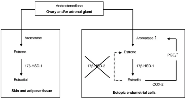
59,64또한 에스트로겐은 자궁내막증 병변의 기질세포에서 강력한 aromatase의 활성 유도제로 알려져 있는 PGE
2의 생성에 관여하는 cyclooxy- genase type-2 (COX-2) 효소의 발현을 증가시키고,
62그 결과 양성 되먹이기 고리 (positive feedback loop) 를 형성하여 병변 조직 자체에서 국소적인 에스트 로겐 생성을 지속시킨다고 한다 (Figure 1).
59,60이 러한 연구들은 자궁내막증 병변에서의 aromatase의 이상 발현이 병변의 생존과 증식을 조장하여 자궁 내막증의 발병과정에 관여할 수 있음을 시사하고 있다.
59에스트론 (estrone, E
1)과 에스트라디올 (estradiol,
E
2)은 17β-hydroxysteroid dehydrogenase (HSD)에 의 해 서로 전환될 수 있는데, 17β-HSD의 두 가지 형 태 중 type 1은 에스트론을 생물학적 활성이 더 강 한 에스트라디올로 전환시키며 type 2는 그 반대의 작용을 한다.
65Zeitoun 등은 자궁내막증 병변에서 는 17β-HSD type 1은 정상적인 발현을 보이나 type 2의 발현은 결여되어 있는 경우가 많아 에스트라 디올의 농도를 저하시키는 방어기전이 소실되어 있다고 하였다 (Figure 1).
66정상 여성의 자궁내막 에서 프로게스테론은 17β-HSD type 2의 활성을 증 가시키나,
68자궁내막증의 병변 조직에서는 활성을 유도하지 못하는 것은 프로게스테론 수용체의 발 현 역시 비정상적이기 때문이라고 한다.
21프로게스테론 수용체는 progesterone receptor (PR)-A와 PR-B의 두 가지 아이소형 (isoform)이 있 으며, 프로게스테론의 작용은 주로 PR-B를 통해 매개되며 PR-A는 PR-B의 작용을 억제한다고 한다.
69,70
프로게스테론에 대한 저항성은 자궁내막증의
Aromatase ↑ Androstenedione
Ovary and/or adrenal gland
Aromatase
Estrone
17β-HSD-1
Estradiol
Skin and adipose tissue
Estrone
Estradiol 17β-HSD-1
Ectopic endometrial cells
17β-HSD-2PGE2↑
COX-2
Figure 1. Estrogen synthesis in extraovarian tissues and endometriotic lesions. Androstenedione is converted by
aromatase to estrone (E
1) in skin, adipose tissue and also in endometriotic lesions. E
1is further converted to estradiol
(E
2) by 17β-hydroxysteroid dehydrogenase (17-βHSD) type 1. In ectopic endometrial cells, E
2induces the production
of PGE
2, which is the most potent stimulator of the aromatase. There is a positive feedback loop for continuous local
estrogen and PGE
2production in endometriotic stromal cells. Endometriotic tissue is often deficient in 17β-HSD type
2, which normally converts the strong estrogen, E
2, into weak estrogen, E
1. COX-2 = cyclo-oxygenase type 2 (Ulukus
M. et al
67).
특징적 소견일 가능성이 있는데 Attia 등은 자궁내 막증 환자의 자궁내막에서는 PR-A와 PR-B가 모두 발현되나 병변 조직에서는 PR-A만 발현되는 것을 관찰하였고,
70Igarashi 등은 자궁내막증 환자의 자 궁내 자궁내막 PR-B/PR-A 비가 정상 여성에 비해 유의하게 낮았다고 하였다.
71결
론어떤 하나의 가설만으로 자궁내막증의 모든 경 우들을 설명할 수는 없지만 월경으로 탈락된 자궁 내막 세포들이 난관을 경유, 복강으로 역류하여 복 막과 난소에 착상하여 성장함으로써 자궁내막증이 발병한다는 이식설이 현재 가장 널리 받아들여지 고 있다. 월경혈의 역류는 가임기 여성의 90%에서 관찰될 정도로 매우 흔하지만 실제로 자궁내막증 은 가임기 여성의 일부에서만 발생한다. 따라서 자 궁내막증의 발병기전에 관여하는 다른 인자들이 반드시 존재할 것으로 생각되고 있으며 유전적 소 인, 환경적 요인, 면역학적 및 내분비적 기능의 변 화 등이 중요한 역할을 할 것으로 알려져 있다.
자궁내막 조직이 복막에 착상되기 위해서는 복 막표면에 부착이 가능해야 하며, 기저막과 세포외 바탕질을 침범하고, 새로운 혈액공급을 획득하여 생존하는 과정이 필요하다. 이러한 일련의 과정들 을 인체를 대상으로 직접 실험할 수는 없지만 자 궁내막증의 여러 가지 질환 모델 체계들을 이용한 많은 in vivo 및 in vitro 연구들이 시행되어 자궁내 막증 환자의 자궁내 자궁내막에서 면역감시체계의 회피, 세포자멸사의 감소, 침습력의 증가, 스테로이 드 및 싸이토카인들의 생성 증가 등과 같은 다양 한 생화학적, 분자생물학적 이상들이 존재함을 보 고하였다. 이들 연구결과들은 자궁내막증의 발병 과정에서 환자의 자궁내막 자체의 이상이 중요한 역할을 담당할 가능성을 강력히 시사하고 있으므 로 향후 보다 체계화된 연구들이 집중되어야 할 것으로 생각된다.
참 고 문 헌
1. Sampson JA. Perforating hemorrhagic (chocolate) cysts of the ovary; their importance and especially their relation to pelvic adenomas of endometrial type (adenoma of the uterus, recto- vaginal septum, sigmoid, etc.). Archives of Surgery 1921; 3:
245-323.
2. Halme J, Hommond MG, Hulka J, Raj SG, Talbert LM.
Retrograde menstruation in healthy women and in patients with endometriosis. Obstet Gynecol 1984; 64: 151-4.
3. Jenkins S, Olive DL, Haney AF. Endometriosis: pathogenetic implications of the anatomic distribution. Obstet Gynecol 1986; 67: 335-8.
4. Ishimura T, Masuzaki H. Peritoneal endometriosis: endome- trial tissue implantation as its primary etiologic mechanism.
Am J Obstet Gynecol 1991; 165: 210-4.
5. TeLinde RW, Scott RB. Experimental endometriosis. Am J Obstet Gynecol 1950; 60: 1147-73.
6. D,Hooghe TM, Bambra CS, Raeymaekers BM, De Jonge I, Lauweryns JM. Koninckx PR. Intrapelvic injection of men- strual endometrium causes endometriosis in baboons (Papio cynocephalus and Papio anubis). Am J Obstet Gynecol 1995;
173: 125-34.
7. Cramer DW, Missmer SA. The epidemiology of endometriosis.
Ann N Y Acad Sci 2002; 955: 396-406.
8. Levander G, Normann P. The pathogenesis of endometriosis.
An experimental study. Acta Obstet Gynecol Scand 1955; 34:
366-98.
9. Suginami H. A reappraisal of the coelomic metaplasia theory by reviewing endometriosis occurring in unusual sites and instances. Am J Obstet Gynecol 1991; 165: 214-8.
10. Ichimiya M, Hirota T, Muto M. Intralymphatic embolic cells with cutaneous endometriosis in the umbilicus. J Dermatol 1998; 25: 333-6.
11. Moore JG, Binstock MA, Growdon WA. The clinical impli- cations of retroperitoneal endometriosis. Am J Obstet Gynecol 1988; 158: 1291-8.
12. Vignali M, Infantino M, Matrone R, Chiodo I, Somigliana E, Busacca M, et al. Endometriosis: novel aetiopathogenetic concepts and clinical perspectives. Fertil Steril 2002; 78: 665 -78.
13. Vigano P, Parazzini F, Somigliana E, Vercellini P. Endome-
triosis: epidemiology and aetiological factors. Best Pract Res Clin Obstet Gynaecol 2004; 18: 177-200.
14. Nisolle M, Donnez J. Peritoneal endometriosis, ovarian endo- metriosis, and adenomyotic nodules of the rectovaginal septum are three different entities. Fertil Steril 1997; 68: 585-96.
15. Vercellini P, Aimi G, Panazza S, Vincentini S, Pisacreta A, Crosignani PG. Deep endometriosis conundrum: evidence in favor of a peritoneal origin. Fertil Steril 2000; 73: 1043-6.
16. Dmowski WP, Ding J, Shen J, Rana N, Fernandez BB, Braun DP. Apoptosis in endometrial glandular and stromal cells in women with and without endometriosis. Hum Reprod 2001;
16: 1802-8.
17. Gebel HM, Braun DP, Tambur A, Frame D, Rana N, Dmowski WP. Spontaneous apoptosis of endometrial tissue is impaired in women with endometriosis. Fertil Steril 1998; 69: 1042-7.
18. Meresman GF, Vighi S, Buquet RA, Contreras-Ortiz O, Tesone M, Rumi LS. Apoptosis and expression of Bcl-2 and Bax in eutopic endometrium from women with endometriosis. Fertil Steril 2000; 74: 760-6.
19. Goumenou A, Panayiotides I, Matalliotakis I, Tzardi M, Koumantakis E. Bcl-2 and Bax expression in human endo- metriotic and adenomyotic tissues. Eur J Obstet Gynecol Reprod Biol 2001; 99: 256-60.
20. Oral E, Arici A. Pathogenesis of endometriosis. Obstet Gynecol Clin N Am 1997; 24: 219-33.
21. Haney AF, Muscato JJ, Weinberg JB. Peritoneal fluid cell populations in infertility patients. Fertil Steril 1981; 35: 696 -8.
22. Koninckx PR. Is mild endometriosis a condition occurring intermittently in all women? Hum Reprod 1994; 9: 2202-5.
23. Speroff L, Fritz MA. Clinical Gynecologic Endocrinology And Infertility. In: Endometriosis. 7th ed. Philadelphia:
Lippincott Williams & Wilkins; 2005. p.1103-33.
24. Semino C, Semino A, Pietra G, Mingari MC, Barrocci S, Venturini PL, et al. Role of major histocompatibility complex class I expression and natural killer-like T cells in the genetic control of endometriosis. Fertil Steril 1995; 64: 909-16.
25. Somigliana E, Vigano P, Gaffuri B, Guarneri D, Busacca M, Vignali M. Human endometrial stromal cells as a source of soluble intercellular adhesion molecule (ICAM)-I molecules.
Hum Reprod 1996; 11: 1190-4.
26. Garcia-Velasco JA, Arici A, Zreik T, Naftolin F, Mor G.
Macrophage derived growth factors modulate Fas ligand
expression in cultured endometrial stromal cells: a role in endometriosis. Mol Hum Reprod 1999; 5: 642-50.
27. Hill JA, Faris HM, Schiff I, Anderson DJ. Characterization of leukocyte subpopulations in the peritoneal fluid of women with endometriosis. Fertil Steril 1988; 50: 216-22.
28. Lebovic DI, Mueller MD, Taylor RN. Immunobiology of endometriosis. Fertil Steril 2001; 75: 1-10.
29. Nap AW, Groothuis PG, Demir AY, Evers J, Dunselman G.
Pathogenesis of endometriosis. Best Pract Res Clin Obstet Gynaecol 2004; 18: 233-44.
30. Kikuchi Y, Ishikawa N, Hirata J, Imaizumi E, Sasa H, Nagata I. Changes of peripheral blood lymphocyte subsets before and after operation of patients with endometriosis. Acta Obstet Gynecol Scand 1993; 72: 157-61.
31. Oosterlynck DJ, Cornillie FJ, Waer M, Vandeputte M, Kon- inckx PR. Women with endometriosis show a defect in natural killer activity resulting in a decreased cytotoxicity to autologous endometrium. Fertil Steril 1991; 56: 45-51.
32. Oosterlynck DJ, Meuleman C, Waer M, Vandeputte M, Kon- inckx PR. The natural killer activity of peritoneal lympho- cytes is decreased in women with endometriosis. Fertil Steril 1992; 58: 290-5.
33. Wu MY, Yang JH, Chao KH, Hwang JL, Yang YS, Ho HN.
Increase in the expression of killer cell inhibitory receptors on peritoneal natural killer cells in women with endometriosis.
Fertil Steril 2000; 74: 1187-91.
34. Maeda N, Izumiya C, Yamamoto Y, Oguri H, Kusume T, Fukaya T. Increased killer inhibitory receptor KIR2DL1 expression among natural killer cells in women with pelvic endometriosis. Fertil Steril 2002; 77: 297-302.
35. Groothius PG, Koks CA, de Goeij AF, Dunselman GA, Arends JW, Evers JL. Adhesion of human endometrial fragments to peritoneum in vitro. Fertil Steril 1999; 71: 1119-24.
36. Koks CA, Groothius PG, Dunselman GA, de Goeij AF, Evers JL. Adhesion of shed menstrual tissue in an in vitro model using amnion and peritoneum: a light and electron micro- scopic study. Hum Reprod 1999; 14: 816-22.
37. Dunselman GA, Groothuis PG, de Goeij AF, Evers JL. The mesothelium, Teflon or Velcro? Hum Reprod 2001; 16: 605-7.
38. Koks CA, Demir Weusten AY, Groothuis PG, Dunselman GA, de Goeij AF, Evers JL. Menstruum induces changes in mesothelial cell morphology. Gynecol Obstet Invest 2000; 50:
13-8.
39. Demir Weusten AY, Groothuis PG, Dunselman GA, de Goeij AF, Arends JW, Evers JL. Morphological changes in meso- thelial cells induced by shed menstrual endometrium in vitro are not primarily due to apoptosis or necrosis. Hum Reprod 2000; 15: 1462-8.
40. Witz CA, Thomas MR, Montoya-Rodriguez IA, Nair AS, Centonze VE, Schenken RS. Short-term culture of peritoneum explants confirms attachment of endometrium to intact peri- toneal mesothelium. Fertil Steril 2001; 75: 385-90.
41. Witz CA, Allsup KT, Montoya-Rodriguez IA, Vaughn SL, Centonze VE, Schenken RS. Culture of menstrual endome- trium with peritoneal explants and mesothelial monolayers confirms attachment to intact mesothelial cells. Hum Reprod 2002; 17: 2832-8.
42. Beliard A, Donnez J, Nisolle M, Foidart JM. Localization of laminin, fibronectin, E-cadherin, and integrins in endometrium and endometriosis. Fertil Steril 1997; 67: 266-72.
43. Witz CA. Cell adhesion molecules and endometriosis. Semin Reprod Med 2003; 21: 173-82.
44. Dechaud H, Witz CA, Montoya-Rodriguez IA, Degraffenreid LA, Schenken RS. Mesothelial cell-associated hyarulonic acid promotes adhesion of endometrial cells to mesothelium.
Fertil Steril 2001; 76: 1012-8.
45. Bruner KL, Matrisian LM, Rodgers WH, Gorstein F, Osteen KG. Suppression of matrix metalloproteinases inhibits esta- blishment of ectopic lesions by human endometrium in nude mice. J Clinic Invest 1997; 99: 2851-7.
46. Spuijbroek MD, Dunselman GA, Menheere PP, Evers JL.
Early endometriosis invades the extracellular matrix. Fertil Steril 1992; 58: 929-33.
47. Osteen KG, Rodgers WH, Gaire M, Hargrove JT, Gorstein F, Matrisian LM. Stromal-epithelial interaction mediates steroidal regulation of metalloproteinase expression in human endome- trium. Proc Natl Acad Sci USA 1994; 91: 10129-33.
48. Osteen KG, Yeaman GR, Bruner-Tran KL. Matrix metallopro- teinases and endometriosis. Semin Reprod Med 2003; 21:
155-64.
49. McLaren J. Vascular endothelial growth factor and endome- triotic angiogenesis. Hum Reprod Update 2000; 6: 45-55.
50. Charnock-Jones DS, Sharkey AM, Rajput-Williams J, Burch D, Schofield JP, Fountain SA, et al. Identification and loca- lization of alternately spliced mRNAs for vascular endothelial growth factor in human uterus and estrogen regulation in
endometrial carcinoma cell lines. Biol Reprod 1993; 48: 1120 -8.
51. Smith SK. Regulation of angiogenesis in the endometrium.
Trend Endocrinol Metab 2001; 12: 147-51.
52. Donnez J, Smoes P, Gillerot S, Cansanas-Rouz F, Nisolle M.
Vascular endothelial growth factor (VEGF) in endometriosis.
Hum Reprod 1998; 13: 1686-90.
53. McLaren J, Prentice A, Charnock-Jones DS, Smith SK.
Vascular endothelial growth factor (VEGF) concentrations are elevated in peritoneal fluid of women with endometriosis.
Hum Reprod 1996; 11: 220-3.
54. Tan XJ, Lang JH, Liu DY, Shen K, Leng JH, Zhu L.
Expression of vascular endothelial growth factor and thrombospondin-1 mRNA in patients with endometriosis.
Fertil Steril 2002; 78: 148-53.
55. Na YJ, Yang SH, Baek DW, Lee DH, Kim KH, Choi YM, et al. Effects of peritoneal fluid from endometriosis patients on the release of vascular endothelial growth factor by neutrophils and monocytes. Hum Reprod 2006; 21: 1846-55.
56. Hull ML, Charnock-Jones DS, Chan CL, Bruner-Tran KL, Osteen KG, Tom BD, et al. Antiangiogenic agents are effective inhibitors of endometriosis. J Clin Endocrinol Metab 2003; 88:
2889-99.
57. Gazvani R, Templeton A. Peritoneal environment, cytokines and angiogenesis in the pathophysiology of endometriosis.
Reprod 2002; 123: 217-26.
58. Rana N, Braun DP, House R, Gebel H, Rotman C, Dmowski WP. Basal and stimulated secretion of cytokines in peritoneal macrophages in women with endometriosis. Fertil Steril 1996; 65: 925-30.
59. Zeitoun KM, Bulun SE. Aromatase: a key molecule in the pathophysiology of endometriosis and a therapeutic target.
Fertil Steril 1999; 72: 961-9.
60. Gurates B, Bulun SE. Endometriosis: the ultimate hormonal disease. Semin Reprod Med 2003; 21: 125-34.
61. Kitawaki J, Noguchi T, Amatsu T, Maeda K, Tsukamoto K, Yamamoto T, et al. Expression of aromatase cytochrome P450 protein and messenger ribonucleic acid in human endometriotic and adenomyotic tissues but not in normal endometrium. Biol Reprod 1997; 57: 514-9.
62. Noble LS, Takayama K, Zeitoun KM, Putman JM, Johns DA, Hinshelwood MM, et al. Prostaglandin E2 stimulates aromatase expression in endometriosis-derived stromal cells. J Clin
Endocrinol Metab 1997; 82: 600-6.
63. Bulun SE, Mahendroo MS, Simpson ER. Polymerase chain reaction amplification fails to detect aromatase cytochrome P450 transcripts in normal human endometrium or decidua. J Clin Endocrinol Metab 1993; 76: 1458-63.
64. Gurates B, Sebastian S, Yang S, Zhou J, Tamura M, Fang Z, et al. WT1 and DAX-1 inhibit aromatase P450 expression in human endometrial and endometriotic stromal cells. J Clin Endocrinol Metab 2002; 87: 4369-77.
65. Andersson S, Moghrabi N. Physiology and molecular genetics of 17 beta hydroxysteroid dehydrogenases. Steroids 1997; 62:
143-7.
66. Zeitoun K, Takayama K, Sasano H, Suzuki T, Moghrabi N, Andersson S, et al. Deficient 17beta-hydroxysteroid dehydro- genase type 2 expression in endometriosis: failure to meta- bolize 17beta-estradiol. J Clin Endocrinol Metab 1988; 83:
4474-80.
67. Ulukus M, Cakmak H, Arici A. The role of endometrium in
endometriosis. J Soc Gynecol Investig 2006; 13: 467-76.
68. Casey ML, MacDonald PC, Andersson S. 17 beta-Hydroxy- steroid dehydrogenase type 2: chromosomal assignment and progestin regulation of gene expression in human endometrium J Clin Invest 1994; 94: 2135-41.
69. Vegeto E, Shahbaz MM, Wen DX, Goldman ME, O'Malley BW, McDonnell DP. Human progesterone receptor A form is a cell- and promoter-specific repressor of human progesterone receptor B function. Mol Endocrinol 1993; 7: 1244-55.
70. Attia GR, Zeitoun K, Edwards D, Johns A, Carr BR, Bulun SE. Progesterone receptor isoform A but not B is expressed in endometriosis. J Clin Endocrinol Metab 2000; 85: 2897-902.
71. Igarashi TM, Bruner-Tran KL, Yeaman GR, Lessey BA, Edwards DP, Eisenberg E, et al. Reduced expression of progesterone receptor-B in the endometrium of women with endometriosis and in cocultures of endometrial cells exposed to 2,3,7,8-tetrachlorodibenzo-p-dioxin. Fertil Steril 2005; 84:
67-74.