• 검색 결과가 없습니다.

Micro-computed tomography analysis of changes in the periodontal ligament and alveolar bone proper induced by occlusal hypofunction of rat molars

N/A
N/A
Protected

Academic year: 2022

Share "Micro-computed tomography analysis of changes in the periodontal ligament and alveolar bone proper induced by occlusal hypofunction of rat molars"

Copied!
5
0
0

로드 중.... (전체 텍스트 보기)

전체 글

(1)

Micro-computed tomography analysis of changes in the periodontal ligament and alveolar bone proper induced by occlusal hypofunction of rat molars

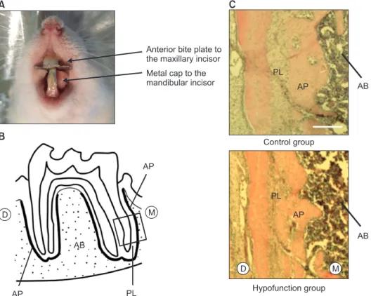
Objective: To three-dimensionally elucidate the effects of occlusal hypofunction on the periodontal ligament and alveolar bone proper of rat molars by micro- computed tomography (micro-CT). Methods: Occlusal function in the molar area was restricted by attaching an anterior bite plate on the maxillary incisors and a metal cap on the mandibular incisors of 5-week-old male Wistar rats for 1 week. The periodontal ligament space and alveolar bone proper around roots of the mandibular first molar were assessed by histology and micro-CT. Results:

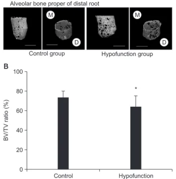
The periodontal ligament space was narrower and the alveolar bone proper was sparser and less continuous in the hypofunction group than in the control group. Further, both the volume of the periodontal ligament and the volumetric ratio of the alveolar bone proper to the total tissue in the region of interest were significantly lower in the hypofunction group ( p < 0.05). Conclusions: Occlusal hypofunction induces atrophic changes in the periodontal ligament and alveolar bone proper of rat molars.

[Korean J Orthod 2014;44(5):263-267]

Key words: Three dimensional scanner, Computed tomography, Histology, Bone biology, Occlusal stimuli, Micro-computed tomography

Yasuhiro Shimizu Jun Hosomichi Saeko Nakamura Takashi Ono

Orthodontic Science, Department of Orofacial Development and Function, Division of Oral Health Sciences, Graduate School, Tokyo Medical and Dental University, Tokyo, Japan

Received November 25, 2013; Revised February 27, 2014; Accepted March 18, 2014.

Corresponding author: Yasuhiro Shimizu.

Clinical Fellow, Orthodontic Science, Department of Orofacial Development and Function, Division of Oral Health Sciences, Graduate School, Tokyo Medical and Dental University, 1-5-45 Yushima, Bunkyo-ku, Tokyo 113-8549, Japan.

Tel +81-3-5803-5528 e-mail [email protected]

*This work was supported by a Grant-in-Aid for Scientific Research (KAKENHI; grant number 24890062) from the Japan Society for the Promotion of Science (JSPS).

© 2014 The Korean Association of Orthodontists.

The authors report no commercial, proprietary, or financial interest in the products or companies described in this article.

This is an Open Access article distributed under the terms of the Creative Commons Attribution Non-Commercial License (http://creativecommons.org/licenses/by-nc/3.0) which permits unrestricted non-commercial use, distribution, and reproduction in any medium, provided the original work is properly cited.

http://dx.doi.org/10.4041/kjod.2014.44.5.263

(2)

INTRODUCTION

The compact bone lining the tooth socket, where periodontal ligament (PDL) fibers attach, is known as bundle bone or alveolar bone proper. It is easily identifiable in radiographs and histological sections. The PDL and alveolar bone proper have a close relationship:

the PDL provides progenitor cells for bone formation and remodeling.

1-4

Occlusal hypofunction leads to atrophic changes in the periodontium.

5-12

These changes include narrowing of the PDL space, vascular constriction, and deformation of the mechanoreceptor structure (Ruffini endings).

Therefore, occlusal stimuli are essential for maintaining the structural integrity of the periodontium.

5-7,11,12

Previous histological studies have shown regressive changes in the periodontium of rat molars after loss of occlusal stimuli,

7,11,12

but three-dimensional morpho- metric analysis has not yet been reported. In this study, we aimed to three-dimensionally elucidate the effects of occlusal hypofunction on the PDL and alveolar bone proper of rat molars by micro-computed tomography (micro-CT).

MATERIALS AND METHODS

Experimental model

Five-week-old male Wistar rats (n = 18) were ran dom ly divided into two equal groups. The control group was not treated. To restrict occlusal function in the hypofunction group, an anterior bite plate was atta ched on the ma xillary incisors and a metal cap constructed from or tho dontic band material (4.6 × 0.13 mm; Rocky Mountain Morita Corp., Tokyo, Japan) was attached on the mandibular incisors by using photopolymerizing composite resin (Clearfil Liner Bond 2; Kuraray Dental, Okayama, Japan) (Figure 1A).

8-10

Body weight was mo nitored daily.

8-10

One week later, the animals were sacri ficed by cervical dislocation under diethyl ether ane sthesia (Wako Pure Chemical Ind. Ltd., Osaka, Ja pan).

8,10

The experimental procedures were reviewed and approved by the Institutional Animal Care and Use Committee of the Tokyo Medical and Dental University (approval number

#0130279A).

Histology

The mandibles were dissected and most soft tissue was removed. The specimens were immediately immersed

Figure 1. A, The experimental model; B, histological observational area (rectangular area); C, representative hematoxylin

and eosin-stained sections (Scale = 250 mm). M, Mesial; D, distal; PL, periodontal ligament; AP, alveolar bone proper; AB,

inter-radicular alveolar bone.

(3)

in 10% buffered formalin (Wako Pure Chemical Ind.

Ltd.; pH 7.0) at 4

o

C overnight and decalcified in 10%

ethylenediaminetetraacetic acid (EDTA) at 4

o

C for 6 weeks. They were embedded in paraffin and cut into 5-mm-thick sagittal sections parallel to the long axis of the roots of the mandibular first molar.

8,10

The sections were finally mounted on glass slides coated with poly-L-lysine (Matsunami Glass Ind. Ltd., Osaka, Japan) and stained with hematoxylin and eosin for histomorphometric examination. The observational area included the dental pulp, PDL, alveolar bone proper, and inter-radicular alveolar bone (Figure 1B).

Micro-CT analysis

The histological observational area was imaged by using a desktop X-ray micro-CT system (SMX- 90CT; Shimadzu Corp., Kyoto, Japan) with a scanning resolution of 20 μm. First, the mesial and distal roots of the mandibular first molar were manually located at 20 mm intervals on individual images. The selected images were compiled into a three-dimensional image by using image-analysis software (TRI/3-D Bon; Ratoc System Engineering Co. Ltd., Tokyo, Japan). The region of interest was enlarged to a 40 mm radius around

the roots.

8-10

The software was then used for data quantification. Tissue volume (TV) was defined as the volume of tissue in the enlarged region of interest. The bone-to-tissue volume (BV/TV) ratio was calculated as the volumetric ratio of bone to total tissue in the enlarged region of interest. Both parameters were evaluated according to the software instructions.

Statistical analysis

Statistical calculations were performed by using StatView 5.0 software (SAS Institute, Cary, NC, USA).

The Mann-Whitney U-test was used for all comparisons at the significance level of p < 0.05.

RESULTS

No significant differences in body weight were noted between the groups at the time of killing.

Histologically, the PDL space in the hypofunction group was narrower than that in the control group (Figure 1C). Furthermore, the alveolar bone proper showed less continuity in the hypofunction group (Figure 1C).

Figure 2A shows that the PDL space around the mesial

Figure 2. A, Three-dimensional reconstructed images of the periodontal ligament (PDL) space (Scale = 1 mm); B, comparison of tissue volume (TV) around the mesial root of the mandibular first molar.

***p < 0.001 by the Mann-Whitney U-test.

M, Mesial; D, distal.

Figure 3. A, Three-dimensional reconstructed images of the periodontal ligament (PDL) space (Scale = 1 mm); B, comparison of tissue volume (TV) around the distal root of the mandibular first molar.

***p < 0.001 by the Mann-Whitney U-test.

M, Mesial; D, distal.

(4)

root was thinner in the hypofunction group than in the control group. Identical narrowing of the PDL space was observed around the distal root in the hypofunction group (Figure 3A). As shown in Figure 4A, the alveolar bone proper around the mesial root was more discontinuous in the hypofunction group. Furthermore, the alveolar bone proper around the distal root was sparser in this group (Figure 5A).

In response to occlusal hypofunction, the PDL volume around both the roots significantly reduced (Figures 2B and 3B). The BV/TV ratio of the alveolar bone proper was also significantly lower around both the roots in the hypofunction group (Figures 4B and 5B).

DISCUSSION

We reduced occlusal function in the molar area by using an anterior bite plate on the maxillary incisors and a metal cap on the mandibular incisors.

8-10

We selected the mandibular first molar as the region of interest because it is exposed to concentrated occlusal stimuli and has been used in previous studies of alveolar bone.

8,10

By micro-CT, we succeeded in building a three-dimensional representation of the PDL space and alveolar bone proper.

Occlusal hypofunction caused the molar PDL space to become significantly narrow, consistent with previous reports

5-12

and supporting the view that occlusal stimuli are important for thickening PDL fibers and maintaining the PDL space.

6,7,11,12

Further, the alveolar bone proper was discontinuous and its BV/TV ratio was significantly reduced. We determined the adaptive threshold of bone by using the “Discriminant Analysis” function of the image-analysis software. Bone images were also visually examined with more than the calculated threshold values to confirm the accuracy of our estimations.

This adaptive threshold was then used to discriminate areas of bone from areas of other tissue and was kept constant across all the examined sections.

Mechanical stimuli are important for maintaining bone.

13

Alveolar bone is particularly responsive because of its high turnover under mechanical stress (e.g., occlusal force) transmitted from the teeth through the PDL.

14,15

When occlusal force is absent, osteoclast numbers are boosted by the expression of the oste oclast differentiation factor, receptor activator of nu clear factor kappa-B ligand (RANKL), which leads to bone resorption.

8,10

Cellular components of the PDL participate in the formation and resorption of periodontal hard tissues.

16-18

Therefore, discontinuity of the alveolar bone Figure 4. A, Three-dimensional reconstructed images of

the alveolar bone proper (Scale = 1 mm); B, comparison of the bone-to-tissue volume (BV/TV) ratio around the mesial root of the mandibular first molar.

*p < 0.05 by the Mann-Whitney U-test.

M, Mesial; D, distal.

Figure 5. A, Three-dimensional reconstructed images of the alveolar bone proper (Scale = 1 mm); B, comparison of the bone-to-tissue volume (BV/TV) ratio around the distal root of the mandibular first molar.

*p < 0.05 by the Mann-Whitney U-test.

M, Mesial; D, distal.

(5)

proper in this hypofunction model may have risen from suppression of bone formation and/or acceleration of bone resorption by cellular components of the PDL.

Further investigation is needed to reveal how osteoblasts and osteoclasts in the PDL affect the alveolar bone proper under conditions of occlusal hypofunction.

CONCLUSION

Occlusal hypofunction induces atrophic changes in the PDL and alveolar bone proper of rat molars. This study is the first three-dimensional evaluation of the PDL and alveolar bone proper by micro-CT under conditions of occlusal hypofunction.

REFERENCES

1. Gault P, Black A, Romette JL, Fuente F, Schroeder K, Thillou F, et al. Tissue-engineered ligament: implant constructs for tooth replacement. J Clin Periodontol 2010;37:750-8.

2. Sebaoun JD, Kantarci A, Turner JW, Carvalho RS, Van Dyke TE, Ferguson DJ. Modeling of trabecular bone and lamina dura following selective alveolar decortication in rats. J Periodontol 2008;79:1679- 88.

3. Devlin H, Sloan P. Early bone healing events in the human extraction socket. Int J Oral Maxillofac Surg 2002;31:641-5.

4. Gay IC, Chen S, MacDougall M. Isolation and charac terization of multipotent human periodontal ligament stem cells. Orthod Craniofac Res 2007;10:

149-60.

5. Hayashi Y, Iida J, Warita H, Soma K. Effects of occlusal hypofunction on the microvasculature and endothelin expression in the periodontal ligaments of rat molars. Orthod Waves 2001;60:373-80.

6. Tanaka A, Iida J, Soma K. Effect of hypofunction on the microvasculature in the periodontal ligament of the rat molar. Orthodontic Waves 1998;57:180-8.

7. Muramoto T, Takano Y, Soma K. Time-related changes in periodontal mechanoreceptors in rat molars after the loss of occlusal stimuli. Arch Histol Cytol 2000;63:369-80.

8. Enokida M, Kaneko S, Yanagishita M, Soma K.

Inference of occlusal stimuli on the remodelling of alveolar bone in a rat hypofunction-recovery model.

J Oral Biosci 2005;47:321-34.

9. Shimomoto Y, Chung CJ, Iwasaki-Hayashi Y, Mura- moto T, Soma K. Effects of occlusal stimuli on alveolar/jaw bone formation. J Dent Res 2007;86:

47-51.

10. Shimizu Y, Hosomichi J, Kaneko S, Shibutani N, Ono T. Effect of sympathetic nervous activity on alveolar bone loss induced by occlusal hypofunction in rats.

Arch Oral Biol 2011;56:1404-11.

11. Motokawa M, Terao A, Karadeniz EI, Kaku M, Kawata T, Matsuda Y, et al. Effects of long-term occlusal hypofunction and its recovery on the mor- pho genesis of molar roots and the periodontium in rats. Angle Orthod 2013;83:597-604.

12. Watarai H, Warita H, Soma K. Effect of nitric oxide on the recovery of the hypofunctional periodontal ligament. J Dent Res 2004;83:338-42.

13. Bourrin S, Palle S, Genty C, Alexandre C. Physical exercise during remobilization restores a normal bone trabecular network after tail suspension- induced osteopenia in young rats. J Bone Miner Res 1995;10:820-8.

14. Vignery A, Baron R. Dynamic histomorphometry of alveolar bone remodeling in the adult rat. Anat Rec 1980;196:191-200.

15. Johnson RB. Effect of altered occlusal function on transseptal ligament and new bone thicknesses in the periodontium of the rat. Am J Anat 1990;187:

91-7.

16. Meeran NA. Cellular response within the periodontal ligament on application of orthodontic forces. J Indian Soc Periodontol 2013;17:16-20.

17. Mayahara K, Yamaguchi A, Takenouchi H, Kariya T, Taguchi H, Shimizu N. Osteoblasts stimulate oste- oclastogenesis via RANKL expression more strongly than periodontal ligament cells do in res ponse to PGE(2). Arch Oral Biol 2012;57:1377-84.

18. Krishnan V, Davidovitch Z. Cellular, molecular, and

tissue-level reactions to orthodontic force. Am J

Orthod Dentofacial Orthop 2006;129:469. e1-32.

수치

Figure 1. A, The experimental model; B, histological observational area (rectangular area); C, representative hematoxylin  and eosin-stained sections (Scale = 250 mm)
Figure 3. A, Three-dimensional reconstructed images of  the periodontal ligament (PDL) space (Scale = 1 mm); B,  comparison of tissue volume (TV) around the distal root  of the mandibular first molar
Figure 5. A, Three-dimensional reconstructed images of  the alveolar bone proper (Scale = 1 mm); B, comparison  of the bone-to-tissue volume (BV/TV) ratio around the  distal root of the mandibular first molar

참조

관련 문서

After choosing the type of bike, the next step is the right bike size. the right size for you from

44 글의 첫 번째 문장인 The most important thing in the Boat Race is harmony and teamwork.을 통해 Boat Race에서 가장 중요한 것은 조 화와 팀워크임을

 The Dutch physicist Pieter Zeeman showed the spectral lines emitted by atoms in a magnetic field split into multiple energy levels...  With no magnetic field to align them,

Modern Physics for Scientists and Engineers International Edition,

If both these adjustments are considered, the resulting approach is called a bootstrap-BC a -method (bias- corrected-accelerated). A description of this approach

③ A student who attended Korean course at KNU Korean Language Program and holds TOPIK Level 3 or a student who completed Korean course Level 4 at the KNU Korean Language

· 50% exemption from tuition fee Ⅱ for the student with a TOPIK score of level 3 or higher or completion of level 4 or higher class of the Korean language program at the

웹 표준을 지원하는 플랫폼에서 큰 수정없이 실행 가능함 패키징을 통해 다양한 기기를 위한 앱을 작성할 수 있음 네이티브 앱과