DOI:10.4078/jkra.2009.16.1.54
<접수일:2009년 1월 2일, 심사통과일:2009년 2월 23일>
※통신저자:전 재 범
서울시 성동구 행당동 17번지
한양대학교 의과대학 내과학교실 류마티스병원 류마티스내과
Tel:02) 2290-9244, Fax:02) 2298-8231, E-mail:[email protected]
전신홍반루푸스에 동반된 IgA 신병증 1예
한양대학교 의과대학 내과학교실 류마티스병원 류마티스내과, 한양대학교 의과대학 신장내과학교실1, 호흡기학교실2, 병리학교실3
방소영ㆍ김근호
1ㆍ신동호
2ㆍ박문향
3ㆍ전재범
= Abstract =
A Case of IgA Nephropathy Associated with Systemic Lupus Erythematosus
So-Young Bang, Gheun Ho Kim1, Dong Ho Shin2, Moon Hyang Park3, Jae-Bum Jun
Division of Rheumatology, Department of Internal Medicine, The Hospital for Rheumatic Diseases, Departments of Nephrology
1, Pulmonary Medicine
2, and Pathology
3,
Hanyang University College of Medicine, Seoul, Korea
Renal involvement is frequently seen in patients with systemic lupus erythematosus (SLE).
The occurrence of non-lupus nephritis, and especially IgA nephropathy, in SLE patients has rarely been reported. We describe here the case of a 30-year-old woman who had systemic lupus erythematosus and nontuberculous mycobacterial lung disease, and her biopsy of a renal lesion was unexpectedly diagnostic of IgA nephropathy. Although both IgA nephropathy and lupus nephritis are immune complex mediated diseases, their laboratory and histopathologic findings and the extra-renal clinical manifestations are different and these all support a different pathogenesis for the 2 diseases. Renal biopsy plays a crucial role in identifying and diagnosing renal lesions, which may have prognostic and therapeutic implications that are distinct from those of lupus nephritis. In conclusion, performing a renal biopsy in SLE patients who have urinary abnormalities is important since a correct diagnosis would permit the most appropriate treatment to be started and so avoid unnecessary immunosuppressive treatments.
Key Words: IgA nephropathy, Systemic lupus erythematosus, Non-tuberculous mycobacterium
서 론
전신홍반루푸스(systemic lupus erythematosus, 루푸 스)에서 면역복합체에 의한 신장의 침범은 흔하다.
루푸스 신염은 World Health Organization classification 의 분류와 그 후 변화를 거쳐 최근 International So- ciety of nephrology/Renal Pathology Society (ISN/RPS) 2003 분류에서 제시한 여섯 가지로 분류되며, 치료방 법의 결정과 예후를 알기 위해 신장 조직검사를 시행 한다 (1-3). 한편 IgA 신병증은 세계적으로 가장 흔한 일차성 사구체신염의 한 형태로, 신생검으로 진단되 는 빈도는 미국에서는 사구체신염의 5∼10%, 유럽에 서는 20%, 우리나라를 포함한 아시아에서는 40% 정 도이다 (4). 루푸스 환자의 신장 조직검사에서 비루푸 스 신염을 동반한 증례들이 드물게 보고되고 있지만 (5), 그 중에서도 IgA 신병증은 매우 드문 경우로 1995년 Mac-Moune Lai 등이 처음으로 3예를 보고한 이후 (6) 국내외적으로 드물게 보고되고 있다 (7,8).
저자들은 비결핵 항산균 폐질환으로 치료중인 전 신홍반루푸스환자에서 IgA 신병증으로 진단받은 1예 를 보고하는 바이다.
증 례
환 자: 30세 여자 주 소: 피로감, 단백뇨
현병력: 13년 전 전신홍반루푸스를 진단받았으며, 내원 5개월 전부터 비결핵 항산균(non-tuberculous my- cobacterium, NTM) 폐질환으로 치료 중에 있던 중, 최 근 피로감과 소변검사에서 단백뇨가 있어 내원하였다.
과거력: 13년 전 구강궤양, 피부발진, 다발성관절 염을 주소로 내원하여 항핵항체 양성, 항 ds-DNA항 체양성, 혈소판감소증 등으로 전신홍반루푸스로 진 단받았다. 5년 전 심한 혈소판 감소증으로 고용량 스테로이드 치료 중 Mycobacterium avium으로 배양 된 NTM 폐질환이 발생하여 항결핵제 치료 후 호전 되었다. 내원 5개월 전 NTM 폐질환이 재활성화되었 고, 객담검사에서 Mycobacterium avium이 배양되어 치료 중이었다. 루푸스에 대해서는 임상소견과 질병 의 활성도가 호전을 보여 현재까지 항말라리아제제
(hydroxychloroquine 400 mg)와 저용량의 스테로이드 (prednisolone 5 mg)로 치료 중이었다.
가족력: 특이사항 없음.
이학적 소견: 내원시 체온 36.9oC, 맥박은 80회/분, 호흡수는 20회/분, 혈압은 130/80 mmHg였다. 피부발 진은 없었고, 경정맥 확장소견은 보이지 않았으며 흉부 청진상 양측 폐야에서 호흡음은 비교적 정상이 었고 심잡음은 청진되지 않았다. 복부에는 특이소견 없었으며, 척추 늑골각 압통은 없었고, 양 하지에서 함요 부종이 관찰되지 않았다. 신경학적 검사에서 이상소견은 없었다.
검사실 소견: 말초 혈액검사에서 백혈구 9,500/mm3 (중성구 57%, 림프구 28%), 혈색소 12.3 g/dL, 혈소 판 282,000/mm3이었다. 생화학 검사에서 나트륨 137 mEq/L, 칼륨 4.0 mEq/L, 총 단백 7.5 g/dL, 알부민 4.1 g/dL, aspartate aminotransferase (AST) 22 IU/L, ala- nine aminotransferase (ALT) 10 IU/L, blood urea nitro- gen (BUN) 16 mg/dL, 크레아티닌 0.8 mg/dL, lactate dehydrogenase 172 U/L, creatine phosphokinase (CPK) 28 U/L, total cholesterol/LDL cholesterol/triglyceride/
HDL cholesterol은 각각 203 mg/dL, 132 mg/dL, 107 mg/dL, 41 mg/dL이었다. 면역학적 검사에서 항핵항 체 양성(homogenous level 4, 1:2,560), 항ds-DNA 항 체 1:320 양성이었다. 혈청 C3는 86 mg/dL (90∼
180 mg/dL)로 약간 감소하였고, C4는 10.2 mg/dL (10∼40 mg/dL), CH50은 46 units/mL (23∼46 units/
mL)로 정상범위 내에 있었다. 적혈구침강속도는 34.0 mm/hr (Wintrobe법), C-반응단백은 <0.3 mg/dL으 로 정상이었고, 류마티스인자, 항CCP항체, ANCA, 항 Ro항체, 항 La항체, 항 RNP항체, 항 Sm항체는 모두 음성이었다. IgA는 522 mg/dL (70∼400 mg/dL), ASO 227 U/mL (<200 mg/dL)이었고, B형 간염, C형 간 염, HIV 항체는 모두 음성이었다.
소변검사에서 비중 1.025, pH 6.5, 단백뇨 3+, 잠 혈 3+, 백혈구 10-19/HPF, 적혈구 many/HPF이었다.
24시간 소변 검사에서 단백질 1,356 mg/day, CCr 79 mL/min/m2이었다.
흉부 단순 방사선 사진에서 5개월 전보다 양측 폐 야에 폐결핵 소견은 다소 호전을 보였다(그림 1). 복 부 초음파 검사에서는 특이 소견없이 우측 신장은 12.8 cm, 좌측 신장은 14.4 cm로 측정되었다. 단백뇨
Fig. 1. Multifocal infiltrations of non-tuberculosis myco- bacterium in both lungs are noted on the chest X-ray.
Fig. 2. The mesangium is mildly expanded with prolife- rations of mesangial cells and expansion of the mesangial matrix. Mild tubular atrophy and inter- stitial fibrosis are seen (PAS, ×400).
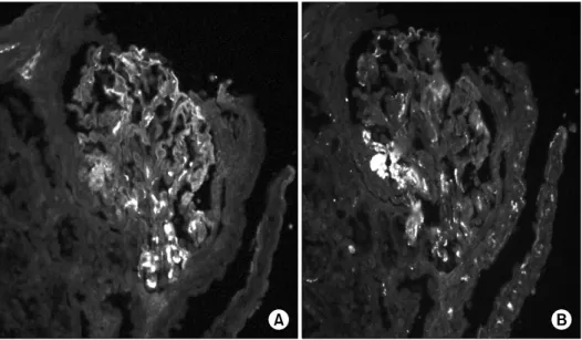
Fig. 3. Direct immunofluorescence microscopy demonstrates positive mesangial staining for IgA (A) and C3 (B) (×400).
와 혈뇨에 대한 진단과 치료방법의 결정을 위해 신 장 조직검사를 시행하였다.
병리 소견: 광학현미경 검사 표본에는 15개의 사구 체가 포함되었다. 대부분의 사구체는 미만성으로 메 산지움이 확장되었고 메산지움 기질(mesangium ma- trix) 내에 약간의 면역물질의 침착이 보였으나, 기저
막의 비후나 모세혈관 내 내피세포(endothelium)의 증식은 관찰되지 않았다(그림 2). 면역 형광현미경 검사에서 메산지움은 분절성으로 IgA와 C3에 대해 면역형광 반응을 관찰할 수 있었고, IgG나 C1q는 분 절성으로 매우 소량 관찰되었으며, IgG와 C4, Fibri- nogen에 대한 반응은 음성이었다(그림 3). 전자현미
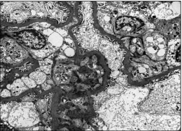
Fig. 4. The electron micrograph demonstrates electron dense deposits in the mesangial and paramesangial areas (×6,000).
경검사에서 메산지움과 그 근위부(paramesangium)를 따라 뚜렷한 전자고밀도 침착물(electron dense depos- it)이 관찰되었다. 내피세포에 소수의 tubulo-reticular 구조가 보였으나 사구체 기저막의 내피세포와 상피 세포 하에서는 전자고밀도 침착물을 관찰할 수 없었 다(그림 4). 상기 병리 소견에 의해 Lee 분류에 따라 IgA 신병증, grade 3으로 진단하였다.
치료 및 경과: 환자는 hydroxychloroquine 400 mg, prednisolone 5 mg를 유지하며, 안지오텐신 전환억제 제(lisinopril 5 mg)를 투여하기 시작하였고, 항결핵제 (clarithromycin, moxifloxacin)를 복용 중이다. 현재 2 개월째 외래 추적 중 단백뇨는 362 mg/day로 호전 되었으며, 환자는 특별한 증상 없이 외래에서 관찰 중이다.
고 찰
IgA 신병증은 면역 반응의 장애로 골수에서 poly- meric IgA1이 과다하게 생산되어 사구체혈관간부에 IgA와 C3가 주로 침착되는 사구체 신염이다 (4). IgA 신병증은 IgA가 과다하게 생산되거나 간에서의 배설 장애를 보이는 경우를 병인으로 설명하고 있다. 원 인질환이 없는 경우가 많지만, 간질환, 강직성 척추 염, 건선, Reiter 증후군, 포도막염, 염증성 장질환, celiac 질환, 후천성 면역결핍증, 그리고 드물게 폐결 핵과 동반된 경우가 보고되고 있다 (4,9).
결핵과 동반되는 IgA 신병증의 발생기전은 결핵균 (Mycobacterium tuberculosis)에 대한 세포 매개에 의 한 면역 반응으로 알려져 있으며, 마이코박테리움 항원(mycobacterial antigen) A-60에 대한 특수한 IgA 생산에 의해 혈청 IgA가 증가되고, 증가된 IgA가 면 역복합체의 생성을 유도하여 신독성을 유발한다고 여겨진다 (9,10). 국외에서 다양한 사구체 신염들과 관련된 활동성 폐결핵이 보고되었는데, IgA 신병증 이 동반된 경우 이에 대한 특별한 다른 치료 없이, 항결핵제의 사용만으로도 단백뇨와 다른 임상 증상 등이 호전되었다 (11). 또한 비결핵 항산균(non-tuber- culous mycobacterium, NTM) 폐질환이 있는 환자에 서 IgA 신병증이 동반된 보고는 없으나, NTM 폐질 환과 사구체 신염이 동반된 국외의 증례보고에서는 비결핵 항산균 치료만으로 신기능이 향상되었다 (12,13).
루푸스에 있어서 신염은 면역복합체의 사구체내 침착을 특징으로 하고 그 임상 양상은 무증상 현미 경적 혈뇨, 단백뇨에서부터 말기 신부전까지 다양하 다. Baranowska-Daca 등은 1988년부터 10년간 루푸 스 환자의 7% (25예/367예)에서 신장조직검사를 통 해 비루푸스 신염을 진단하였다 (7). 이처럼 적은 수 이지만 루푸스 환자에서 신조직 검사에서는 다양한 비루푸스 신염이 보일 수 있다. 루푸스 신염에 있어 서는 주로 IgG와 보체의 전형적 경로(classical path- way)의 산물인 C4, C1q의 침착을 보이나 IgA 신병 증은 혈관 간부(mesangium)에 IgA와 교체경로 (alter- native pathway)의 산물인 C3가 주된 침착을 보인다 는 것이 차이점이나, 공통적으로 C1q 에 대한 항체 가 발견되고, 보체 활성 산물이 두 질병의 병인에 연관되었다는 가설이 제시되고 있다 (14,15).
따라서, 본 증례를 통해서 루푸스 환자에서 소변 검사에서 단백뇨 등의 이상이 나타날 때, 드물지만 루푸스 신염 이외에도 IgA 신병증을 포함한 비루푸 스 신염의 가능성이 있으므로 신장조직검사없이 루 푸스 신염만을 생각하여 불필요한 면역억제제 치료 를 하지 말아야 한다는 교훈을 얻을 수 있었다.
요 약
전신홍반루푸스환자에서 소변검사에서 이상소견이
발생한 경우에는 루푸스 신염뿐만 아니라 드물게 IgA 신증을 포함한 비루푸스 신염의 경우도 있으므로, 이를 염두에 두어 신장 조직검사로 정확한 진단을 하여 적절한 치료방법을 결정하는 것이 중요하겠다.
참고문헌
1) Kim HW, Kang SW, Choi KH, Lee HY, Han DS, Kie JH, et al. A case of IgA nephropathy with systemic lupus erythematosus. Korean J Med 2004;66:190-94.
2) Han KW, Lee YK, Lee HR, Hwang SI, Kim SG, Oh JE, et al. A case of IgA nephropathy with systemic lupus nephritis. Korean J Nephrol 2005;24:326-31.
3) Park MH. International society of nephropathy/renal pathology society 2003 classification of lupus ne- phritis. Korean J Pathol 2006;40:165-75.
4) Galla JH. IgA nephropathy. Kidney Int 1995;47:377- 87.
5) Baranowska-Daca E, Choi YJ, Barrios R, Nassar G, Suki WN, Truong LD. Nonlupus nephritides in patients with systemic lupus erythematosus: a com- prehensive clinico-pathologic study and review of the literature. Hum Pathol 2001;32:1125-35.
6) Mac-Moune Lai F, Li EK, Tang NL, Li PK, Lui SF, Lai KN. IgA nephropathy: a rare lesion in systemic lupus erythematosus. Mod Pathol 1995;8:5-10.
7) Baranowska-Daca E, Choi YJ, Barrios R, Cartwigh J, Sheik-Hamad D, Truong LD. Non-lupus nephritis in patients with SLE. Lab Invest 1999;79:154A.
8) Corrado A, Quarta L, Di Palma AM, Gesualdo L, Cantatore FP. IgA nephropathy in systemic lupus ery- thematosus. Clin Exp Rheumatol 2007;25:467-9.
9) De Siati L, Paroli M, Ferri C, Muda AO, Bruno G, Barnaba V. Immunoglobulin A nephropathy compli- cating pulmonary tuberculosis. Ann Diagn Pathol 1999;3:300-3.
10) Alifano M, Sofia M, Mormile M, Micco A, Mormile AF, Del Pezzo M, et al. IgA immune response against the mycobacterial antigen A60 in patients with active pulmonary tuberculosis. Respiration 1996;63:292-7.
11) De Siati L, Paroli M, Ferri C, Muda AO, Bruno G, Barnaba V. Immunoglobulin A nephropathy compli- cating pulmonary tuberculosis. Ann Diagn Pathol 1999;3:300-3.
12) Chen SJ, Wen YK, Chen ML. Rapidly progressive glomerulonephritis associated with nontuberculous mycobacteria. J Chin Med Assoc 2007;70:396-9.
13) Wen YK, Chen ML. Crescentic glomerulonephritis associated with non-tuberculous mycobacteria infec- tion. Ren Fail 2008;30:339-41.
14) Gunnarsson J, Ronnelid J, Lundberg I, Jacobson SH.
Occurence of anti-C1q antibodies in IgA nephropathy.
Nephrol Dial Transplant 1997;12:2263-8.
15) Topaloglu R, Bakkaloglu A, Slingsby JH, Mihatsch MJ, Pascual M, Norsworthy P, et al. Molecular basis of hereditary C1q deficiency associated with SLE and IgA nephropathy in a Turkish family. Kidney Int 1996;50:635-42.