第23卷 第2號 2015年 2月 20日
I. 서 론
* 교신저자 : 진미림, 대전대학교 한의과대학 약리학교실 E-mail : [email protected] Tel : 042-280-2633 투고일 : 2014년11월11일 수정일 : 2014년12월12일 게재일 : 2014년12월12일
사상자탕(蛇床子湯)은 사상자(蛇床子), 고삼(苦 蔘), 당귀(當歸), 위령선(威靈仙)으로 구성되어 있는 처방으로 전통 한의서인 外科正宗에 “腎 囊風乃肝經風濕而成. 其患作癢, 喜浴熱湯, 甚者 疙瘩頑麻, 破流脂水, 宜蛇床子湯熏洗二次卽愈”
사상자탕과 가미사상자탕의 항염증 및 항산화 효과 비교 연구
최정은1․박보경1․진미림1 *9)
Anti-inflammatory and Antioxidant Effects of Water Extracts of Sasangja-tang(SSJ) and Gami-sasangja-tang(GSJ)
Choi Jung –eun1 ・ Park Bo-kyung1 ・ Jin Mirim1 *
1Laboratory of Pharmacology, College of Korean Medicine, Daejeon University
Objectives : The anti-inflammatory and antioxidant effects of water extracts of Sasangja-tang(SSJ) and Gami-sasangja-tang(GSJ) were investigated. The effects of SSJ and GSJ were compared.
Methods : We performed cell viability assay in HaCaT cells and RAW 264.7 cells using 3- (4,5-dimethylthiazol-2-yl)-2, 5 diphenyltetrazolium bromide(MTT) assay. We measured chemokines( regulated on activation normal T-cell expression and secreted ; RANTES/CCL5, interferon-inducible protein;
IP-10/CXCL10, macrophage-derived chemokine; MDC/CCL22) in HaCat cells, also we measured cytokines (tumor necrosis factor-α; TNF-α, interleukin-6; IL-6) and nitric oxide(NO) production in RAW 264.7 cells using enzyme-linked immunosorbent assay(ELISA) and NO assay. Western blot assay was used to evaluate the expression for inducible nitric oxide synthase(iNOS) in RAW 264.7 cells.
Results : SSJ and GSJ did not affect the cell viability at the concentrations treated (0-800 µg/ml). As a result of SSJ and GSJ treatment in HaCat cells stimulated by TNF-α(10 ng/ml) and interferon(IFN)-γ(10 ng/ml), the production of RANTES and IP-10 was inhibited significantly. However there was no significant difference in the secretion of MDC. And in RAW 264. 7 cells stimulated by lipopolysaccharide(LPS, 1 μg/ml), SSJ and GSJ treatment significantly inhibited the secretion of TNF-α and IL-6 and the production of NO. The expression of iNOS was also decresed by SSJ and GSJ treatment in RAW 264. 7 cells. Compared with SSJ, GSJ was superior to SSJ in inhibition of RANTES, IP-10, TNF-α, IL-6 and NO production at the concentration of 200 µg/ml.
Conclusion : Both SSJ and GSJ have anti-inflammatory and antioxidant effects. And GSJ has better effects than SSJ.
Key Words : Sasangja-tang(SSJ), Gami-sasangja-tang(GSJ), anti-inflammatory effect, antioxidant effect, cytokine, chemokine
라고 기록되어 있으며 오랫동안 주로 염증성 피 부질환에 사용되어 왔다1). 가미사상자탕(加味蛇 床子湯)은 사상자탕 처방에 백부근(百部根)과 부 평초(浮萍草)를 가미한 처방으로, 백부근(百部 根)은 항균효과와 면역조절작용에 대해 보고된 바 있으며2),3) 부평초(浮萍草)는 항알러지 효과가 있다고 알려져 있다4).
HaCaT 세포는 표피의 대부분을 차지하는 각 질형성세포로서, 다양한 염증성 사이토카인 (cytokine)과 케모카인(chemokine)을 생성하여 피부의 면역반응실험에 주로 사용되고 있고5), RAW 264.7 세포는 대식세포로 초기 염증성 사 이토카인 분비 및 nitric oxide(NO)를 생성하는 염증성 세포로서 염증매개질환 연구에 주로 사용 되고 있다6). 활성화된 HaCaT 세포에서 발현되 는 regulated on activation normal T-cell expression and secreted(RANTES/CCL5), interferon-inducible protein-10(IP-10/CXCL10), macrophage-derived chemokine(MDC/CCL22) 등 의 케모카인은 백혈구의 이동과 활성을 조절하는 인자로서 염증세포의 조직 침윤을 유도한다7). 활 성화된 RAW 264.7 세포에서 발현되는 interleukin(IL)-6, tumor necrosis factor-alpha (TNF-α) 등의 사이토카인은 pro-inflammatory cytokine으로서 inducible nitric oxide synthase (iNOS)와 cyclooxygenase(COX)-2를 합성하여 nitric oxide(NO) 생성을 촉진시키고 염증 반응 을 유도한다8). 유도된 NO는 염증상태에서 부종 등을 일으키고 염증매개체의 생합성을 촉진시켜 염증을 심화시킬 수 있다9). 이러한 연구 결과를 토대로, 본 실험에서는 사상자탕과 가미사상자탕 의 염증성 피부질환에 대한 효능 평가를 하기 위 해, 각질형성세포인 HaCaT 세포와 대식세포인 RAW 264.7 세포를 이용하여 사상자탕 물추출물 (SSJ)과 가미사상자탕 물추출물(GSJ)의 항염증 및 항산화 효과를 실험하였다10).
Ⅱ. 재료 및 방법
1. 재료 및 추출물 제조
사상자(蛇床子), 고삼(苦蔘), 당귀(當歸), 위령 선(威靈仙), 백부근(百部根), 부평초(浮萍草)를 동경제약(주)(Seoul, Korea)에서 구입하여 사상 자탕(1:1:1:1) 또는 가미사상자탕(1:1:1:1:1:1) 처방에 따라 무게를 재고 사상사탕 80 g, 가미사 상자탕 120 g에 증류수 1ℓ를 넣어 100℃에서 2 시간동안 끓였다. 끓인 약재를 Whatman No. 2 filter(Maidstone, UK)에 걸러주고 모은 여액을 감압증류기를 사용하여 농축하고 동결건조하여 사상자탕 8.3 g(10.4 %), 가미사상자탕 12.6 g(10.5%)의 분말을 얻었다. 얻어진 분말은 –8 0℃에 나누어 분주하여 보관하였고 사용하기 전 에 증류수로 희석하여 필터 후 사용하였다.
Table 1. Prescription of Sasangja-tang(SSJ)
Herb Scientific name Amount(g)
蛇床子 Cnidii fructus 20
苦蔘 Sophora flavescens root 20 當歸 Angelica gigas root 20
威靈仙 Clematidis radix 20
Total amount 80
Table 2. Prescription of Gami-sasangja-tang(GSJ)
Herb Scientific name Amount(g)
蛇床子 Cnidii fructus 20
苦蔘 Sophora flavescens
root 20
當歸 Angelica gigas root 20
威靈仙 Clematidis radix 20
百部根 Stemonae radix 20
浮萍草 Spirodelae herba 20
Total amount 120
2. 세포배양 및 시약
실험에 사용한 RAW 264.7 세포는 한국세포주 은행(KTCC, Seoul, Korea)에서 분양받았으며 HaCat 세포는 현진원 교수님(Cheju National University, Korea)으로부터 분양 받아 사용하 였다. RAW 264.7 세포와 HaCat 세포는 100 units/ml penicilin/streptomycin과 10% fetal bovine serum(FBS)이 함유된 DMEM 배지를 사 용하여 37℃, 5% CO2 인큐베이터에서 배양하였 다. Lipopolysaccharide(LPS), cyclosporin A(CsA), 3- (4, 5-dimethylthiazol-2-yl)-2, 5 diphenyltetrazolium bromide(MTT), dimethylsulfoxide(DMSO)는 시그마알드리치코리 아(Yong-in, Korea)에서 구입하였고, iNOS monoclonal antibody(mAb), β-actin mAb, TNF- α, interferon(IFN)-γ는 Santa Cruz Biotechnology 사(Santa Cruz, CA)에서 구입하였다.
3. MTT assay(세포독성실험)
RAW 264.7 및 HaCaT 세포를 5x10⁵ cells/ml 로 96 well에 분주하고 24시간 동안 안정화 시 킨다. 시료를 농도별로 처리하고 24시간 동안 CO2 배양기에 배양하였다. 배양액을 버리고 PBS 로 세척한 후 MTT 시약(0.1 μg/ml)을 처리하고 4시간 동안 CO2 배양기에 두었다. MTT 시약을 버리고 DMSO를 처리한 뒤 30분 동안 CO2 배양 기에서 배양한 후 540 nm 파장에서 마이크로 스 펙트로 포토미터(Molecular Device, Sunnyvale, CA, USA)로 분석하였다.
4. Enzyme-linked immunosorbent assay (ELISA, 사이토카인 및 케모카인 측정) RAW 264.7 세포를 2×10⁵ cells/ml 로 48 well에 분주하고 24시간 동안 안정화 시킨 후, 시료를 처리하고 한 시간 뒤에 LPS (Lipopolysaccharide, 1 µg/ml)로 활성화 시킨 다음, 이후 18시간 동안 CO2 배양기에서 배양하 였다. 또 HaCaT 세포를 2×10⁵ cells/ml 로 48 well에 분주하고 24시간 동안 안정화 시킨 후, 시료를 처리하고 한시간 뒤에 TNF-α(10
ng/ml)+IFN-γ(10 ng/ml)로 활성화 시킨 다음, 이후 18시간 동안 CO2 배양기에서 배양하였다.
배양액을 얻어 원하는 농도로 희석한 뒤 IL-6, TNF-α(eBioscience, San Diego, CA, USA), RANTES, IP-10, MDC(R&D systems Inc., Minneapolis, USA) ELISA Kit를 이용하여 제조사 의 지시에 따라 ELISA를 시행하고 450 nm 파장에 서 마이크로 스펙트로 포토미터로 분석하였다.
5. NO assay(Nitric Oxide 측정)
RAW 264.7 세포를 2×10⁵ cells/ml 로 48 well에 분주하고 24시간 동안 안정화 시킨 후, 시료를 처리하고 한 시간 뒤에 LPS(1 µg/ml)로 활성화 시킨 다음, 이후 24시간 동안 CO2 배양 기에서 배양하였다. 배양액을 얻어 NO assay Kit(iNtRON Biotechnology, Sungnam, Korea) 를 사용하여 제조사의 지시에 따라 NO assay를 시행하고 540 nm 파장에서 마이크로 스펙트로 포토미터로 분석하였다.
6. Western Blots(타겟 단백질 측정)
RAW 264.7 세포를 2×10⁵ cells/ml 로 6 well에 분주하고 24시간 동안 안정화 시킨 후, 시료를 처리하고 한 시간 뒤에 LPS(1 µg/ml)로 활성화 시킨 후 18시간 동안 CO2 배양기에서 배 양하였다. 배양액을 버리고 PBS로 세척한 후 세 포를 회수하여 용해물을 얻었다. 이 용해물을 SDS-page Gel에 분리하고 멤브레인에 옮겨서 blocking한 후 1차 antibody를 16시간 동안 4℃
에서 반응시킨 후, 2차 antibody를 1시간 동안 실온에서 반응시키고 Chemi Doc 이미지 분석기 기(Bio-Rad Laboratories, Hercules, CA)로 현 상하여 분석하였다.
7. 통계 처리
실험 결과는 mean ± standard deviation (SD)으 로 기록되었으며 SPSS 프로그램을 이용하여 일원 배치 분산분석 (ANOVA)으로 유의성을 검증하였 다. P<0.05 일 때 유의한 것으로 판단하였다.
Ⅲ. 결 과
1. 세포 독성에 미치는 영향
RAW 264.7 세포와 HaCat 세포(5×10⁵ cells/ml)에 사상자탕 추출물(SSJ)과 가미사상자 탕 추출물(GSJ)을 농도별로(0-800 µg/ml) 처리 하고 24시간 배양 후 MTT assay로 측정한 결과 모든 농도에서 독성을 나타내지 않았다(Table 1, Table 2).
Table 3. Effects of various extracts on cell viability in HaCaT cells.1)
Concentration (µg/ml)
Cell viability (%)
SSJ GSJ
0 100.00±1.90 100.00±1.90
25 106.45±2.55 100.95±4.86
50 106.13±1.00 113.88±6.91
100 111.66±5.28 119.48±20.15
200 113.63±9.30 131.42±7.61
400 109.33±5.66 125.18±4.91
800 115.29±2.60 125.08±8.23
Table 4. Effects of various extracts on cell viability in Raw 264.7 cells.2)
Concentration (µg/ml)
Cell viability (%)
SSJ GSJ
0 100.00±3.73 100.00±3.73
25 93.71±7.48 104.02±3.69 50 103.06±7.34 124.14±8.06 100 108.82±14.57 119.94±5.66 200 97.19±5.93 124.10±3.97 400 106.22±10.32 126.95±11.24 800 111.80±6.36 130.82±9.33
1) HaCaT cells were treated with various concentrations of extracts for 24 hr, and then cell viability was measured by MTT assay. The data represent the mean ± SD of triplicate determinations.
2) Raw 264.7 cells were treated with various concentrations of extracts for 24 hr, and then cell viability was measured by MTT assay. The data represent the mean ± SD of triplicate determinations.
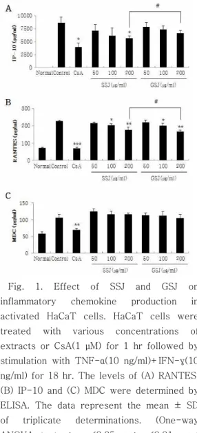
2. 케모카인(RANTES, IP-10, MDC) 생성에 미치는 영향
사상자탕 추출물(SSJ)과 가미사상자탕 추출물 (GSJ)이 HaCaT 세포에서 발현되는 RANTES와 IP-10 생성을 억제하는 것으로 확인되었으며 SSJ보다 GSJ의 효과가 더 큰 것으로 나타났다 (Fig. 1A, 1B). 그러나 MDC 생성에는 영향을 미 치지 않은 것으로 나타났다(Fig. 1C). 양성대조 군으로서 CsA(1 µM)를 사용하였다.
Fig. 1. Effect of SSJ and GSJ on inflammatory chemokine production in activated HaCaT cells. HaCaT cells were treated with various concentrations of extracts or CsA(1 µM) for 1 hr followed by stimulation with TNF-α(10 ng/ml)+IFN-γ(10 ng/ml) for 18 hr. The levels of (A) RANTES, (B) IP-10 and (C) MDC were determined by ELISA. The data represent the mean ± SD of triplicate determinations. (One-way ANOVA test *; p <0.05, **; p<0.01, ***;
p<0.001 vs. Control, #; p <0.05 vs. SSJ)
3. 사이토카인(IL-6, TNF-α) 생성에 미치는 영향
사상자탕 추출물(SSJ)과 가미사상자탕 추출물 (GSJ)이 RAW 264.7 세포에서 발현되는 IL-6와 TNF-α 생성을 억제하는 것으로 확인되었으며 SSJ보다 GSJ의 효과가 더 큰 것으로 나타났다 (Fig. 2). 양성대조군으로서 CsA(1 µM)를 사용 하였다.
Fig. 2. Effect of SSJ and GSJ on inflammatory cytokine production in activated Raw 264.7 cells. Raw 264.7 cells were treated with various concentrations of extracts or CsA(1 µM) for 1 hr followed by stimulation with LPS(1 µg/ml) for 18 hr. The levels of (A) TNF-α and (B) IL-6 were determined by ELISA. The data represent the mean ± SD of triplicate determinations.
(One-way ANOVA test *; p <0.05, **;
p<0.01, ***; p<0.001 vs. Control, #; p <0.05 vs. SSJ)
4. Nitric Oxide(NO) 생성에 미치는 영향 사상자탕 추출물(SSJ)과 가미사상자탕 추출물 (GSJ)이 RAW 264.7 세포에서 발현되는 Nitric
Oxide 생성을 억제하는 것으로 확인되었으며 SSJ보다 GSJ의 효과가 더 큰 것으로 나타났다 (Fig. 3).
Fig. 3. Effect of SSJ and GSJ on NO production in activated Raw 264.7 cells. Raw 264.7 cells were treated with various concentrations of extracts or CsA(1 µM) for 1 hr followed by stimulation with LPS(1 µg/ml) for 24 hr. The level of NO in the cell culture supernatant was measured by NO detection Kit. The data represent the mean
± SD of triplicate determinations. (One-way ANOVA test ***; p<0.001 vs. Control, #; p
<0.05 vs. SSJ)
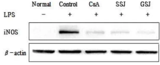
5. iNOS 발현에 미치는 영향
사상자탕 추출물(SSJ)과 가미사상자탕 추출물 (GSJ)이 RAW 264.7 세포에서 iNOS 발현을 감 소시키는 것으로 확인되었으며 SSJ보다 GSJ의 효과가 더 큰 것으로 나타났다(Fig. 4).
Fig. 4. Effect of SSJ and GSJ on iNOS expression in activated Raw 264.7 cells. Raw 264.7 cells were treated with SSJ(200 µg/ml), GSJ(200 µg/ml) or CsA(1 µM) for 1 hr followed by stimulation with LPS(1 µg/ml)
for 24 hr. The total cell lysates(18 hr) were isolated and analyzed by western bolt analysis using iNOS antibody. β-actin was used as loading controls. The data presented are representative of three independent experiments.
Ⅳ. 고 찰
사상자탕(蛇床子湯)에 관하여서는 外科正宗
에 음낭습진의 일종인 신낭풍(腎囊風)을 치료한 다고 기록되어 있으며, 임상에서도 주로 염증성 피부질환에 많이 사용되고 있다. 본 실험에서는 사상자(蛇床子), 고삼(苦蔘), 당귀(當歸), 위령선 (威靈仙)에 백부근(百部根)과 부평초(浮萍草)를 추가하여 가미사상자탕(加味蛇床子湯) 처방을 구성하고 이 처방의 추출물(GSJ)과 사상자탕 추 출물(SSJ)의 항염증 및 항산화 효과를 실험하고 그 효과를 비교하였다.
HaCaT 세포를 이용하여 케모카인(RANTES, IP-10, MDC)을, RAW 264.7 세포를 이용하여 사이토카인(TNF-α, IL-6)과 NO를 측정하였다.
케모카인은 염증 질환, 알러지 질환 등과 밀접한 관련이 있으며 백혈구들의 이동을 조절한다고 알려져 있는데, RANTES는 화학주성에만 제한 되지 않고 백혈구의 활성인자로도 작용하고 있음 이 보고되었다11),12). 또 IP-10은 염증 반응에서 호중구와 단핵구의 화학주성에 직접 관여하고 있
음이 알려져 있으며13,14) MDC는 단핵구
(monocyte)와 단핵구 유래 수지상 세포(denritic cell) 및 자연 살해세포(natural killer cell)에 대 한 이동에 중요하게 관여하는 케모카인이다12),15).
HaCaT 세포를 이용하여 이들 케모카인을 측 정한 결과, 사상자탕 추출물(SSJ)과 가미사상자 탕 추출물(GSJ)은 RANTES와 IP-10 생성을 모 두 억제하는 것으로 나타났으며, 농도 200 µg/ml에서 SSJ보다 GSJ의 효과가 더 큰 것으로 확인되었다(Fig. 1A, 1B). 그러나 MDC 분비에는 유의적인 차이가 없었다(Fig. 1C).
인체에 병원체가 침입했을 때 가장 먼저 대응
하는 세포들은 호중구(neutrophil), 단핵구 (monocyte) 및 대식세포(macrophage) 같은 탐 식세포들로 이들은 선천면역 반응의 주요 세포군 을 형성하고 있다. 이중 대식세포는 호중구와 함 께 생체방어에 있어 최초 대응세포로서 항원제시 세포로서의 기능도 수행한다. 대식세포가 활성화 되면 TNF-α, IL-6 등의 초기 염증성 사이토카 인을 분비하는데 TNF-α는 감염 초기에 호중구 와 단핵구가 감염 부위로 이동하는 것에 관여하 며 이들을 활성화시켜 항원을 제거하도록 하는 중요한 인자이다16),17). 또 IL-6는 감염이나 손상 으로 인한 급성 반응을 보이며 염증 반응이 일어 나는 동안 일정하게 생성이 된다18),19). 이러한 염 증매개 물질의 형성은 iNOS와 COX-2의 합성을 촉진시켜 NO를 생성한다. NO는 불안정한 기체 상태의 free radical로서 체내 여러 생리 및 병리 적 환경에서 중요한 역할을 한다. 생리적 조건에 서 NO는 종양이나 기생충, 세균 등의 방어에 관 여하는 반면 과도하게 생성된 NO는 toxic radical로 작용하여 세포나 조직의 손상을 일으
킨다20),21). 이를 토대로 세포에 독성이 없는 농도
를 확인한 후 LPS로 염증 반응이 유도된 RAW 264.7 세포에 사상자탕 추출물(SSJ)과 가미사상 자탕 추출물(GSJ)을 처리하여 사이토카인을 측 정한 결과, TNF-α와 IL-6 모두 농도 의존적으 로 유의성 있게 감소되었으며, 농도 200 µg/ml 에서 GSJ가 SSJ보다 TNF-α와 IL-6를 유의적으 로 감소시켰다(Fig. 2). 그리고 Nitric oxide를 측정한 결과 SSJ와 GSJ 모두 RAW 264.7 세포 에서 발현되는 Nitric Oxide 생성을 억제하는 것 으로 확인되었으며 SSJ보다 GSJ의 효과가 더 큰 것으로 나타났다(Fig. 3). 또한, SSJ와 GSJ는 NO 의 생성을 유도하는 iNOS도 감소시키는 효과를 나타냈으며 GSJ의 효과가 SSJ보다 더 큰 것으로 확인되었다(Fig. 4).
결론적으로, 사상자탕 추출물(SSJ)과 가미사상 자탕 추출물(GSJ)은 염증반응이 유도된 HaCaT 세포에서 RANTES, IP-10 분비를 억제하였고, RAW 26.47 세포에서 TNF-α, IL-6 분비 및 NO 생성을 억제하였다. 따라서 두 처방 모두 항
염증 및 항산화 효과가 있다고 할 수 있으며, 두 처방의 비교에서는 가미사상자탕 추출물(GSJ)이 사상자탕 추출물(SSJ)보다 더 효과가 좋은 것으 로 나타났다. 본 실험의 결과들을 고려해 봤을 때, 이들 약물 각각의 효과 및 기전에 대하여 추 가적인 연구가 이루어져야 할 것으로 생각되며, 적용 가능한 다양한 염증성 질환이나 알러지 질 환 중에서 타겟 질환을 선정한 뒤 동물 실험을 수행하면 좋은 결과를 얻을 수 있을 것으로 사료 된다.
Ⅴ. 결 론
본 실험에서는 사상자탕 추출물(SSJ)과 가미사 상자탕 추출물(GSJ)의 항염증 및 항산화 효과를 연구하고 비교한 결과 다음과 같은 결론을 얻었 다.
1. 세포 독성 실험 결과, 사상자탕 추출물(SSJ) 과 가미사상자탕 추출물(GSJ)은 0-800 µg/ml 농도에서 독성을 나타내지 않았다.
2. HaCat 세포에서 케모카인을 측정한 결과, 사상자탕 추출물(SSJ)과 가미사상자탕 추출 물(GSJ)은 RANTES와 IP-10 생성을 농도 의존적으로 억제하였으나 MDC 생성에는 영향을 미치지 않은 것으로 나타났다.
3. RAW 264.7 세포에서 사이토카인을 측정한 결과, 사상자탕 추출물(SSJ)과 가미사상자 탕 추출물(GSJ)은 TNF-α와 IL-6의 생성을 농도 의존적으로 억제하는 것으로 나타났다.
4. RAW 264.7 세포에서 NO 생성과 iNOS 발 현 측정 결과, 사상자탕 추출물(SSJ)과 가미 사상자탕 추출물(GSJ)은 NO 생성과 iNOS 발현을 농도 의존적으로 억제하는 것으로 나타났다.
5. 사상자탕 추출물(SSJ)과 가미사상자탕 추출 물(GSJ)의 효과를 비교한 결과, 가미사상자
탕 추출물(GSJ)이 사상자탕 추출물(SSJ)보 다 농도 200 µg/ml에서 RANTES, IP-10, TNF-α, IL-6 그리고 NO를 억제하는 효과 가 더 우수한 것으로 나타났다.
결론적으로, 사상자탕 추출물(SSJ)과 가미사상 자탕 추출물(GSJ) 모두 항염증 및 항산화 효과가 있다고 할 수 있으며, 두 처방의 비교에서는 가미 사상자탕 추출물(GSJ)이 사상자탕 추출물(SSJ) 보다 효과가 더 우수한 것으로 사료된다.
감사의 글
본 논문은 산업통상자원부 지정 대전대학교 난 치성 면역질환의 동서생명 의학연구 지역혁신센 터의 지원에 의한 것임.
참고문헌
1. 陳實功. 外科定宗. 북경, 인민위생출판사, p.244, 1983.
2. Zhang YZ1, Xu GB, Zhang T. Antifungal stilbenoids from Stemona japonica. J Asian Nat Prod Res. 10(7-8):639-44, 2008.
3. 허민, 오영선, 조현경, 유호룡, 김윤식, 설인 찬. 백부근(百部根) 약침(藥鍼)의 천식억제 (喘息抑制) 및 면역조절효과에 대한 실험적 연구. 동의생리병리학회지. 20(3):
609-616. 2006.
4. 노태석, 임낙철, 노석선. 數種의 韓藥 抽出 物의 抗알레르기 效果에 關한 實驗的 硏究.
대한외관과학회지. 14(2): 21-34. 2001.
5. Adeline F. Deyrieux, V. G. Wilson. In vitro culture conditions to study keratinocyte differentiation using the HaCaT cell line. Cytotechnology. 54(2):
77-83. 2007.
6. YS Chi, BS Cheon, HP Kim. Effect of wogonin, a plant flavone from
Scutellaria radix, on the suppression of cyclooxygenase-2 and the induction of inducible nitric oxide synthase in lipopolysaccharide-treated RAW 264.7 cells. Biochemical Pharmacology.
61(10): 1195–1203. 2001.
7. Andrew D Luster. The role of chemokines in linking innate and adaptive immunity. Current Opinion in Immunology. 14(1): 129–135. 2002.
8. Linda R. Watkins, Steven F. Maier, Lisa E. Goehler. Immune activation: the role of pro-inflammatory cytokines in inflammation, illness responses and pathological pain states. Pain. 63(3):
289–302. 1995.
9. Matthew B. Grisham , David Jourd’Heuil , David A. Wink. Physiological chemistry of nitric oxide and its metabolites: implications in inflammation. American Journal of Physiology - Gastrointestinal and Liver Physiology. 276(2): 315-321. 1999.
10. 이혜자, 강경진, 윤원종, 강희경, 김영석, 김소미, 유은숙. RAW 264.7 및 HaCaT Cell에서 당유자 미숙과의 염증억제 효과.
생약학회지 , 37(2): 74-80. 2006.
11. Appay V, Rowland-Jones SL. RANTES:
a versatile and controversial chemokine. Trends in Immunology.
22(2): 83–87. 2001.
12. Lim HS, Ha H, Lee MY, Jin SE, Jeong SJ, Jeon W1, Shin NR, Sok DE, Shin HK. Saussurea lappa alleviates inflammatory chemokine production in HaCaT cells and house dust mite-induced atopic-like dermatitis in Nc/Nga mice. Food and Chemical Toxicology. 63: 212-20. 2014.
13. J M Farber. Mig and IP-10: CXC
chemokines that target lymphocytes.
Journal of Leukocyte Biology. 61(3):
246-257. 1997.
14. Villagomez MT, Bae SJ, Ogawa I, Takenaka M, Katayama I. Tumour necrosis factor-alpha but not interferon-gamma is the main inducer of inducible protein-10 in skin fibroblasts from patients with atopic dermatitis. British Journal of Dermatology. 150(5): 910-6. 2004.
15. Alberto Mantovani, Patrick A. Gray, Jo Van Damme, Silvano Sozzani.
Macrophage-derived chemokine (MDC). Journal of Leukocyte Biology.
68(3): 400-404. 2000.
16. Feuerstein GZ1, Liu T, Barone FC.
Cytokines, inflammation, and brain injury: role of tumor necrosis factor-alpha. Cerebrovascular & Brain Metabolism Reviews. 6(4): 341-60.
1994.
17. Choi YH, Kim GY, Lee HH.
Anti-inflammatory effects of cordycepin in lipopolysaccharide -stimulated RAW 264.7 macrophages through Toll-like receptor 4-mediated suppression of mitogen-activated protein kinases and NF-κB signaling pathways. Drug Design Development and Therapy. 16(8): 1941-1953. 2014.
18. 윤석빈, 한효상, 이영종. 황금이 LPS로 유 발된 RAW 264.7 cell의 염증 인자에 미치 는 영향. 대한본초학회지. 26(2): 45-81.
2011.
19. Seo KH, Park MJ, Ra JE, Han SI, Nam MH, Kim JH, Lee JH, Seo WD.
Saponarin from barley sprouts inhibits NF-κB and MAPK on LPS-induced RAW 264.7 cells. Food & Function.
22;5(11): 3005-3013. 2014.
20. Ryu JH, Ahn H, Kim JY, Kim YK.
Inhibitory activity of plant extracts on nitric oxide synthesis in LPS-activated macrophages. Phytotherapy Research.
17(5): 485-9. 2003.
21. Mu MM1, Chakravortty D, Sugiyama T, Koide N, Takahashi K, Mori I, Yoshida T, Yokochi T. The inhibitory action of quercetin on lipopolysaccharide -induced nitric oxide production in RAW 264.7 macrophage cells. Journal of Endotoxin Research .7(6): 431-8.
200.