* Corresponding author
Phone: +82-51-999-5459, Fax: +82-51-999-5176 E-mail: [email protected]
해양 미생물 Algibacter lectus AS-3 으로부터 agarase의 분리 및 특성
정일선․최영주*
신라대학교 의생명과학대학 식품영양학과
Purification and Characterization of Agarase from Marine Bacterium, Algibacter lectus AS-3. Il Sun Jung and Young Ju Choi*. Department of Food and Nutrition, College of Medical Life Science Silla University, Busan 617-736, Korea
Abstract An agar-degrading marine bacterium, strain AS-3 was isolated from the seawater. The strain AS-3 was identified as Algibacter lectus AS-3 by 16S rDNA sequence. The optimum medium for agarase activity of the isolated strain was determined to be marine medium, marine broth 2216 containing 0.1% agar as carbon source. An extracellular agarase was purified 6.9-fold from the culture supernatant by ammonium sulfate precipitation, ion exchange chromatography and gel filtration methods. The optimum pH and temperature for this enzyme were 7.0 and 40-50℃, respectively. Antioxidative activity of the strain AS-3 was 62.4% in the supernatant cultured for 12 h.
Key words : Marine bacteria, agarase, Algibacter lectus, agarooligosaccharides, purification
서 론
Agar는 보통 적조류 Gelidium과 Gracilaria로부터 만들어지며, agarose와 agaropectin로 구성되어 있다.
Agarose는 3-O-linked β-D-galactopyranose와 4-O- linked 3,6-anhydro-α-L-galactopyranose로 번갈아 정 열된 직쇄상 다당체 중의 하나이다. Agaropectin은 agarose와 같은 기본 골격을 가지고 있지만 sulfate, pyruvate, methoxy와 같은 치환기를 함유하고 있다.
Agarase는 agar의 가수분해를 촉매 하는데, agar- ooligosaccharides를 생산하기 위해서 α-1,3 결합을 절 단하는 α-agarase [29]와 neoagarooligosaccharides를 생산하기 위해 β-1,4 결합을 절단하는 β-agarase [6]
로 분류된다.
β-agarase에 의한 agar 및 agarose의 가수분해 산물 인 neooligosaccharides는 다양한 화학적 특성과 생물 학적 활성을 가지고 있다. Neooligosaccharides는 세 균의 성장을 저해하거나, starch의 분해를 느리게 하 거나, 식품의 칼로리를 감소시킬 뿐만 아니라 항암
효과 [8]나 항산화 활성 [27,30]을 나타낸다. 또한 난 소화성으로 당뇨병, 비만, 변비 등의 치료에도 효과 가 있는 것으로 알려져 있다 [1,16]. Neoagarobiose는 피부 보습효과 및 melanoma cell에 미백 효과가 있는 것으로 보고되었으며 [7,13], 최근에 β-agarase가 agarose를 가수분해하여 주요 분해산물로서 neo- agarobiose, neoagarotetrose, neoagarohexose를 생산하 는 것으로 밝혀졌다 [7,13,20].
한천 올리고당은 한천의 산 가수분해 또는 효소분 해에 의해 생성되지만, 산 가수분해시에는 한천에 함유되어 있는 고유의 비타민이나 무기질 등이 다량 파괴되고, 올리고당의 기능성 및 안정성 유지에 문 제가 제기되므로 효소분해에 의한 방법이 보다 유용 한 것으로 알려져 왔다 [21]. 따라서 한천 올리고당 의 대량 생산 또는 산업적 이용을 위해서는 우선적 으로 높은 활성을 지닌 한천 분해효소 및 이를 이용 한 agarooligosaccharides의 대량생산에 관한 연구가 많이 수행되고 있다.
Agarase를 생성하는 bacteria는 agar에 대한 작용기
작에 따라 지난 수년 동안 수종의 agarase 생성균주 가 Pseudoalteromonas [5,25], Pseudomonas [11,28], Streptomyces [4], Alteromonas [26], Microbulbifer [18,19]. Vibrio [21,22], Cytophaga [24], Agarivorans [20], 및 Acinetobacter [14] 등의 해양 미생물을 중심 으로 분리되어 한천분해 효소의 특성에 관한 연구가 활발히 수행되어 왔다.
저자들은 수종의 agar-softening 균주와 agar-lique- fying 균주를 분리하여 agar를 가수분해하는 ex- tracellular agarase의 특성 연구를 수행하여 왔으며, 본 논문에서는 해양으로부터 한천 분해능이 뛰어난 Algibacter lectus AS-3을 분리․동정하고 정제하여 효소의 특성과 agar 분해산물의 항산화활성을 연구 하였다.
재료 및 방법 시약 및 기자재
균주의 탐색을 위해 사용한 배지 marine broth 2216은 Difco 사 (Detroit, USA)에서 구입하여 사용하 였다. Agarase를 정제하기 위해 사용한 ammonium sulfate는 USB 사 (Cleveland, USA) 제품을 사용하였 다. Ion exchange chromatography에 사용된 HiPrepTM 16/10 DEAE FF와 gel filtration에 사용된 HiPrepTM 26/60 SephacrylTM S-200HR은 Amersham Biosciences 에서 구입하여 사용하였다. Protein 정제를 위해 사 용한 기기는 AKTA Prime (Amersham Biosciences)이 었고, UV-Vis spectrophotometer는 Ultrospec® 3000을 사용하였다. 항산화활성 측정은 1,1-diphenyl-2-pic- rylhydrazyl (DPPH)는 Sigma-Adrich사 제품을 사용하 였고, 그 외의 기타 시약은 특급을 사용하였다.
Agarolytic bacteria의 분리 및 동정
Agarolytic bacteria의 탐색을 위해 해양으로부터 해수 sample을 채취하여 희석한 후, marine broth (MB) 2216 agar plate에 도말하여 27℃에서 24시간 동안 배양하였다. 배양 후, agar plate 표면의 colony 를 중심으로 움푹 파이거나 clear zone을 형성하는 균주를 분리하였다. 분리된 균주를 AS-3으로 명명 하였으며 균주의 동정은 16S rDNA sequence로 확인 하였다.
분리된 균주의 생육특성 조사
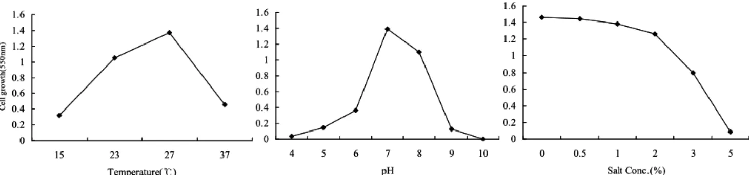
분리된 균주의 배양조건에 따른 균주의 특성은 온 도, 염 농도, pH를 달리하여 27℃에서 24시간 동안 진탕배양한 후 550 nm에서 흡광도를 측정하였다. 배 양조건은 온도 (15, 23, 27, 37℃), 염 농도 (0.5, 1, 2, 3, 5%), pH (4.0. 5.0, 6.0, 7.0, 8.0, 9.0, 10.0)에 따른 생육 특성을 조사하였다.
Agarase 정제
Agarase 정제를 위한 모든 실험은 4℃에서 수행하 였다. 균주는 27℃에서 72시간동안 180 rpm의 조건 에서 진탕 배양하였다. 배양액을 6,000×g에서 30분 동안 원심분리한 상등액을 조효소액으로 사용하였 다. 분리된 상등액은 70% ammonium sulfate로 포화 시킨 후 24시간 동안 침전시킨 다음 6,000×g에서 30 분 동안 원심분리 하였다. 침전물을 25 mM Tris-HCl buffer (pH 8.0)로 녹인 후 동일한 buffer로 투석하였 다. 투석된 용액은 HiPrep 16/10 DEAE FF column에 loading하고 25 mM Tris-HCl buffer (pH 8.0)로 평형 화하였으며, 유속은 1 ml/min으로 조절하였다. 단백 질은 NaCl gradient (0~1.0 M)를 포함하는 동일한 buffer로 용출한 후 fraction을 회수하여 효소활성을 측정하였다. 효소활성이 높은 fraction을 회수한 다 음 HiPrepTM 26/60 SephacrylTM S-200 HR column에 loading하여 25 mM Tris-HCl buffer (pH 8.0)로 평형 화하였으며, 유속은 0.5 ml/min으로 조절하였다. 활 성이 높은 fraction을 회수하여 단백질 전기영동으로 효소정제를 확인하였으며, agarase 효소 특성 조사를 위한 정제효소로 사용하였다.
Agarase 활성 측정 및 단백질 정량
Agarase 활성 측정은 0.1% agarose 용액을 기질로 하여 Leon [15]의 방법에 따라 한천 분해효소의 반응 산물인 환원당을 DNS (3,5-Dinitrosalicylic acid)법 [17]에 의해 측정하였다. 한천 분해효소의 활성은 1 분당 1 μmol 의 galactose를 생산하는 효소의 양을 1 unit로 나타내었다. 단백질 정량은 UV-Vis spec- trophotometer (Ultrospec® 3000)를 이용하여 280 nm 에서의 흡광도를 측정하였으며, 표준 단백질로 bo- vine serum albumin을 사용하였다.
Agarase 활성에 대한 최적조건 및 안정성 Agarase 활성에 대한 pH 영향은 0.1% agarose가 함 유된 기질용액 1 ml에 각 pH에 따른 완충용액 (0.1 M sodium acetate buffer; pH 4.0-6.0, 0.1 M Tris-HCl buffer; pH 7.0-8.0, sodium carbonate buffer; pH 9.0-10.0) 1 ml를 혼합한 용액에 효소액을 가하여 40
℃에서 30분간 반응시킨 후 환원당을 측정하여 효소 활성에 미치는 pH의 영향을 조사하였다.
효소활성의 최적온도는 0.1% agarose가 함유된 기 질용액 1 ml에 정제된 효소용액을 첨가하여 0.1 M Tris-HCl buffer (pH 7.0)에서 20~70℃까지 온도를 변화시키면서 30분간 반응시킨 다음 효소활성에 미 치는 온도의 영향을 조사하였다.
온도 안정성은 정제효소 1 ml를 20~70℃의 온도 에서 30분간 반응시킨 후 0.1% 기질용액 1 ml를 혼 합하여 40℃에서 30분간 반응시킨 후 환원당을 측정 하여 잔존활성을 확인하였다.
DPPH 라디칼을 이용한 전자 공여능 측정 Agar 분해산물인 oligosaccharides의 DPPH (1,1- Diphenyl-2-picrylhydrazyl) 라디칼소거 활성은 Blois [3]방법에 따라 다음과 같이 측정하였다. 균주를 0.1% agar를 함유한 marine broth에 배양한 후 DPPH 용액 1 ml에 배양액 200 μl를 혼합하여 37℃에서 30 분간 반응하였다. 반응물을 4℃, 12,000 rpm에서 3분 간 원심분리 한 후 525 nm에서 흡광도를 측정하였 다. 항산화 활성은 전자공여능 (electron donating ability, EDA)으로 표기하였으며, 시료 첨가구와 무 첨가구의 흡광도차를 백분율 (%)로 표시하였다.
EDA (%) = [1- (시료 첨가구/시료 무첨가구)] × 100 으로 계산하였다.
결과 및 고찰 균주배양조건
배양온도, pH 및 염 농도에 따른 최적 배양조건을 구하기 위해 UV-Vis Spectrophotometer를 사용하여 550 nm에서 흡광도를 측정하였다 (Fig. 3). 온도 15~ 37℃의 범위에서 배양한 결과, Algibacter lectus AS-3 는 27℃에서 cell growth가 가장 높았으며, pH 4~10 의 범위에서 배양한 결과, AS-3는 pH 7에서 cell growth가 가장 높았다. 그리고 염 농도 0.5~5%의
Fig. 1. Nucleotide sequences of 16S rDNA Algibacter lectus AS-3.
――― 0.01
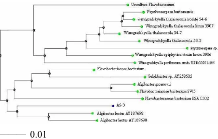
Fig. 2. Phylogenetic relationships between strain AS-3 and other bacteria based on 16S rDNA sequences. The phyloge- netic tree was constructed based on the alignment of complete 16S rDNA sequences. The scale bar indicates one nucleotide substitution per 100 nucleotides. The tree was created using the ClustalX program.
범위에서 배양한 결과, Algibacter lectus AS-3은 salt 를 첨가하지 않은 배지에서 가장 잘 성장하였다. 일 반적으로 해양 미생물의 성장은 주로 25 ℃에서 최 적온도를 나타내며 pH는 7.0-7.5 범위 내에서 배양되 고 있다.
균주의 16S rDNA sequence analysis
선별된 균주 AS-3는 VITEK 2 compact Version으 로 분석하였으나 낮은 유사성으로 인해 16S rDNA sequence로 동정하였다. Agar 분해 미생물 AS-3의 phylogenetic 위치를 결정하기 위해서 1.4 kb의 16S
0 0.2 0.4 0.6 0.8 1 1.2 1.4 1.6
15 23 27 37
Temperature(℃)
Cell growth(550nm)
0 0.2 0.4 0.6 0.8 1 1.2 1.4 1.6
4 5 6 7 8 9 10
pH
0 0.2 0.4 0.6 0.8 1 1.2 1.4 1.6
0 0.5 1 2 3 5
Salt Conc.(%) 0
0.2 0.4 0.6 0.8 1 1.2 1.4 1.6
15 23 27 37
Temperature(℃)
Cell growth(550nm)
0 0.2 0.4 0.6 0.8 1 1.2 1.4 1.6
4 5 6 7 8 9 10
pH
0 0.2 0.4 0.6 0.8 1 1.2 1.4 1.6
0 0.5 1 2 3 5
Salt Conc.(%)
Fig. 3. Growth conditions on Algibacter lectus AS-3. (A) Temperature : 15, 23, 27, 37(℃), (B) pH : 4, 5, 6, 7, 8, 9, 10, (C) NaCl concentration : 0.5, 1, 2, 3, 5(%)
rDNA 단편을 polymerase chain reaction (PCR)에 의하 여 증폭하였다. 1373 bp의 sequence가 결정되었으며 알려진 16S rDNA sequence에 대하여 comparative se- quence 분석을 실시한 결과, 분리균주의 16S rDNA sequence는 Algibacter lectus와 93%의 상동성을 나타 내었다. 분리균주의 phylogentic 위치를 정하기 위하 여 Bioedit 프로그램을 사용하였고 ClustalX로 계통 수를 결정하였다 (Fig. 1, Fig. 2).
Agarase의 정제
Algibacter lectus AS-3 균주는 MB 배지에서 27℃, 180 rpm에서 3일간 배양한 후 배양액을 70% ammo- nium sulfate에 침전시킨 후, HiPrep 16/10 DEAE FF column으로 ion exchange chromatography 및 HiPrepTM 26/60 SephacrylTM S-200 HR column으로 gel filtration chromatography하여 효소를 정제하였다.
Algibacter lectus AS-3의 ion exchange chromatog- raphy 결과는 Fig. 4에서와 같이 fraction number 10에 서 높은 agarase activity를 나타내었다. Algibacter lec- tus AS-3의 gel filtration chromatography한 결과는 Fig. 5에 나타난 바와 같이 fraction number 12에서 agarase activity가 가장 높게 나타났다.
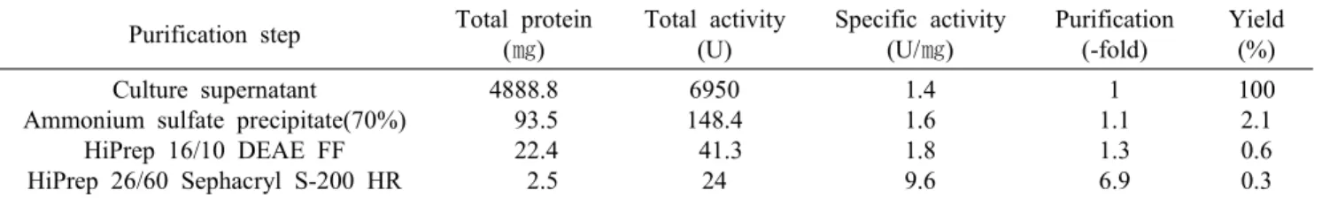
Algibacter lectus AS-3가 생산하는 한천 분해효소 의 정제 단계에 대한 결과는 Table 1에 나타내었다.
수율은 0.3%였으며, 정제도는 6.9배 정도 증가하여 최종적으로 9.6 U/㎎의 specific activity를 지닌 정제 된 agarase을 얻을 수 있었다.
효소활성 최적조건 및 안정성
Agarase의 활성에 미치는 온도의 효과를 조사하기 위하여 반응 온도를 20~70℃까지 변화시키면서 효
Fig. 4. Ion-exchange chromatography of agarase of Algibacter lectus AS-3 on DEAE FF column. The 25 mM Tris-HCl buf- fer (pH 8.0) containing gradient NaCl rising from 0 to 1.0M at a flow-rate of 1 ml/min was used wash out the sample.
Fractions were measured at 280 nm for protein content.
Fig. 5. Gel-filtration chromatography of agarase of Algibacter lectus AS-3 on Sephacryl S-200 HR. The 25 mM Tris-HCl buffer (pH 8.0) at a flow-rate of 0.5 ml/min was used to wash out the sample. Fractions were measured at 280 nm for protein content.
소활성을 측정하였다. 본 연구에서 분리된 Algibacter lectus AS-3에서 생산된 agarase는 40~50℃에서 최대 활성을 나타내었다 (Fig. 6). Kang 등[12]은 해양 미생 물인 Pseudomonas sp.에서 agarase 효소 최적 활성은
Table 1. Purification of agarase from Algibacter lectus AS-3 Purification step Total protein
(㎎)
Total activity (U)
Specific activity (U/㎎)
Purification (-fold)
Yield (%) Culture supernatant
Ammonium sulfate precipitate(70%) HiPrep 16/10 DEAE FF HiPrep 26/60 Sephacryl S-200 HR
4888.8 93.5 22.4 2.5
6950 148.4 41.3 24
1.4 1.6 1.8 9.6
1 1.1 1.3 6.9
100 2.1 0.6 0.3
30℃였으며, 40℃ 이상에서 급격히 효소활성이 감소 하였다고 보고하였으며, Ohta 등[18,19]은 해양 미생 물인 Microbulbifer salipaluids로부터 온도에 안정한 β-agarase 효소를 분리하였으며 최적 효소활성은 5 5℃였으며, 60 ℃ 이상에서 급격히 효소활성이 감소 하는 것으로 보고하였다. 대부분의 agarolytic 균주의 agarase의 최적 활성은 30-40℃ 사이에 존재하였으며, 본 연구에서 분리된 균주는 온도에 비교적 높은 안정 성을 나타내고 있는 것으로 생각된다.
Algibacter lectus AS-3로부터 분리된 효소 활성의 최적 pH를 실험한 결과 (Fig. 7), pH 7.0에서 가장 높은 활성을 보였다. pH에 대한 효소의 활성은 6.0 이하와 pH 8.0 이상의 산, 알칼리 영역에서는 효소활 성이 크게 감소하였다. Suzuki 등 [23]은 Bacillus sp.
에서 최적 pH는 7.6이었고 효소의 안정성도를 조사 한 결과, pH 7.0-8.0 사이에서 안정한 것으로 나타났 다. 일반적으로 agarase의 최적 활성 pH는 7.0 부근이 었으며, 안정성도 pH 6.0-8.0 사이에서 비교적 효소 활성이 안정한 것으로 밝혀졌다 [10,18].
DPPH법에 의한 항산화활성 측정
DPPH 라디칼은 alcohol 용액 내에서는 DPPH의 질 소 원자와 alcohol 간에 수소결합이 형성되기 때문에 다른 유리 라디칼보다 비교적 안정하고, 항산화 활 성을 갖는 물질과 반응하면 진보라색의 DPPH의 색 깔이 점점 옅어져 흡광도가 감소한다. DPPH 라디칼 소거법은 흡광도의 감소를 측정함으로서 radical 소 거 활성을 쉽게 측정할 수 있다는 장점을 가지고 있 어 항산화 활성 측정의 척도로 많이 활용되고 있다.
Algibacter lectus AS-3의 배양 상등액에 대한 DPPH 라디칼 소거 활성을 시간대 별로 측정한 결과 (Fig.
8), Algibacter lectus AS-3은 EDA (%)가 4시간 배양 후 44.5%로 본 실험실에서 분리하여 보고한 Sphingomonas paucimobilis AS-1 [9] 11.4% 보다 높게 나타났다. Algibacter lectus AS-3를 8시간에서 24시
0 20 40 60 80 100 120
20 30 40 50 60 70
Temperature(℃)
Relative activity (%) .
Fig. 6. Effects of temperature on the agarase activity.
Temperature profiles were checked at different temperatures (20-70℃) in 0.1 M Tris-HCl buffer (pH 8.0). The enzyme solution was preincubated at various temperature for 30min and residual activity was measured
0 20 40 60 80 100 120
4 5 6 7 8 9 10
pH
Relative activity (%) .
Fig. 7. Effect of pH on the agarase activity. The activity was determined at 40℃ in different buffer (pH 4.0-10.0): 0.1 M sodium acetate buffer (pH 4.0-6.0), 0.1 M Tris-HCl buffer (pH 7.0-8.0), sodium carbonate buffer (pH 9.0-10.0). The en- zyme solution was placed at various pH for 30min and re- sidual activity was measured at 40℃.
간 배양하는 동안 항산화 활성을 조사한 결과 약 60%의 라디칼 소거능을 나타내었다. 한편, AS-1의 경우, 8시간 배양 후 라디칼 소거능이 51.2%로 증가 하였으며, 12시간 배양 후 72.5%로 최고 활성을 나 타내었지만 24시간 배양 후에는 38.1%로 점차 감소 하는 경향을 보였다. 이러한 결과는 생성된 oligo- saccharides의 형태에 따른 결과로 생각되며 어떤 종
0 10 20 30 40 50 60 70
4 8 12 24 48
Time(h)
EDA(%)
Fig. 8. DPPH radical scavenger activity in different culture time of production of agar by agarase. Each value represents means of triplicates.
류의 oligosaccharides가 생성되는지 추가 실험이 요 구된다. 최근 해양미생물 유래의 agarase에 의하여 생산된 oligosaccharides가 항산화활성 [10,27,30]을 나타내는 것으로 보고되었고, Araki 등 [2]은 생물학 적 활성을 가진 기능성 물질 추출을 위한 유용한 tool 로서 agarase의 중요성을 보고하였다. 또한 식품, 화 장품 및 의약산업에 oligosaccharides를 이용하기 위 하여 새로운 agarase 효소에 대한 연구도 활발히 진 행되고 있다.
요 약
Agar로부터 oligosaccharides를 제조하기 위하여 효소 (agarase)를 이용한 분해법이 시도되면서 agar- ase의 균주의 분리 및 유전자에 대한 많은 연구가 수행되고 있다. 본 연구에서는 해양으로부터 agar- ase를 생성하는 균주를 분리․동정하고 균주의 특 성을 조사하였다. 분리된 균주는 AS-3균주는 16S rDNA 염기서열분석에 의하여 Algibacter lectus AS-3로 동정되었다. 정제된 효소의 최적 활성 조건 및 agar 분해산물의 항산화활성을 조사하였다. 분리 된 균주의 최적배양조건은 marine broth 2216에서 온도 27℃, pH 7일 때 가장 균주의 생육이 높았다.
Agarase 효소는 salt 침전, ion exchange와 gel filtra- tion chromatography에 의해 9.6 units/mg으로 6.9배 정제되었다. 최적 효소활성은 온도 40~50℃, pH 7 일 때 나타났다. Agar 첨가한 배지에 agar 분해균을 접종한 후 분해산물의 시간에 따른 항산화활성은 12시간 배양 후 62%의 가장 높은 전자소거능 (EDA)을 나타내었다.
참 고 문 헌
1. Andrykovitch, G. and Maex, I. 1988. Isolation of a new polysaccharide -degrading bacterium from a salt marsh.
Appl. Environ. Microbiol. 54, 1061-1062.
2. Araki, T., Hayakawa, M., Lu, Z., Karita, S. and Morishita, T. 1998. Purification and characterization of agarases from a marine bacterium, Vibrio sp. PO-303.
J. Mar. Biotechnol. 6, 260-265.
3. Blois, M. S. 1958. Antioxidant determination by the use of a stable free radical. Nature 26, 1199-1200.
4. Buttner, M. J., Feamley, I. M. and Bibb, M. J. 1987.
The agarase gene(dagA) of Streptomyces coelicolor A3(2) nucleotide sequence and transcriptional analysis.
Mol. Gen. Genet. 209, 101-109.
5. Chiura, H. X. and Tsukamoto, K. 2000. Purification and characterization of novel agarase secreted by marine bac- terium, Pseudoalteromonas sp. strain CKT1. Microb.
Environ. 15, 11-22.
6. Duckworth, M. and Turvey, J. R. 1969. The action of bacterial agarase of agarose, porphyran and alkali treated porphyran. Biochem. J. 113, 687-692.
7. Fu, X. Lin, T., H. and Kim, S. M. 2007. Purification and characterization of novel β-agarase, agaA34, from Agarivorans albus YKW-34. Appl. Microbiol.
Biotechnol. in press.
8. Fernandez, L. E., Valiente, O. G., Mainardi, V., Beillo, J. L., Velez, H. and Rosado, A. 1989. Isolation and char- acterization of an antitumor activity agar-type poly- saccharde of Gracilaria dominguensis. Carbohydr. Res.
190, 77-83.
9. Jung, I. S., Kim, Y. J., Song, H. S., Gal, S. W. and Choi, Y. J. 2008. Purification and properties of a novel ex- tracellular agarase fron marine bacterium, Sphingomonas paucimobilis AS-1. J. Life Science 18, 103-108.
10. Giordano, A., Andreotti, G. Tramice, A. and Trincone, A. 2006. Marine glycosyl hydrolases in the hydrolysis and synthesis of oligosaccharides. Biotechnol. J. 1, 511-530.
11. Kirimura, K., Masuda, N., Iwasaki, Y., Nkagawa, H., Kobayashi, R. and Usami, S. 1999. Purification and char- acterization of a novel β-agarase from an alkalophilic bacterium, Alteromonas sp. E-1. J. Biosci. Bioeng. 87, 436-441.
12. Kang, N. Y., Choi, Y. L., Cho, Y. S., Kim, B. K., Jeon, B. S., Cha, J. Y., Kim C. H. and Lee, Y. C. 2003.
Cloning, expression and characterization of a β-agarase gene from a marine bacterium, Pseudomonas sp. SK38.
Biotechnology Letters 25, 1165-1170.
13. Kobayashi, R., Takisada, M., Suzuki, T., Kirimura, K.
and Usami, S. 1997. Neoagarobiose as a novel moistur- izer with whitening effect. Biosci. Biotechnol. Biochem.
61, 162-163.
14. Lakshmikanth, M., Manohar, S., Souche, Y. and Lalitha, J. 2006. Extracellular β-agarase LSL-1 producing neo- agarobiose from a newly isolated agar-liquefying soil bacterium, Acinetobacter sp., AG LSL-1. World J.
Microbiol. Biotechnol. 22, 1087-1094.
15. Leon, O., Quintana, L. Peruzzo, G. and Slebe, J. C. 1992.
Purification and properties of an extracellular agarase from Alteromonas sp. strain C-1. Appl. Environ.
Microbiol. 58, 4060-4063.
16. Macmillan, J. D., Phaff, H. J. and Vaughn, R. H. 1964.
The Pattern of action of an exopolygalacturonic acid-trans-eliminase from Clostridium multifermentans.
Biochemistry 3, 572-578.
17. Miller, G. L. 1959. Use of dinitrosalicylic acid regent for determination of reducing sugar. Anal. Chem. 31, 426-428.
18. Ohta, Y., Nogi, Y. Myazaki, M., Li, Z., Hatada, Y. Ito, S. and Horikoshi, K. 2004. Enzymatic properties and nu- cleotide and amino acid sequences of a thermostable β -agarase from the novel marine isolate, JAMB-A94.
Biosci. Biotechnol. Biochem. 68, 1073-1081.
19. Ohta Y., Hatada, Y. Nogi, Y., Li, Z., Ito, S. and Horikoshi K. 2004. Cloning, expression, and character- ization of a glycoside hydrolase family 86 β-agarase from a deep-sea Microbulbifer-like isolate. Appl.
Microbiol. Biotechnol. 66, 266-275.
20. Ohta, Y., Hatada, Y., Ito, S. and Horikoshi, K. 2005.
High-level expression of a neoagarobiozse-producing be- ta-agarase gene from Agarivorans sp. JAMB-A11 in Bacillus subtilis and enzymic properties of the recombi- nant enzyme. Biotechnol. Appl. Biochem. 41, 183-191.
21. Sugano, Y., Terada, I., Arita, M., Noma M. and Matsumoto. 1993. Purification and characterization of a new agarase from a marine bacterium, Vibrio sp. strain JT0107. Appl. Environ. Microbiol. 59, 1649-1554.
22. Sugano, Y., Kodama, H., Terada, I., Yamazaki, Y. and Noma, M. 1994. Purification and characterization of a novel enzyme, α-neoagarooligosaccharide hydrolase,
from a marine bacterium, Vibrio sp. strain JT0107. J.
Bacteriol. 176, 6812-6818.
23. Suzuki, H., Sawai, Y., Suzuki T. and Kawai, K. 2003.
Purification and characterization of an extracellular β -agarase from Bacillus sp. MK03. J. Biosci. Bioeng. 95, 328-334.
24. van der Meulen, H. and Harder, W. 1975. Production and characterization of the agarase of Cytophaga flevensis. Antonic Leeuwenhoek, 41, 431-447.
25. Vera, J., Alvarez, R., Murano, E., Slebe J. C. and Leon, O. 1998. Identification of a marine agarolytic Pseudoalteromonas isolate and characterization of its ex- tracellular agarase. Appl. Environ. Microbiol. 64, 4378-4383.
26. Wang, J. X., Mou, H. J., Jiang X. L and Guan, H. S.
2006. Characterization of a novel β-agarase from marine Alteromonas sp. SY37-12 and its degrading products.
Appl. Microbiol Biotechnol. 71, 833-839.
27. Wang, J. X., Jiang, X. L., Mou, H. J. and Guan, H. S.
2004. Anti-oxidation of agar oligosaccharides produced by agarase from a marine bacterium. J. Appl. Phycol.
16, 333-340.
28. Yamaura, I., Matsumoto, T., Funatsu, M., Shigeiri, H.
and Shibata, T. 1991. Purification and some properties of agarase from Pseudomonas sp. PT-5. Agric. Biol.
Chem. 55, 2531-2536.
29. Young, K. S., Bhattacharjee, S. S. and Yaphe, W. 1978.
Enzymic cleavage of the α-linkages in agarose, to yield agaro-oligosaccharides. Carbohydr. Res. 66, 207-211.
30. Zhang, Q. B., Li, N., Zhou, G. F., Lu, X. L., Xu, Z.
H. and Li, Z. 2003. In vivo antioxidant activity of poly- saccharides fraction from Porphyra haitanesis (Rhodephyta) in aging mice. Pharmacol. Res. 48, 151-155.