카베디롤
7
0
0
전체 글
(2) 최근 고혈압, 협심증, 심부전 등 각종 심혈관계 질환. 중막의 혈관평활근세포를 1,000 rpm으로 원심분리하여. 에서 신경-호르몬 작용의 변화(neurohormonal alt-. 침전시키고 얻어진 세포는, 가열하여 불활성화시킨 10%. 5)6). erations)의 중요성은 이미 여러 보고. 에 의해 증명. fetal calf serum(FCS)과 1 mM의 비필수아미노산, 50 μg/ml의 페니실린, 그리고 50 μg/ml의 스트렙토마이. 되었다. β 차단제의 경우 첫째, 심부전의 소견이 있는 일부 심. 신을 함유한 DMEM에서 5% CO2, 37℃의 가습한 대. 근 경색 환자에서 β 차단제 치료로 사망률 감소가 입증. 기조건하에 유지, 배양하였고 배지는 2일 간격으로 교체. 되었으며, 둘째, 심부전 환자에서 신경-호르몬 변화의 중. 하여 주었다.. 요성과 여기에 영향을 줄 수 있는 β 차단제의 효용성으. 1차배양에 성공한 혈관평활근세포의 계대배양을 반복. 로 인해 기존의 심부전의 치료에 금기시 되어 온 β 차. 하여, 4대에서 8대까지 계대배양한 세포를 얻어 본 연구. 7)8). 단제 사용의 가능성이 연구되어 왔다.. 특히 카베디롤. 에 사용하였다.. 의 경우는 최근 사망률의 의미있는 감소를 예측케 하는 결과가 보고되었고9)10) 이에 대한 장기추적 연구가 진. 혈관평활근세포 확인 본 연구에 사용된 세포들이 혈관평활근세포임을 확인. 행 중이다. 카베디롤(carvedilol)은 고혈압, 협심증, 심부전 환자. 하기 위하여 현미경하에서 혈관평활근세포의 형태학적인. 의 치료에 있어서 최근 FDA공인을 받은 3세대 β 차. 특징을 확인하였고, 면역세포화학적 방법을 이용하여 평. 단제로서 β 차단제이면서 α1수용체를 동시에 차단하여. 활근 α-액틴(α-actin)에 대한 특이 단일클론성항체. 11)12). 고농도에서 칼슘이온통로 길항. (Sigma사)를 결합시킨 후 ExtrAvidin-Peroxidase 시. 작용을 하며, 항산화효과가 강력하고,13) 유리산소기 발생. 약(Sigma사)으로 염색하였다. 실험 종결 후 현미경으로. 을 억제하며, 풍선도자로 손상 받은 동맥의 신생내막 형. 혈관평활근세포의 형태학적 변형 유무를 관찰하였다.. 혈관을 확장시키고,. 14). 성을 억제하는 사실 이 증명되었다. 이에 본 연구에서는 카베디롤이 배양한 혈관평활근세. PDGF-BB 및 카베디롤 투여효과. 포의 증식에 어떠한 영향을 미치는 작용을 알아보기 위. 혈관평활근세포의 증식을 촉진하는 성장인자로 1 nM. 하여 흰쥐 대동맥 혈관평활근세포의 배양을 시행한 후. platelet-derived growth factor-BB(human recom-. 3. [ H]-thymidine incorporation study를 시행하였다.. binant PDGF-BB, Sigma사)를 사용하였고, 성장인자를 사용하지 않은 군과 그 결과를 비교하였다.. 대상 및 방법. 위의 혈관평활근세포들을 six-well Costar dish에서 10% FCS를 함유한 DMEM을 배지로 하여 계속 배양하. 혈관평활근세포의 배양. 였고, 70~80%의 합류상태(confluent state)에 이른 후. 생후 3개월 된 Wistar 흰쥐의 흉부대동맥을 적출하여. 0.4% FCS를 함유한 DMEM에서 48시간동안 배양하여. Hank’s balanced salt solution에서 지방조직을 제거하. 성장을 정지시킴으로써 휴지상태(quiescent state)로 만. 고 1 mg/ml 콜라게나아제(collagenase, CLS Ⅰ, Wor-. 들었다.. thington사) 용액에 넣고 37℃에서 30분간 반응시킨 후. 카베디롤의 효과는 1 μM에서 10 μM의 범위에 속. 내막 및 외막을 분리해내고 10% fetal calf serum(FCS). 하는 농도에서 시행되었고, 다른 β 수용체 차단제의 효. 을 포함하는 Dulbecco’s modified Eagle medium. 과와 비교하기 위해 프로프라놀롤(propranolol)을 사용. (DMEM, Gibco사)에 담근 후 세포배양기(5% CO2 inc-. 하였다.. ubator)에서 보관했다. 익일 DMEM에서 세척하고 0.5. 실험군은 모두 네 군으로, 대조군(PDGF군), 1 μM 카. mg/ml 콜라게나아제(CLS Ⅱ, Worthington사)와 2 mg/. 베디롤(PDGF+C1군), 10 μM 카베디롤(PDGF+C10. ml의 엘라스타제(elastase, Worthington사)가 함유된. 군), 프로프라놀롤 10 μM(PDGF+prop10군)으로 분. DMEM에서 약 60분간 반응시키고 20% FCS-DMEM. 류하였고, 네 군 모두에 platelet-derived growth factor. 으로 반응을 종결시켰다. 복합소화법으로 분리되어 나온. (PDGF)-BB를 투여하였다.. 1584. Korean Circulation J 1998;28(9):1583-1589.
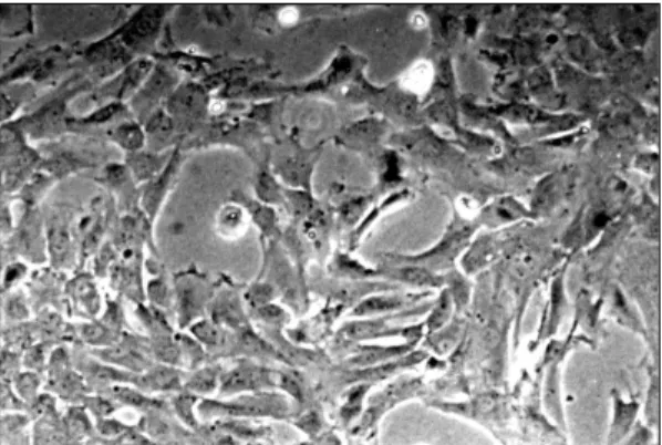
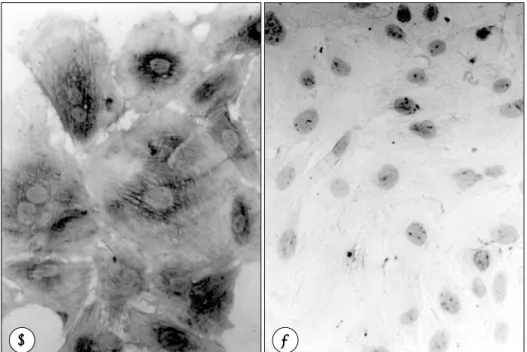
(3) [3H]-thymidine incorporation study 배양액을 흡인해내고 1 μCi/ml의 [3H]-thymidine (464 mCi/mmol, New England Nuclear사)을 함유하 는 신선한 DMEM으로 교체하여 24시간 동안 세포배양 기에서 혈관평활근세포를 성장시킨 후 배양액을 흡인하 였다. 혈관평활근세포를 4℃ 세척용액(10 mM Tris, 150 mM Nacl, pH 7.4)으로 세척한 후 10% trichloroacetic acid로 고정하였다. 95%로 다시 세척하여 건조시킨 후 0.2 N NaOH를 사용하여 DNA를 추출하였으며, 액체 섬광계수기(liquid scintillation spectrometry)로 incorporated radioactivity를 측정하였다.. Fig. 1. 70-80% confluent state of vascular smooth muscle cells. Cells assumed a flattened, will spread morphology.. 통 계 측정된 성적은 평균과 표준편차로 표시하였고, % 억 제율(% inhibition)은 [1-(PDGF-BB와 약물을 투여한 경우의 incorporated radioactivity(cpm))] PDGF-BB만 투여한 경우의 incorporated radioacitivity(cpm). ×100. 을 이용하여 얻은 값을 사용하였다. 검정은 Student’s t-test에 의하여 p value<0.05에서 통계적으로 유의하 다고 판정하였다.. 결. 과. 혈관평활근세포 배양결과 순수 배양한 혈관평활근세포를 0.25% trypsin-ED TA 용액으로 처리하고 1:4 비율로 계대배양을 시행 하여 배양 1일 경과시 혈관평활근세포들이 배양접시에 붙어있는 상태가 확인되었다. 별모양으로 붙고 나서 혈관. Fig. 2. Typical “hill and valley” morphology of vascular smooth muscle cells. Upon reaching confluence, cells formed dense multilayers having the characteristic appearance of cultured arterial smooth muscle cells.. 포들의 상태를 도립현미경(inverted microscope)으로 관찰한 결과, 대조군과 비교하여 형태학적으로 유의한 차 이는 발견되지 않았다.. PDGF-BB의 성장촉진 효과. 평활근세포의 증식이 진행되어 70~80% 합류상태(Fig.. PDGF-BB의 성장촉진효과를 확인하기 위해 PD GF-. 1)에 이르게 되었고, 이 상태의 혈관평활근세포들을 본. BB를 투여한 군과 투여하지 않은 군간의 [3H]-thymi-. 연구에 사용하였다. 한편 이 세포가 순수한 혈관평활근. dine 섭취정도를 조사한 결과, PDGF-BB를 투여한 군. 세포임을 확인하기 위하여 일부 세포를 계속 증식시켜. 에서 [3H]-thymidine 섭취정도가 83% 정도 증가하였. 완전한 합류상태에 이르게 하면 세포의 분화가 진행되어. 다(대조군 532,678±36,340 cpm, PDGF-BB군 975,. 평활근세포의 특징인 hills and valleys형태가 나타났. 781±92,853 cpm)(Fig. 5).. 다(Fig. 2). 그리고 면역세포화학적 방법으로 평활근 α-액틴에 대한 특이 단일클론성 항체를 결합시킨 후 염색한 결과 세포질에서 풍부한 α-액틴을 확인하였다 (Fig. 3). 카베디롤(Fig. 4)을 투여하고 3일 후에 혈관평활근세. 카베디롤의 혈관평활근세포에 대한 증식억제효과 PDGF-BB로 성장을 자극시킨 혈관평활근세포에 카 베디롤 1 μM, 10 μM을 투여한 군들과 PDGF-BB만 투여한 대조군과의 [3H]-thymidine 섭취정도를 조사 1585.
(4) A. B. Fig. 3. A:Immunochemical staining of vascular smooth muscle cells (VSMC) using anti-smooth muscle α-actin monoclonal antibody. VSMC were clearly distinguished by brownish staining of actin fibers running the entire length of each cell. B:Negative control showed no cytoplasmic staining.. Fig. 4. Chemical structure of carvedilol. The asterisk denotes the point of asymmetry.. 한 결과, 1 μM을 투여한 군은 유의한 억제를 보이지 않. Fig. 5. Effect of platelet derived growth factor (PDGF-BB) on vascular smooth muscle cell growth. PDGF-BB (1 nM) significantly stimulated [3H]-thymidine incorporation.. 았지만, 10 μM을 투여한 군은 대조군에 비해 약 30% 정도의 유의한 [3H]-thymidine 섭취억제를 보였다(p<. 우 8%의 [3H]-thymidine 섭취 억제 효과가 있었지만. 0.05)(PDGF군 975,781±92,853 cpm, PDGF+C1군. 통계적으로 유의하지 않았고, PDGF-BB로 자극한 혈관. 908,696±77,043 cpm, PDGF+C10군 757,045±62,. 평활근세포에 10 μM 농도의 카베디롤을 함께 투여한 경. 187 cpm)(Fig. 6).. 우는 24%로 프로프라놀롤을 투여한 군에 비해 24시간. PDGF-BB로 성장을 자극한 혈관평활근세포에 10 μ. [3H]-thymidine 섭취는 유의하게 감소하였다(p<0.05). M의 카베디롤과 동일한 농도의 프로프라놀롤을 투여하. (PDGF+C10군 757,045±62,187 cpm, PDGF+pr-. 고, % 억제율을 조사한 결과, 프로프라놀롤을 투여한 경. op10군 922,457±128,365 cpm)(Fig. 7).. 1586. Korean Circulation J 1998;28(9):1583-1589.
(5) llaborative Group의 보고10)에서는 적은 용량의 카베디 롤에서도 심부전 환자의 사망률이 25% 감소하였다. 이 런 효과는 적어도 혈관 α1수용체의 차단을 통해서 혈 관 확장 작용을 유발하며, β 수용체를 차단하여 반사 빈 맥(reflex tachycardia)을 유발하지 않기 때문일 것이다. 카베디롤과 대사물들은 노에피네프린(norepinephrine)과 이의 독성 중간대사산물의 산화를 억제하며, 심근 에서 산소유리기(oxygen free radicals)의 생성을 억제 한다.18) 이러한 항산화 효과로 심근 손상과 심장개형(cardiac remodeling)에 연관된 유전자의 발현을 차단하고, apoptosis(programmed cell death)를 억제하는 것으로 알려져 있다.19) Fig. 6. Effect of carvedilol on [3H]-thymidine uptake of PDGF-stimulated vascular smooth muscle cells.. 카베디롤의 여러 작용 중 혈관평활근세포의 증식에 관 여하는 기전에 대해 여러 가설이 대두되었으나 확실히 밝 혀진 것은 없는 상태이다. 본 연구에서는 혈관평활근세포를 배양하기 위해 성장 인자로 PDGF-BB를 사용하였다. PDGF는 혈관평활근 세포의 증식에 작용하는 중요한 성장인자 중의 하나3)로 PDGF-AA, -AB, -BB 등의 세 가지 아형20)이 있으며 각각 그 작용이 다른 것으로 알려져 있다. 혈관평활근세 포는 이중에서 PDGF-AA를 생성하나 PDGF-AA는 혈관평활근세포에는 세가지 아형 중 가장 약한 성장인자 로 작용하며 [3H]-thymidine incorporation을 유발하 지는 않는 것으로 알려져 있다. 이에 비해 PDGF-BB 는 세 가지 아형 중에서 가장 강력하게 혈관평활근세포 의 증식을 자극하는 것으로 알려져 있다.21) 본 연구에서. Fig. 7. Percent inhibition of [3H]-thymidine uptake after administration of carvedilol and propranolol.. 혈관평활근세포의 성장인자로 PDGF-BB를 선택하였고 PDGF-BB의 성장촉진효과를 확인하기 위해 PDGF-BB 를 투여한 후 [3H]-thymidine 섭취정도를 조사한 결과,. 고. 찰. PDGF-BB를 투여하지 않은 군에 비해 [3H]-thymidine incorporation이 83% 정도 증가함을 확인하였. β 차단제는 혈압을 하강시키고, 좌심실비대를 억제하 여 뇌졸중 등 심혈관계 합병증을 감소시킴으로 인해 그 동안 고혈압의 일차치료제로 널리 이용되어 왔다. 일반. 다(Fig. 5). 본 연구의 조건에서 카베디롤은 PDGF-BB에 자극된 혈관평활근세포의 증식을 억제하였다(Fig. 6).. 적으로 심부전 환자는 교감신경이 항진되어 있어15) 이 경. 본 연구에서는 β 차단제로 프로프라놀롤을 투여하여. 우에 β 차단제가 도움이 될 것이라는 가설하에16)17) 현. 혈관평활근세포의 증식을 유의하게 억제하지 못하였음을. 재 심부전 치료제로서도 대규모의 연구가 진행되고 있다.. 확인하였다(Fig. 7). 프로프라놀롤의 증식 억제효과가 유. 최근 US Carvedilol Heart Failure Study Group의 보. 의하지 않은 것을 볼 때, 카베디롤의 항증식작용이 단순. 고9)에 의하면 카베디롤은 심부전 환자의 사망률을 65%. 히 항 β 수용체 작용에 의한 것이 아님을 알 수 있다.. 감소시키고, 입원횟수를 27% 감소시킨 것으로 나타났고,. 이전의 연구 결과에서 카베디롤의 항 증식작용이 칼슘. Australia-New Zealand Heart Failure Research Co-. 이온 통로의 억제에 의한 것이라는 가설이 있으나 Hir1587.
(6) ata 등의 연구22)에 의하면, 고농도(1~10 μM)의 니. 며 추후 이에 대한 연구가 더욱 더 진행되어야 할 것이다.. 3. 페디핀(nifedipine)이 [ H]-thymidine incorporation. 요. 을 억제하였고, 그 이하의 농도에서는 억제효과가 나타. 약. 나지 않는 것으로 되어 있어, 카베디롤의 약한 항 칼슘이 온 통로 효과를 고려해 보면 이 약물의 항 증식작용이 항 칼슘이온 통로 효과에 의한 것일 가능성은 희박하다.. 연구배경: 최근 고혈압 및 심부전 환자의 치료에서도 FDA공인. 혈관평활근세포에 대한 카베디롤의 증식억제 효과는 카. 을 받은 3세대 β 차단제인 카베디롤은 β 차단제이면. 베디롤의 고농도 투여에 의한 세포 독성작용에 의한 것일. 서 α1 수용체를 동시에 차단하여 혈관을 확장시키고, 독. 수도 있으며, 이러한 세포 독성작용은 Trypan blue 배. 특하게도 항산화효과가 강력하며 유리산소기 발생을 줄. 출능력과 락트산탈수소효소(lactic dehydrogenase)의. 이는 동시에 풍선도자로 손상받은 동맥의 신생내막 형성. 상승 여부, 현미경하의 세포 변형 등을 확인하여 알 수 있. 을 억제하는 사실이 증명되었다. 본 연구에서는 카베디. 다. 본 연구에서는 Trypan blue 배출능력과 락트산탈. 롤이 배양한 혈관평활근세포의 증식에 어떠한 영향을 미. 수소효소의 상승을 측정하지 않았으나, 카베디롤 10 μ. 치는지 알아보기 위하여 [3H]-thymidine incorporation. M 투여 전 후의 형태학적 비교를 하였고 세포 변형이 없. study를 시행하였다.. 음을 확인하여 카베디롤의 항 증식 작용이 단순히 세포 독성에 의한 것이 아님을 알 수 있었다. 이상과 같이 카베디롤이 PDGF-BB에 의해 자극된 혈 관평활근세포의 증식을 억제하는 것을 볼 때, 병적 환경. 방 법: 혈관평활근세포는 흰쥐 대동맥의 중막에서 효소소화 방 법으로 분리하여 1차 배양에 성공한 후 계대배양을 계 속하여 4~8세대의 세포들을 사용하였다.. 에 있는 혈관평활근세포의 이상증식을 억제하는데 카베. 결 과:. 디롤이 이상적인 약제일 수 있다는 것을 추측케 한다.23). 1) 세포를 휴지상태에서 PDGF-BB로 자극한 결과. 심근세포에 대한 β 차단제의 항허혈 작용은 이미 확 립되어 있는 상태로 특히 내인성 교감성 활성도(intr-. 24시간 [3H]-thymidine uptake는 대조군에 비해서 약 70~100% 증가하였다.. insic sympathomimetic activity)가 없는 β 차단제는. 2) 10 μM 농도의 카베디롤을 함께 투여했을 때 PD-. 심박수와 수축력을 낮추므로 심근의 산소 요구량을 줄. GF-BB로 자극한 24시간 [3H]-thymidine uptake 억. 일 수 있다. 카베디롤은 혈관 확장성이 있는 3세대 β 차. 제효과는 유의하게 감소하였다.. 단제이면서 심근에 항허혈작용을 하는 항산화제로 최근. 3) 반면에 상기 조건에서 프로프라놀롤을 함께 투여. 미국에서 고혈압, 협심증, 심부전 등의 치료제로 인정을 받. 했을 때에는 [3H]-thymidine uptake 억제효과가 나타. 았다. 또 신경-호르몬계 차단효과(neurohormonal ant-. 나지 않았다.. agonist effect)와 항산화 작용이 있어 이를 통해 심부전. 결 론:. 의 치료효과가 있을 것으로 생각된다.. 이상의 결과로 보아 α1 및 β 수용체를 동시에 차. 그러나, 현재 수많은 화학주성인자(chemotactic fac-. 단하는 카베디롤은 본 연구의 조건에서 혈관평활근세포. tor)와 유사분열물질(mitogen)들이 혈관평활근세포의. 의 DNA합성을 유의하게 억제하였다. 카베디롤의 이러한. 성장에 관여하는 것으로 알려져 있고, 따라서 비정상적. 작용은 임상적으로 혈관보호 효과에 일부 관여할 것으로. 인 혈관평활근세포의 성장을 억제하기 위해서는 이러한. 간주된다.. 여러 인자들이 작용하는 공통 생화학 경로(common biochemical pathway)에 작용하는 약제가 필요하며, PD-. 중심 단어:카베디롤・혈관평활근세포・세포증식.. GF나 트롬빈(thrombin)과 같은 성장인자를 한가지만 억. REFERENCES. 제하는 것으로는 그 효과가 미흡할 것이다. 따라서 이. 1) Ross R. The pathogenesis of atherosclerosisan update. N. 에 대한 많은 연구가 진행되어야 할 것이다. 혈관평활근 세포에 대한 카베디롤의 증식억제 기전은 단순히 β 교 감신경계 차단이나 칼슘 통로의 차단에 의한 것은 아니 1588. Engl J Med 1986;314:488-500.. 2) Schwartz SM, Reidy MA. Common mechanisms of proliferation of smooth muscle in atherosclerosis and hypertension. Hum Pathol 1987;18:240-7.. Korean Circulation J 1998;28(9):1583-1589.
(7) 3) Grainger DJ, Witchell CM, Weissberg PL, Metcalfe JC.. 4). 5) 6) 7). 8) 9). 10). 11). 12) 13). Mitogens for adult rat aortic vascular smooth muscle cells in serum-free primary culture. Cardiovasc Res 1994;28: 1238-42. Unterberg C, Meyer T, Wiegand V, Kreuzer, Buchwald AB. Proliferative response of human and minipig smooth muscle cells after coronary angioplasty to growth factor and platelets. Basic Res Cardiol 1996;91(6):407-17. Francis GS. Neurohormonal mechanisms involved in congestive heart failure. Am J Cardiol 1985;55:15A-21A. Packer M. The neurohormonal hypothesis: A theory to explain the mechanism of disease progression in heart failure. J Am Coll Cardiol 1992;20:248-54. Waagstein F, Bristow MR, Swedberg K, Camerini F, Fowler MB, Johnson M, et al. Beneficial effects of metoprolol in idiopathic dilated cardiomyopathy. Lancet 1993; 342:1441-6. CIBIS Investigators and Committees. A randomized trial of blockade in heart failure: The Cardiac Insufficiency Bisoprolol Study (CIBIS). Circulation 1994;90:1765-73. Packer M, Bristow MR, Cohn JN, Colucci WS, Fowler MB, Gilbert EM, et al. for the US Carvedilol Heart Failure Study Group. The effect of carvedilol on morbidity and mortality in patients with chronic heart failure. N Engl J Med 1996;334:1349-55. Australia-New Zealand Heart Failure Research Collaborative Group. Randomised, placebo-controlled trial of carvedilol in patients with congestive heart failure due to ischaemic heart disease. Lancet 1997;349:375-80. Nichols AJ, Sulpizio AC, Ashton D, Hieble JP, Ruffolo RR J. In vitro pharmacologic profile of the novel betaadrenoceptor antagonist and vasodilator, carvedilol. Pharmacology 1989;39:327-36. Ruffolo RRJ, Gellai M, Hieble JP, Willette RN, Nichols AJ. The pharmacology of carvedilol. Eur J Clin Pharmacol 1990;38(suppl 2):S82-8. Yue TL, Wang X, Gu JL, Ruffolo RRJ, Feuerstein GZ. Carvedilol, a new vasodilating beta-adrenoreceptor bloc-. 14). 15). 16) 17) 18). 19) 20). 21) 22) 23). ker, inhibits oxidation of low-density lipoproteins by vascular smooth muscle cells and prevents leukocyte adhesion to smooth muscle cells. J Phar & Exp Ther 1995;273 (3):1442-9. Ohlstein EH, Douglas SA, Sung CP, Yue TL, Louden C, Arleth A, et al. Carvedilol, a cardiovascular drug, prevents vascular smooth muscle cell proliferation, migration, and neointimal formation following vascular injury. Proc Natl Acad Sci USA 1993;90:6189-93. Cohn JN, Levine TB, Olivari MT, Garberg V, Tura D, Francis GS, et al. Plasma norepinephrine as a guide to prognosis in patients with chronic congestive heart failure. N Engl J Med 1984;311:819-23. Bristow MR. Pathophysiologic and pharmacologic rationale for clinical management of chronic heart failure with beta-blocking agents. Am J Cardiol 1993;71:12C-22C. Bristow MR. Mechanism of action of beta-blocking agents in heart failure. Am J Cardiol 1997;80(11A):26L-40L. Feuerstein GZ, Yue TL, Cheng HY, Ruffolo RRJ. Myocardial protection by the novel vasodilating beta-blocker, carvedilol: Potential relevance of antioxidant activity. J Hypertens 1993;11(suppl 4):S41-8. James TN. Normal and abnormal consequences of apoptosis in the human heart: From postnatal morphogenesis to paroxysmal arrhythmias. Circulation 1994;90:556-73. Kondo T, Konishi F, Inui H, Inagami T. Differing signal transductions elicited by three isoforms of platelet derived growth factor in vascular smooth muscle cells. J Biol Chem 1993;268(6):4458-64. Ross R. The biology of platelet-derived growth factor. Cell 1986;46:155-69. Hirata Y, Takagi Y, Fukuda Y, Marumo F. Endothelin is a potent mitogen for rat vascular smooth muscle cells. Arteriosclerosis 1989;78:225-8. Feuerstein GZ, Ruffolo RRJ. Carvedilol, a novel vasodilating beta-blocker with potential for cardiovascular organ protection. Eur Heart J 1996;17(suppl B):24-9.. 1589.
(8)
수치
![Fig. 6. Effect of carvedilol on [ 3 H]-thymidine uptake of PDGF-stimulated vascular smooth muscle cells](https://thumb-ap.123doks.com/thumbv2/123dokinfo/5457594.655949/5.892.135.433.180.441/effect-carvedilol-thymidine-uptake-stimulated-vascular-smooth-muscle.webp)
관련 문서
Bulletin of Food Technology 혈관재형성(Vascular Remodeling)에 미치는 Chemokines의 역할.. 롭게도 smooth muscle cell(SMC) 에서 발현되는 MCP-1 은 neointima
In this study, we investigated the effect of luteolin on the proliferation of primary cultured rat aortic vascular smooth muscle cells induced by 5% fetal bovine serum..
tenuates platelet-derived growth factor-induced contraction in aortic smooth muscle through in hibition of protein tyrosine kinase(s). : Tyrosine kinase inhibitors
Keywords: contraction; hypertension; myosin light chain (MLC); RhoA; translationally controlled tumor protein (TCTP); vascular smooth muscle cell
Concentrations of transforming growth factor-alpha (TGF- α), vas- cular endothelial growth factor (VEGF), platelet-derived growth factor-AB/BB (PDGF-AB/BB), PDGF-AA, and
Effect of angiotensin Ⅱ receptor blocker on angiotensin Ⅱ stimulated DNA synthesis of cultured human aortic smooth muscl cells.. Freeman EJ, Chisolm GM, Ferrario CM,
Background: Vascular endothelial growth factor (VEGF) plays an important role in angiogenesis, including stimulating the proliferation and migration of vascular smooth muscle
Heat shock protein 90 inhibitor AUY922 attenuates platelet-derived growth factor-BB-induced migration and proliferation of vascular smooth muscle cells Jisu Kim1,#, Kang Pa Lee2,#,
