Antioxidants Inhibit Smooth Muscle Cell Proliferation in vitro and Neointimal Hyperplasia in vivo after Carotid Artery Injury in the Rat
Hyuck Kim, MD
1, Chan-Kum Park, MD
2and Kyung-Soo Kim, MD
31
Division of Thoracic surgery,
2Pathology Division and
3Cardiology Division, College of Medicine, Hanyang University, Seoul, Korea
ABSTRACT
Background : Smooth muscle cell proliferation and neointimal hyperplasia are major components in in-stent restenosis. In smooth muscle cells, reactive oxygen species (ROS) have been shown to mediate cell proliferation.
We investigated whether antioxidants inhibit smooth muscle cell proliferation and neointimal hyperplasia after carotid artery injury in a rat model. Methods : Rat aortic smooth muscle cells were stimulated by PDGF (80 ng/mL) with or without N-acetylcysteine (NAC) 1 mM. Intracellular ROS levels were measured by carboxyl-2’, 7’-dichlorodihydrofluorescein diacetate confocal microscopy, and cellular proliferation was evaluated by cell counting and XTT assay. Rat carotid arteries were injured with a balloon and with NAC 100 mM, pyrrolidine- dithiocarbamate (PDTC) 100 uM, catalase 5000 u/mL, or superoxide dismutase (SOD) 2,000 u/mL. All agents were applied in pluronic gel on the periadventitial side of the injured artery. At 21 days after, the intima/media (I/M) ratios were measured. Results : In rat aortic smooth muscle cells culture, NAC inhibited PDGF-induced increase of ROS by 77% and PDGF-induced cellular growth by 45%. In balloon injured rat carotid artery, PDTC showed the most prominent effect and reduced the I/M ratio by 51% (0.94±0.32 vs. 1.96±0.14, p<0.05) versus the control. Catalase, SOD, and NAC treatments also reduced the I/M ratio (1.08±0.43, 1.30±0.31, 1.43±0.34, respectively, versus the control (1.96±0.14), all p<0.05). Conclusions : Antioxidant treatment may inhibit the proliferation of RASMC stimulated by PDGF by reducing intracellular ROS in vitro and neointimal hyperplasia after balloon injury to the rat carotid artery. (Korean Circulation J 2004;34 (1):69-75)
KEY WORDS : Antioxidants; Myocytes, smooth muscle; Restenosis.
Introduction
Restenosis after coronary intervention causes serious clinical and economical problems. The proliferation of smooth muscle cell plays important roles in restenosis after coronary angioplasty by producing neointimal hype- rplasia.
1)Smooth muscle cells are the major cellular component of neointimal lesions. In this stent era, renewed
interest has been focused on inhibiting smooth muscle cell proliferation, because in-stent restenosis is almost entirely dependent on smooth muscle cell proliferation, manifesting as neointimal hyperplasia, after coronary in- tervention.
2)For many years, reactive oxygen species (ROS), like the superoxide anion ( O
2-) and hydrogen peroxide ( H
2O
2) , have been believed to be the toxic byproducts of aerobic life. But recently, a plethora of evidence has suggested that ROS are intimately involved in signal transduction in mammalian cells.
3)In vascular smooth muscle cells, ROS was found to mediate platelet derived growth factor ( PDGF ) - or angiotensin II- induced cell proliferation.
4-6)Moreover, the production of ROS in
Received:May 27, 2003Accepted:July 30, 2003
Correspondence:Kyung-Soo Kim, MD, Cardiology Division, College of Medicine, Hanyang University, 17 Haengdang-dong, Sungdong-gu, Seoul 133-792, Korea
Tel:82-2-2290-8312, Fax:82-2-2298-9183 E-mail:[email protected]
blood vessels was found to be enhanced in experimental models of hypercholesterolemia, hypertension, diabetes, and of balloon injury to the coronary arteries.
7-10)These findings suggest that ROS can mediate common mecha- isms of diseases characterized by their dependences upon vascular smooth muscle cell proliferation.
Recently, treatments with antioxidants such as pyrro- idinedithiocarbamate ( PDTC ) or N-acetylcysteine ( NAC ) , and overexpression of catalase have been shown to inhibit the proliferation of vascular smooth muscle cells by reducing the level of intracellular ROS.
11)12)Moreover, these strategies also reduced preformed ROS levels.
In this study, we examined whether antioxidants can inhibit the proliferation of rat aortic smooth muscle cells ( RASMCs ) stimulated by PDGF by reducing intrace- llular ROS levels in vitro, and inhibit neointimal hyperp- lasia after balloon injury in the rat carotid artery.
Methods
Cell cultures
We used primary cultures of RASMCs as an in vitro model for growth factor-induced ROS production and cell growth. RASMCs were obtained as a primary culture of thoracic aorta of a 3-month-old Sprague-Dawley rat by using the cell explant method described previously.
13)Cells were maintained in DMEM ( Gibco BRL, Grand Island, NY, U.S.A. ) containing 10% fetal calf serum ( Gibco BRL, Grand Island, NY, U.S.A. ) in a humidified atmosphere containing 5% CO
2at 37℃. Before drug treatment, RASMCs were pre-cultured in a medium with the same constitution but with 3% fetal bovine serum for 24 hr, because this was the lowest concentration that showed a linear growth pattern ( data not shown ) .
To select a growth factor for RASMC stimulation, we were examined angiotensin II, basic FGF, thrombin, and PDGF. PDGF was chosen because it most prominently induced ROS and cell growth. At 80 ng/mL, PDGF ( BB isoform )( Upstate Biotechnology, Waltham, MA, U.S.A. ) maximally induced ROS and cell growth, and therefore, we used this concentration for the experiment. N-ace-
tylcysteine ( Sigma, St. Louis, MO, U.S.A. ) was selected, because we observed that PDTC and catalase induced significant cellular toxicity by trypan blue exclusion in preliminary experiments ( data not shown ) , and we used 1 mM of NAC, because at this concentration, NAC maximally inhibited the ROS level and cell growth.
Assessment of intracellular ROS and RASMC proli- feration
Antioxidants were added with PDGF and their anti- oxidant effects were observed.
Levels of intracellular ROS were measured 12 hr after PDGF stimulation with or without NAC. The intra- cellular generation of ROS was detected using 5- ( and 6 ) -carboxyl-2’, 7’-dichlorodihydrofluorescein diacetate ( carboxyl-DCFH-DA )( Molecular Probes Inc., Eugene, OR, U.S.A.).
14-17)Carboxyl-DCFH-DA fluoresces green when oxidized by superoxide radical or H
2O
2. Fluore- scence was detected by confocal laser scanning micro- scope using excitation and emission wavelengths of 488 and 525 nm, respectively, after incubating cells for 5 min with 10 ug/mL of carboxyl-DCFHDA, as previously described.
6)The levels of carboxyl-DCFHDA fluore- scence shown represent the values from at least 100 random cells ( means±SD ) , based on an arbitrary scale of fluorescence intensity.
6)RASMC proliferation was evaluated at 72 hr after PDGF stimulation with or without NAC by two methods, cell number counting using trypan blue dye exclusion or XTT assay. For cell number counting, only viable cells by trypan blue exclusion were counted by a hemo- cytometer. A “Cell Proliferation Kit II ( XTT ) ” ( Boe- hringer Mannheim Corp., Mannheim, Germany ) was used for the XTT assay. XTT labeling mixture ( 50 uL ) was added to each well of a 96-well plate and readings were taken 24 hr later at 492 nm, and corrected against < what at > 690 nm.
Arterial injury model
Adult male 8-week-old Sprague-Dawley rats ( 200 -
250 g ) were subjected to balloon angioplasty of the left
common carotid artery using a 2 Fr Fogarty catheter. All
animals were anesthetized beforehand with an intraperi- toneal injection of ketamine ( 50 mg/kg ) and xylazine ( 2 mg/kg ) . The distal left common carotid artery, and the internal and external carotid arteries were exposed through a midline incision in the neck. A 2 Fr Fogarty catheter was passed three times with the balloon distended sufficiently with saline to generate slight resistance:
this technique produced distension of the carotid artery.
In initial experiments, the extent of endothelial denudation was confirmed 2 days after balloon injury by Evans blue staining ( data not shown ) . All animal experiments were carried out according to the guidelines of the Interna- tional Committee for Thrombosis and Hemostasis, and approved by the Hanyang University Ethical Committee for Animal Experimentation.
After arterial injury has been administered, the carotid artery was carefully examined and blood pulsation was checked distally. In each rat 200 uL of pluronic gel ( 25%
w/v in phosphate buffered solution, pH 7.4 )( Sigma, St.
Louis, MO, U.S.A. ) or 200 uL of pluronic gel containing various antioxidants was applied to the periadventitial side of the injured artery. The concentrations of the antioxidants used were as follows; N-acetyl cysteine 100 mM ( Sigma, St. Louis, MO, U.S.A. ) , pyrrolidinedithi- ocarbamate ( PDTC ) 100 uM ( Sigma, St. Louis, MO, U.S.A. ) , catalase 5000 u/mL ( Sigma, St. Louis, MO, U.S.A. ) , or superoxide dismutase ( SOD ) 2,000 u/mL ( Sigma, St. Louis, MO, U.S.A. ) . The pluronic gel rem- ains liquid at 4℃, but rapidly solidifies at 37℃, when in contact with living tissues.
In the rat carotid artery injury model, with peria- dventitial application of the mentioned drugs in Pluronic gel, they were found to be present in the arterial tissue up to 2nd week after application with peak concentrations at ca. 24 hrs.
18)The external carotid artery was ligated after removing the catheter, and the neck incision was closed.
Histological analysis
Three weeks after surgery, the animals were eutha- nized after being anaesthetized with an intraperitoneal
injection of ketamine & xylazine. Carotid arteries and aortas were dissected free from the surrounding tissues, and 4% formaldehyde was infused through the aortic root at a perfusion pressure of 120 mmHg for 5 minutes.
A 10 mm section of both carotid arteries ( both left ( surgi- cally treated ) and right ( control ) arteries from the same rat ) were then removed and bathed in the same forma- ldehyde solution for > 2 hours. The mid 5 mm of each injured artery was embedded in paraffin and cut into 5- um sections. Twelve randomly chosen sections were stained with hematoxylin & eosin and Massons’s tri- chrome ( 6 sections each ) . Under an Olympus BH-2 microscope, each digital image was captured and ana- lyzed using a personal computer running Metavue image software ( ver. 4.6r5 ) . The internal elastic lamina, external elastic lamina, and luminal areas were measured, and the intimal area, the medial area, and the intima/media area ratio ( I/M ratio ) were calculated.
Statistical analysis
All values are expressed as means±SD. Statistical significance was determined by performing ANOVA with the multiple range test ( LSD ) .
Results
In Vitro
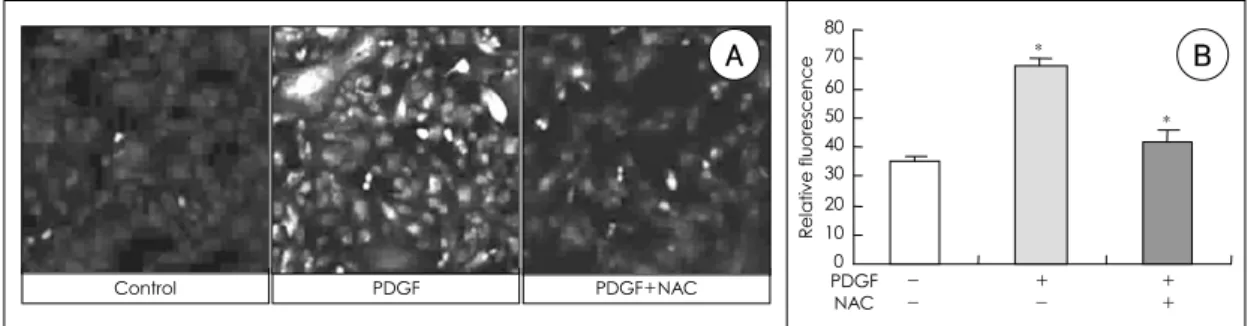
After stimulating with 80 ng/mL PDGF for 12 hr the
levels of intracellular ROS increased ca 2-fold ( Figure
1 ) , and with 80 ng/mL PDGF for 72 hr, the cell number
as counted by trypan blue dye exclusion, increased about
1.6-fold versus the control ( Figure 2A ) . The XTT assay
showed a similar result with a 1.8-fold increase in
absorbance ( Figure 2B ) . We then assessed the effects of
NAC. Consistent with previous studies, which demo-
nstrated that ROS scavengers reduced proliferation by
reducing intracellular ROS levels,
19)20)treatment with
NAC 1 mM was found to down-regulate the intracellular
ROS ( Figure 1 ) and cell proliferation ( Figure 2 ) induced
by PDGF.
In Vivo
Seventy rat carotid arteries were injured and 12 were excluded from the analysis, because of arterial thro- mbosis, wound infection, and death. Major excluded cases developed early one third periods of surgical proce- dures. For the analysis, 58 carotid arteries ( 8 pluronic gel only, 10 SOD in pluronic gel, 9 NAC in pluronic gel, 10 catalase in pluronic gel, 12 PDTC in pluronic gel, and 9 balloon injury only ) were used. On the 21st day after carotid arterial injury, no difference was observed in the intimal areas ( 0.18±0.02 vs. 0.18±0.03 mm
2, p = 0.41 ) of the pluronic gel only and the balloon injury only groups, but the I/M ratio was greater in the pluronic
gel only group ( 1.96±0.14 vs. 1.82±0.21, p = 0.18 ) . No significant histological changes, such as fibroplasia in the adventitia due to pluronic gel application, were obs- erved. To negate the effect of any possible antioxidants in pluronic gel, we used the pluronic gel only group as a control.
On the 21st day after carotid arterial injury, all antio- xidant treatments showed reduced intimal areas and I/M ratios versus the control ( Figure 3, 4 ) . Though the effects of the antioxidants were not significantly different, PDTC showed the most prominent effect, i.e., a 49% reduction in intimal area versus the control ( 0.09±0.03 vs. 0.18
±0.02 mm
3, p < 0.05 ) , and a 51% decrease in the I/M
Figure 1. Effects of PDGF and NAC on the levels of ROS in rat aortic smooth muscle cells. A: representative images obtained by carboxyl-DCFH-DA confocal laser microscopy. control (control), cells incubated with PDGF 80 mg/mL for 12 hours (PDGF) and cells co-incubated with PDGF 80 mg/mL and NAC 1 mM for 12 hours (PDGF+NAC). B: levels of carboxyl-DCFH-DA fluorescence measured by confocal laser microscopy. cells were incubated with PDGF 80 mg/mL (PDGF) or NAC 1 Mm (NAC) for 12 hours. *: p<0.05 PDGF (-) NAC (-) vs PDGF (+) NAC (-), PDGF (+) NAC (-) vs PDGF (+) NAC (+), carboxyl-DCFH-DA: 5- (and 6)-carboxyl-2’,7’-dichlorodihydrofluorescein diacetate, NAC: N-acetylcysteine, PDGF: platelet derived growth factor, ROS: reactive oxygen species.0.00 0.25 0.50 0.75 1.00 1.25
- -
+ -
+ + Cell count (×105/mL)
PDGF NAC
*
*
0.00 0.25 0.50 0.75 1.00
- -
+ -
+ +
Absorbance (A492)
PDGF NAC
*
*
A B
Figure 2. Effects of PDGF and NAC on the proliferation of rat aortic smooth muscle cells. A: effects of PDGF and NAC on cell numbers. B: effects of PDGF and NAC on relative absorbance at 492 nm on XTT assay. cells were incubated with PDGF 80 mg/mL (PDGF) or NAC 1 mM (NAC) for 72 hours. *: p<0.05 PDGF (-) NAC (-) vs PDGF (+) NAC (-), PDGF (+) NAC (-) vs PDGF (+) NAC (+), NAC: N-acetylcysteine, PDGF: platelet derived growth factor.
Control PDGF PDGF+NAC
0 10 20 30 40 50 60 70 80
Relative fluorescence
PDGF
*
*
NAC - + +
- - +
A B
ratio ( 0.94±0.32 vs. 1.96±0.14, p < 0.05 ) , respectively.
The other antioxidants showed similar effects. In terms of intimal area, catalase, SOD, and NAC treatments showed 0.11±0.04, 0.13±0.04, and 0.14±0.04 mm
3,
versus a control of 0.18±0.02 mm
3( all p < 0.05 ) . And, for I/M ratio the corresponding values were 1.08±0.43, 1.30±0.31, and 1.43±0.34, versus a control of 1.96±
0.14 ( all p < 0.05 ) .
Normal Control PDTC
NAC CAT SOD
Figure 3. Reduction in neointimal hyperplasia by antioxidants after carotid artery injury in the rat. Representative images of hematoxylin & eosin stained sections at 21 days after balloon injury. The arteries shown are non-injured (normal), injured without antioxidant (control), injured with the periadventitial application of PDTC 100 uM (PDTC), NAC 100 mM (NAC), CAT 5000 u/mL (CAT), or SOD 2,000 u/mL (SOD). CAT: catalase, NAC: N-acetyl cysteine, PDTC:
pyrrolidine-dithiocarbamate, SOD: superoxide dismutase.
0 0.0 0.1 0.1 0.2 0.2
PG Balloon SOD NAC CAT PDTC
*
*
*
* Intimal area (mm3)
0 0.5 1 1.5 2 2.5
PG Balloon SOD NAC CAT PDTC
*
*
* * I/M ratio
A B
Figure 4. Reduction in the intimal areas (A) and intima/media ratios (B) by antioxidants at 21 days after carotid artery injury in the rat. The arteries shown were injured only (control), injured with the periadventitial application of PG only (PG), injured with the periadventitial application of various antioxidants in PG: PDTC 100 uM (PDTC), NAC 100 mM (NAC), CAT 5000 u/mL (CAT), SOD 2,000 u/mL (SOD). *: p<0.05 compared with PG, CAT: catalase, NAC: N- acetyl cysteine, PDTC: pyrrolidine-dithiocarbamate, PG: pluronic gel, SOD: superoxide dismutase.
Discussion
This study suggest that antioxidants do inhibit the proli- feration of rat aortic smooth muscle cells ( RASMC ) stimulated by PDGF, and reduce intracellular ROS and neointimal hyperplasia after balloon injury in the rat carotid artery.
ROS plays an important role in signal transduction in many cells.
3)21-23)In particular, growth factors, like PDGF, epidermal growth factor, and angiotensin II, trigger ligand mediated ROS production. In addition, ROS can stimu- late the proliferation of vascular smooth muscle cells,
5)and in RASMCs, PDGF was found to stimulate H
2O
2production and DNA synthesis; moreover, both of these responses were blocked by antioxidant treatment.
6)These reports and the results of our study, suggest that endo- genously produced ROS has an important role in the regulation of vascular smooth muscle cell growth, and that antioxidants could inhibit the proliferation of vas- cular smooth muscle cells-the major mechanism of instent restenosis.
In the present study, these in vitro effects of antio- xidants on smooth muscle cell proliferation were obse- rved in an in vitro model of rat carotid artery balloon injury.
In a previous study, orally or intraperitoneally admi- nistrated antioxidants, i.e., Ginkgo biloba extract,
24)pro- bucol,
25)26)vitamin E,
27)and Salvia miltiorrhiza,
28)were found to inhibit neointimal hyperplasia after arterial balloon injury in several animal models. In addition, clinical evidence indicates that antioxidants, like, pro- bucol reduce the restenosis rate after PTCA.
29)The findings of the above studies are wholly compatible with our results.
In particular, in the present study shows that all of the antioxidants examined, effectively inhibited neointimal hyperplasia after rat carotid artery balloon injury. This finding supports the notion that antioxidants inhibit neoi- ntimal hyperplasia. In addition, we believe that the peri- adventitial pluronic gel drug delivery system used in this study has advantages over the previously used systemic
method, because it directly affects drug delivery and substantially excludes systemic effects. This periadventi ( periadventitial ) antioxidant delivery has limitations in terms of its appli-cation in clinical situations in which systemic or endolu-minal drug delivery is used, but may be useful in vascular surgery.
The inhibition of smooth muscle cell proliferation and neointimal hyperplasia by antioxidants, but this inhibition was not complete. Moreover, previous studies and the present study, have shown a 20% to 50% reduction in the intimal/medial ratio.
24-28)30)These findings suggest that ROS may not be the only common pathway to smooth muscle cell proliferation.
In the present experiment, PDTC and catalase showed cell toxicity in vitro, which might have played a role in the inhibition of neointimal hyperplasia. Moreover, such cytotoxic effects could cause inflammation and neointi- mal hyperplasia in the long term. These issues were not addressed in the present study.
The antioxidant used showed different effectivenesses at inhibiting neointimal hyperplasia. Although these di- fferences were statistically insignificant, PDTC most prominently inhibited neointimal hyperplasia. In this study, we did not examine different antioxidant conce- ntrations, and their optimal concentrations were not determined. Perhaps this explains the different degrees of neointimal hyperplasia inhibition observed.
In conclusion, this study shows that antioxidants can inhibit neointimal hyperplasia after arterial injury. Mo- reover, antioxidants may be useful in the clinical setting, to prevent or inhibit restenosis after coronary intervent- ion, where the proliferation of vascular smooth muscle cells plays a key role.
■
Acknoewledgments
This work was supported by the Korean Society of Circu-lation. (Industrial-educational cooperation 2000-3).
REFERENCES
1)
Clowes AW, Reidy MA, Clowes MM. Kinetics of cellular proliferation after arterial injury: I. smooth muscle growth in the absence of endothelium. Lab Invest 1983;49:327-33.
2)
Kearney M, Pieczek A, Haley L, Losordo DW, Andres V,
Schainfeld R, et al. Histopathology of in-stent restenosis in patients with peripheral artery disease. Circulation 1997;
95:1998-2002.
3)
Finkel T. Oxygen radicals and signaling. Curr Opin Cell Biol 1998;10:248-53.
4)
Ushio-Fukai M, Griendling KK, Becker PL, Hilenski L, Halleran S, Alexander RW. Epidermal growth factor receptor transactivation by angiotensin II requires reactive oxygen species in vascular smooth muscle cells. Arterioscler Thromb Vasc Biol 2001;21:489-95.
5)
Rao GN, Berk BC. Active oxygen species stimulate vascular smooth muscle cell growth and proto-oncogene expression.
Circ Res 1992;70:593-9.
6)
Sundaresan M, Yu ZX, Ferrans VJ, Irani K, Finkel T.
Requirement for generation of H2O2 for platelet-derived growth factor signal transduction. Science 1995;270:296-9.
7)
Ohara Y, Peterson TE, Sayegh HS, Subramanian RR, Wilcox JN, Harrison DG. Dietary correction of hypercho- lesterolemia in the rabbit normalizes endothelial superoxide anion production. Circulation 1995;92:898-903.
8)
Grunfeld S, Hamilton CA, Mesaros S, McClain SW, Domi- niczak AF, Bohr DF, et al. Role of superoxide in the depre- ssed nitric oxide production by the endothelium of genetically hypertensive rats. Hypertension 1995;26:854-7.
9)
Langenstroer P, Pieper GM. Regulation of spontaneous EDRF release in diabetic rat aorta by oxygen free radicals. Am J Physiol 1992;263:H257-65.
10)
Nunes GL, Robinson K, Kalynych A, King SB 3rd, Sgoutas DS, Berk BC. Vitamins C and E inhibit O2- production in the pig coronary artery. Circulation 1997;96:3593-601.
11)
Brown MR, Miller FJ Jr, Li WG, Ellingson AN, Mozena JD, Chatterjee P, et al. Overexpression of human catalase inhibits proliferation and promotes apoptosis in vascular smooth muscle cells. Circ Res 1999;85:524-33.
12)
Tsai JC, Jain M, Hsieh CM, Lee WS, Yoshizumi M, Patterson C, et al. Induction of apoptosis by pyrrolidinedi- thiocarbamate and N- acetylcysteine in vascular smooth muscle cells. J Biol Chem 1996;271:3667-70.
13)
Fang X, Kaduce TL, Weintraub NL, van Rollins M, Spector AA. Functional implications of a newly characterized pa- thway of 11,12- epoxyeicosatrienoic acid metabolism in arterial smooth muscle. Circ Res 1996;79:784-93.
14)
Speir E, Shibutani T, Yu ZX, Ferrans V, Epstein SE. Role of reactive oxygen intermediates in cytomegalovirus gene expre- ssion and in the response of human smooth muscle cells to viral infection. Circ Res 1996;79:1143-52.
15)
Bass DA, Parce JW, Dechatelet LR, Szejda P, Seeds MC, Thomas M. Flow cytometric studies of oxidative product formation by neutrophils: a graded response to membrane stimulation. J Immunol 1983;130:1910-7.
16)
Ohba M, Shibanuma M, Kuroki T, Nose K. Production of hydrogen peroxide by transforming growth factor-beta 1 and its involvement in induction of egr-1 in mouse osteoblastic cells. J Cell Biol 1994;126:1079-88.
17)
Zhu H, Bannenberg GL, Moldeus P, Shertzer HG. Oxidation pathways for the intracellular probe 2’, 7’- dichlorofluore- scein. Arch Toxicol 1994;68:582-7.
18)
Ishizaka N, Taguchi J, Kimura Y, Ikari Y, Aizawa T, Togo M, et al. Effects of a single local administration of cilostazol on neointimal formation in balloon-injured rat carotid artery.
Atherosclerosis 1999;142:41-6.
19)
Kyaw M, Yoshizumi M, Tsuchiya K, Kirima K, Suzaki Y, Abe S, et al. Antioxidants inhibit endothelin-1(1-31)-induced proliferation of vascular smooth muscle cells via the inhi- bition of mitogen-activated protein(MAP) kinase and acti- vator protein-1(AP-1). Biochem Pharmacol 2002;64:1521-31.
20)
Okuyama H, Shimahara Y, Kawada N, Seki S, Kristensen DB, Yoshizato K, et al. Regulation of cell growth by redox- mediated extracellular proteolysis of platelet-derived growth factor receptor beta. J Biol Chem 2001;276:28274-80.
21)
Rhee SG. Redox signaling: hydrogen peroxide as intrace- llular messenger. Exp Mol Med 1999;31:53-9.
22)
Finkel T. Redox-dependent signal transduction. FEBS Lett 2000;476:52-4.
23)
Lander HM. An essential role for free radicals and derived species in signal transduction. FASEB J 1997;11:118-24.
24)
Lin SJ, Yang TH, Chen YH, Chen JW, Kwok CF, Shiao MS, et al. Effects of Ginkgo biloba extract on the proliferation of vascular smooth muscle cells in vitro and on intimal thick- ening and interleukin- 1 beta expression after balloon injury in cholesterol-fed rabbits in vivo. J Cell Biochem 2002;85:
572-82.
25)
Inoue K, Cynshi O, Kawabe Y, Nakamura M, Miyauchi K, Kimura T, et al. Effect of BO-653 and probucol on c-MYC and PDGF-A messenger RNA of the iliac artery after balloon denudation in cholesterol-fed rabbits. Atherosclerosis 2002;
161:353-63.
26)
Jackson CL, Pettersson KS. Effects of probucol on rat carotid artery responses to balloon catheter injury. Atherosclerosis 2001;154:407-14.
27)
Upston JM, Witting PK, Brown AJ, Stocker R, Keaney JF Jr.
Effect of vitamin E on aortic lipid oxidation and intimal proliferation after arterial injury in cholesterol-fed rabbits.
Free Radic Biol Med 2001;31:1245-53.
28)
Chen YL, Yang SP, Shiao MS, Chen JW, Lin SJ. Salvia milti- orrhiza inhibits intimal hyperplasia and monocyte chemotactic protein-1 expression after balloon injury in cholesterol- fed rabbits. J Cell Biochem 2001;83:484-93.
29)
Tardif JC, Gregoire J, Schwartz L, Title L, Laramee L, Reeves F, et al. Effects of AGI-1067 and probucol after pe- rcutaneous coronary interventions. Circulation 2003;107:
552-8.
30)