Bulletin of Food Technology Vol. 22, No. 4, pp. 721-730
혈관재형성 (Vascular Remodeling) 에 미치는 Chemokines 의 역할
성 미 정
바이오제론 연구단
Role of Chemokines in Vascular Remodeling
Mi-Jeong Sung
Biogeron Food Technology Research Group
서 론
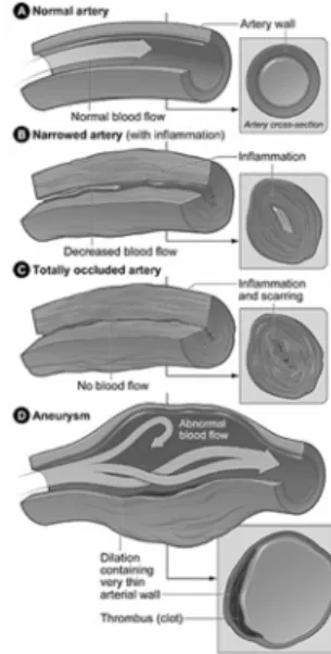
혈관은 혈액이 흐르는 통로로써 동맥
,
정맥,
모세혈관이 있으며 혈관벽에 의해 둘러싸여 있 다
.
그림1
에서 보는 것과 같이 정상 동맥의 혈관벽은 내막
(intima tunica: endothelial cell),
중막
(media tunica: smooth muscle cell),
그리고외막
(adventitia tunica: fibroblast cell)
으로 구성되어 있다
.
그러나 이러한 혈관이 동맥경화(artherosclerosis),
폐고혈압(pulmonary arterial hypertention),
경피경 개입술(percutaneous inter- vention),
심장이식(cardiar allograft vascularpathy)
등과 같은 질병에서
shear stress, hypoxia,
면역학적 또는 물리적인 손상 등의 자극을 받게 되 면 혈관의 크기나 내막넓이
(luminal width)
에 변화를 가져오는 혈관재형성
(vascular remodeling)
이 일어나게 된다
.
혈관재형성은 혈관을 가로로 잘랐을 때 보이 는 혈관 직경이나 두께에 영향을 미치는 것으 로 주된 원인은 관류저하
(hypoperfusion)
로 인해서 나타난다
.
혈관재형성은 임상의학적으로 혈류역학
(hemodynamic)
스트레스,
물리적인 손상,
염증
,
저산소증상태에서 나타나고,
형태학적으로는 동맥벽의
3
개의 층인 신생혈관의 과다형성
(neointimal hyperplasia),
중간층의 비대(medial thickening),
그리고 외피섬유화(adven-
titial fibrosis)
로 백혈구 응집(leukocyte recruit-
ment),
평활근세포 축적(smooth muscle cell
(SMC) accumulation),
그리고 내피세포 회복(endothelial cell recovery)
과 관련하여 나타나는것을 일컬으며
,
분자생물학적으로chemokine
은손상부위로 순환하는 단핵구세포
(mononuclear cell)
의 안내나휴지기 혈관세포(resident vascular
cell)
의 활성화를 통해서 이루어지는 것을 말하는것으로다양한형태의
chemokine
과chemokine receptor
의 발현은동맥재형성(arterial remodeling)
의 모든 과정에서 중심적인 역할을 하는 것으
로 이것은 질병을 치료에
target
으로 이용될 수있을 것이다
.
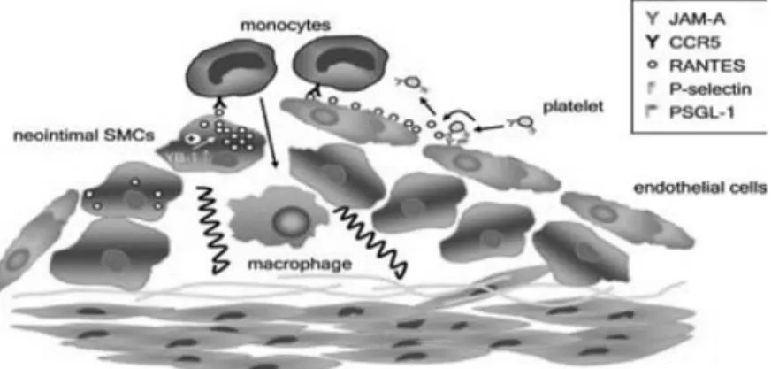
여기에서는 다양한 혈관질환에서 염증세포 응 집
(imflammatory cell recruitment),
평활근의 증식
(proliferation of SMC),
혈관전구세포의 응집(recruitment of vascular progenitor cell),
그리고 혈관내피세포 회복
(endothelial cell recovery)
에 의한 혈관 재형성을
chemokine
을 매개로 하여 그림
2
에 요약해서 나타내었으며 이것을 설명하고자 한다
.
본 론
1. 혈관재형성에 MCP-1(CCL2)/CCR2의 역할
Neointima formation
의 실험모델에서 monocyte chemokine protein-1(MCP-1)/CC motif ligand 2(CCL2)
은 가장 넓게연구되고 있는chemokine
으로써급성 손상
(acute mechanical injury)
시동맥벽
(arterial wall)
이나 혈액 순환 안에서mRNA MCP-1
가 발현되고 뒤따라 거의4
시간 안에 단백질의발현까지 급격한증가가 나타난다
.
흥미그림 1. 혈관벽의구조와혈관벽의구조적인변화
Bulletin of Food Technology 혈관재형성(Vascular Remodeling)에 미치는 Chemokines의 역할
롭게도
smooth muscle cell(SMC)
에서 발현되는MCP-1
은neointima formation
을 촉진시키는PDGF(platelet-derived growth factor)
와 동맥손상초기에 관여하는
thrombin
의 발현을 증가시킨다
.
그러나 혈관 벽에서 손상으로 인한MCP-
1
발현의 증가는 일시적인 현상으로 손상 후3~4
일 후에 원래의 수치로 되돌아가 유지된다.
다양한 동물모델의
arterial injury
에서MCP-1
의 억제는
neointimal hyperplasia
를 일정하게감소시킨다는 그림
3
을통해서 확인할수 있다. Periarterial cuff plasement
또는hyperlipidemic
동물모델에서 혈관내피세포의 박피와 같은 손 상 후 현저하게 염증반응이 증가하는 것을 보 아
MCP-1
을 매개로 한neointimal formation
은marcrophage infiltration
을 통해서 이루어진다는것을 알 수 있다
.
그러나non-hyperlipidemic
동물에서 혈관손상시에는 단지 약간의
macrophage infiltration
이 일어날 뿐이다.
ApoE
유전자 결핍mice
에 전기적인 자극을주어 유도한 경동맥
(carotid artery)
손상모델에서초기의
monocyte adhesion
에MCP-1
의 역할과 작용기작 연구를 시작하였다
.
이 연구를 통해 손상된 혈관에서 증가된
MCP-1
발은 순환하는
MCP-1
의 양에서 더 증가된 양과 관련성을 가지는 것으로 손상부위의 혈소판에
MCP- 1
의 부착을 유도한다.
또한ex-vivo
상태인perfusion
연구에서monocyte
는 내피세포의 손상 후 손상부위에 빠르고 확실하게 부착된다
.
그러나 증가한
monocyte adhesion
은 중화된MCP-1
의 투여시 유의하게감소된다.
이러한 결과는
MCP-1
이 내피세포 손상 후 초기monocyte recruitment
에aarest
에 의해 유도된chemokine
으로써 중요한 기능을 가진다는 것을설명해 준다
.
그러나 이것은 염증부위와 이차임파선으로
leukocyte recruit
하는MCP-1
의 고도의 특이적인 역할과는 확연하게 다르다는 것 그림 2. Arterial remodeling을위한 3가지구성요소
(출처: Schober A et al., Thromb Haemost, 2007)
을 알 수 있다
.
즉,
혈관 손상시MCP-1
의 발현이 일시적으로 증가하는 것은 초기
monocyte recuitment
것과neointimal hyperplasia
에 결정적인 역할을 하는 것으로써 혈관손상 후 혈관재 형성에 중요한 기작으로 작용된다
.
혈관 손상 후
re-endothelialization
은 촉진되는혈관손상 후 내피세포의 회복
(endothelial cell recovery)
시에 나타나는neointimal growth
을 유의하게 감소시키는 것을 일컫는 것으로 동맥재 형성에보호 인자로써 나타나는 특징이다
.
혈관손상 후 혈액순환으로
endothelial progenitor cell(EPC)
의mobilization
과 재생하는 내피세포라인으로
recruitment
은 내피세포회복에 크게 영향을 미친다
.
그러나 재생하는 내피세포에EPC
가 미치는 영향은
EPC
의 치료적인 투여, G- CSF
나statin
과 같은 전처리를 통한 투여의 동그림 3. 다양한동물모델에서혈관재형성시 MCP-1/CCR2의억제효과
Bulletin of Food Technology 혈관재형성(Vascular Remodeling)에 미치는 Chemokines의 역할
반 없이는
10%
로 매우 낮게 나타난다.
2. 혈관재형성에 SPC에서 분비되는 SDF-1a (CXCL12)/ CXCR4 역할
BM
에서기원된순환하는SPC(SMC progenitor cell)
는 손상된 혈관벽을recuitment
과neointimal SMC accumulation
에 영향을 주는 것으로 줄기세포의
migration
과mobilization
은CXC che- mokine stromal cell -derived factor(SDF)-1
a에 의해서 조절이 된다. SPC
에서 유도된SDF-1
a은
neointimal formation
을 이끌게 된다.
물리적인 혈관 벽 손상 후 순환하는
SPC
의 활동을통해서 일시적으로
SDF-1
a은 혈중과SMC
에서발현이 증가한다
.
전기적인 손상을 준apoE
유전자 결핍
mice
에서SDF-1
a의 차단은BM
에서기원된
SPC
의accumulation
을 억제함으로써neointimal area
의 유의한감소를 일으키게 되고이것은
neointimal SMC content
까지 감소시키는 것을 확인하였다
.
이때SPC recuitment
시 발현된
SDF-1
a은BM
에서 발현되는SDF-1
a 수용체인
CXCR4
와 결합하여 반응을 나타낸다.
SDF-1
a는SPC
에서 뿐만 아니라 혈소판의 표면에서도 결합을한다
.
손상된 혈관에서 증가하는
p-selectin
의 농도에 따라progenitor cell
들도유의하게 증가한다
.
그러므로 혈소판은SDF-1
a를 제공하는
source
로써 작용을 하게 된다.
따라서
SDF-1
a의 분비는arterial thrombosis
가 형성될 때
progenitor cell recuitment
을 유발하게되는 것이다
.
대부분의lin-/ sca-1+SPC
에서PDGFR
b가 발현된다.
이렇게 발현된PDGFR
b는
BM progenitor cell
에서SMC
의 분화를 유도한다
.
따라서lin-/ sca-1+ peripheral cell
에서PDGFR
b 발현은SDF-1
a에 의존적인neointimal formation
을 포함하는SPC subpopulation
으로특징지을 수 있다
.
Arterial
조직에서 손상에 의해 유도된SDF- 1
a의 발현과neointimal SPC recuitment
정도는손상의 유형에 따라서 달라질 수 있다
.
in vivo에서
carotid ligation, periarterial cuff placement
과 비교 했을 때 전기적인 손상이 가장 많이
SDF-1
a 발현을 생산한다.
또한 in vitro 실험시 에는SMC
에서scratch
에 의해 유발된apoptosis
는 손상받지 않은
SMC
에서SDF-1
a의 발현을 촉진한결과로 나타난다. neointima formation
을위한
carotid ligation
동물모델에서SDF-1
a 발 현은entothelial nitric oxide synthase
유전자가결여된
mice
모델에서 더욱 증가하였다.
이것은
progenitor cell mobilization
과adventitial recruitment
와 상관관계가있다는 것을 가리킨다.
따라서 혈관재형성은 혈관내피세포의 손상 후 발현되는
SDF-1
a을조절함으로써나타낼수있다. Hypoxia
에서SDF-1
a 전사인자 조절을 할 수있다
. hypoxia-induced factor(HIP)-1
a의 활성화 는 혈관내피세포에서SDF-1
apromoter
에 있는HIP-1
a 특이 반응 자리와 결합을 함으로써SDF-1
a 발현을 유도한다.
혈관 손상시SMC
에서도
HIP-1
a가 유도 되었다.
따라서HIP-1
atranscription
을 억제하면neointimal SDF-1a
발현과
neointima
성장을 줄일 수 있다.
결국 이러한 결과는 혈관 손상 후
HIP-1
a가SDF-1
a발현을 위한
upstream regulator
로써 중요한 역할을 한다는 것을 제시해 준다
.
그러므로 손상후 발현되는
SDF-1
a은SPC recruitment
를 통해서 혈관
repair
와remodeling
을 하는데 관여한다(
그림4).
3. 혈관재형성에 RANTES(CCL5)/ CCR1 CCR5의 역할
CC-chemokine
RANTES(regulated upon activa- tion, normally T-expressed, and presumably secreted)
는 혈소판에 저장이 되고 혈소판이 활성화되면 분비된다
. Flow
상태에서,
혈소판에서유래된
RANTES
는 혈소판에서 나온p-selectin
에 의해 혈관내피세포를 활성화시킨다
.
활성화된 혈관내피세포는 다시
monocyte
를adhesion
시킴으로써
artherogenesis
를 유발한다. RANTES
는 또한 혈관내피세포에서 분비되는 것으로 CC motif recepto
r(CCR)-1
과CCR5
와 결합을 하여neointima formation
과macrophage infiltration
에 관여한다
.
혈소판에 있는
P-selectin
과 함께junctional adhesion molecule(JAM)-A
도내피세포의RANTES deposition
에 관여를 한다. JAM-A
는IgG super-
family
로써 내피세포와 외피세포가 결합하는데관여하는 물질로써
leukocyte, monocyte, neutro- phil
의 내피세포로의 이동을 조절할 때 사용된다
. Hyperlipidemic
한 상태인ApoE
유전자 결여된
mouse
의 혈관내피세포에서는JAM-A
의발현이증가하며 이것은
arthelogenic leukocyte
침전을 야기시킨다
.
또한JAM-A
는cytokine
에 의그림 4. 혈관재형성에 SPC에서발현되는 SDF-1a/CXCR4의역할 (출처: Schober A, Arterioscler Thromb Vasc Biol, 2008)
Bulletin of Food Technology 혈관재형성(Vascular Remodeling)에 미치는 Chemokines의 역할
해 자극된혈관내피세포에 혈소판이
adhesion
하는데 중요한 역할을 하기도 한다
.
흥미롭게도JAM-A
유전자 결핍, apoE
유전자 결핍mouse
에 전기에 의해 유도된
carotinoid
손상 모델에서
luminal RANTES
의 발현은 감소되었고monocyte
의neointimal
침전과neointimal
성장도 감소하였다
.
즉, luminal RANTES
발현 감소는
JAM-A
부재시 혈소판에서 유래된RANTES
의 혈관내피세포로의deposition
이 부족하기 때문이다
.
Cardiac allograft vasculopathy(CAV)
에서coro- nary artery
의infiltration
되는mononuclear cell, microvessel
의 내피세포,
그리고intimal SMC
에서
RANTES
발현이 증가하였다. CAV
에서RANTES receptor
인CCR1
과CCR4
의 발현은neointimal
성장과mononuclearcell
의 혈관벽으로의 침투를감소시키는 역할을하였다
.
따라서, CAV
발병시RANTES
의receptor
인CCR1
과CCR5
를 차단함으로써CAV
를 감소시킬 수 있으며 이는 또한 부분적으로 이식생존율을 증가 시킬 수 있을 것이다
(
그림5).
4. Fractalkine/CX3R1의 역할
Fractalkine
은 구조적으로chemokine
과 비슷한것으로
membrane bound
형태와soluble
형태로나뉜다
. Transmembrane protein
은integrin
의존적으로
leukocyte adhesion
의 역할을 하고soluble protein
은chemo-attractant
특성을 가진다
.
이러한 특징은CXC3R
과 결합하여 반응을나타낸다
.
in vitro 상에서fractlkine
은NF-kB
기전을 통해서 활성화던 혈관내피세포와
SMC
에서 증가하고 이렇게 증가한
fractalkine
의 발현은
monocyte adhesion
을 유발한다.
물리적인손상 후 불완전한
re-endothelialization
때문에fractalkine
을 발현되는neointimal SMC
에서는만성적인
monocyte recruitment
가 촉진될 것이다
. Femoral artery denudation
모델에서frac-
그림 5. RANTES/CCR1/CCR5의역할 (출처: Schober A, Arterioscler Thromb Vasc Biol, 2008)
talkine
은intimal SMC
와endothelial cell
에서발현이 증가되었다
. CX3CR1
유전자 결핍mouse
에서neointimal hyperplasia
는 대조군에비해 유의하게 감소되었다
. CXC3R1
은 손상된혈관으로의
monocyte infiltration
과SMC proliferation
의 감소에 밀접한 관련이있다는 것을 나타내는 것이다
.
그러나 물리적인 혈관손상에 반해
pulmonary hypertension
에서fractalkine
은
perivsscular inflammatory cell
에서 나타났고CX3CR
은medial SMC
에서 발현되었다.
5. CXCL1(KC/GRO-a)와 CXCR2에 의한 endothelial recovery
Atherosclerosis
에서CXC
motif receptor 2(CXCR2)
발현은monocyte
의intimal accumu-
lation
을 위해 형성되는 것으로 대부분이 활성화된 혈관내피세포에서
keratinocyte-derived chemokine(KC)/growth-related oncogene(GRO)-
a의 기능이 억제되기 때문에 나타난다
. ApoE
유전자 결핍
mouse
에서carotid
전기에 의한 손상모델에
neutraling KC
항체 투여는KC
발현을억제시켜서
neointimal area
의 증가와 혈관내피세포
recovery
장애를 유발시켰다.
그러나 그에반해
neointimal macrophage
와SMC con- tent
는KC
항체에 영향을 받지 않았다. CXCR2
는 in vivo에서는 재생되는 혈관내피세포에서
발현되고 in vitro에서는 내피세포의
wound
healing
에KC
에 의해 유도되었다.
그러므로 분비된
KC
에 의해 형성된neointimal macro- phage
는 혈관내피세포recovery
을 유도해서 혈관을 보호하게 된다
.
그러므로chemokine
은vascular remodeling
시작에 기여할뿐만 아니라,
혈관내피세포
recovery
에 의한vascular healing
에도 관여한다
.
결 론
Chemokine
은 기능적으로 다양한chemokine
과의협력을 통해서
arterial remodeling
과정을조절한다
(
그림5).
이러한 과정을 통해chemokine
은
artherogenesis
와non-artherogenic arterial remodeling
사이에 다양한 작용을 할 수 있다.
그림 6. 혈관재형성시 chemkine과 chemokine receptor의특이반응
Bulletin of Food Technology 혈관재형성(Vascular Remodeling)에 미치는 Chemokines의 역할
임상에서
chemokine receptor
는 치료제로 이용될 가능성이 제시되고 있다
.
첫 번째로, stent
이식후 협착
,
심장이식 등의 혈관질환 치료제로서 이용될 가능성이 가장 크다
.
이것은 질병부위에 직접적으로 타겟한 것으로 비교적 정확한 발병원인과 질병을 유발하는 부위에 직접 작용 할 수 있다는 장점을 가지고 있기 때문이다
.
다른 가능성으로는
signal transducton
의 억제제인transcription factor decoy
나siRNA
을 통해서chemokine
의 활성 또는 발현을 간접적으로 억제하여 치료할 수 있을 것이다
.
참고문헌
1. Braunersreuther V, Zernecke A, Steffens S, et al., Ccr5 but notCcr1 deficiency reduces development of diet-induced atherosclerosis in mice, Arterioscler Thromb Vasc Biol, 188, 51-58, 2006 2. Charo IF, Ransohoff RM, The many roles of
chemokines and chemokine receptors in inflammation, N Engl J Med, 354, 610-621, 2006 3. Combadiere C, Potteaux S, Gao JL, et al.,
Decreased atherosclerotic lesion formation in CX3CR1/apolipoprotein E double knockout mice, Circulation, 107, 1009-1016, 2003
4. Damas JK, Smith C, Oie E, et al., Enhanced expression of the homeostatic chemokines CCL19 and CCL21 in clinical and experimental atherosclerosis. Possible pathogenic role in plaque
destabilization, Arterioscler Thromb Vasc Biol,
27, 614-620, 2007
5. Demicheva E, Hecker M, Korff T, Stretch-induced activation of the transcription factor activator protein-1 controls monocyte chemoattractant protein-1 expression during arteriogenesis, Circ Res., 103(5), 477-484, 2008
6. Flanagan K, Moroziewicz D, Kwak H, et al., The lymphoid chemokine CCL21 costimulates naive T cell expansion and Th1 polarization of non- regulatory CD4+ T cells, Cell Immunol, 231, 75- 84, 2004
7. Glass CK, Witztum JL, Atherosclerosis, The road ahead. Cell, 104, 503-516, 2001
8. Greaves DR, Hakkinen T, Lucas AD, et al., Linked chromosome 16q13 chemokines, macrophage-derived chemokine, fractalkine, and thymus- and activationregulated chemokine, are expressed in human atherosclerotic lesions, Arterioscler Thromb Vasc Biol, 21, 923-929, 2001 9. www.hper.txstate.edu
10. www.daviddarling.info
11. Hansson GK, Inflammation, atherosclerosis, and coronary artery disease, N Engl J Med, 352, 1685-1695, 2005
12. Hansson GK, Libby P, The immune response in atherosclerosis: a double-edged sword, Nat Rev Immunol, 6, 508-519, 2006
13. Lesnik P, Haskell CA, Charo IF, Decreased atherosclerosis in CX3CR1-/- mice reveals a role
for fractalkine in atherogenesis, J Clin Invest, 111, 333-340, 2003
14. Libby P, Inflammation in atherosclerosis, Nature,
420, 868-874, 2002
15. Marsland BJ, Battig P, Bauer M, et al., CCL19 and CCL21 induce a potent proinflammatory differentiation program in licensed dendritic cells, Immunity, 22, 493-505, 2005
16. Newby AC, Dual role of matrix metallo- proteinases (matrixins) in intimal thickening and atherosclerotic plaque rupture, Physiol Rev, 85, 1- 31, 2005
17. Schober A, Chemokines in vasculasr dysfunction and remodeling Arterioscler Thromb Vasc Biol,
28(11), 1950-1959, 2008
18. Teupser D, Pavlides S, Tan M, et al., Major
reduction of atherosclerosis in fractalkine (CX3CL1)-deficient mice is at the brachiocephalic artery, not the aortic root, Proc Natl Acad Sci USA, 101, 17795-17800, 2004
19. Trogan E, Fayad ZA, Itskovich VV, et al., Serial studies of mouse atherosclerosis by in vivo magnetic resonance imaging detect lesion regression after correction of dyslipidemia, Arterioscler Thromb Vasc Biol, 24, 1714-1719, 2004
20. Wong BW, Wong D, McManus BM, Charac- terization of fractalkine (CX3CL1) and CX3CR1 in human coronary arteries with native atherosclerosis, diabetes mellitus, and transplant vascular disease, Cardiovasc Pathol, 11, 332-338, 2002
성미정 의학박사
⋅ 소속 한국식품연구원 바이오제론연구단
⋅ 전문분야 기능성식품 항염증 기전 연구
⋅ E-mail [email protected]
⋅ TEL 031-780-9316