안지오텐신
7
0
0
전체 글
(2) 압과 심박출량을 증가시키는 등 다양한 생리적 반응들 2-4). 에서 밤새 보관한다. 익일 DMEM에서 세척하고 0.5 mg/ml collagenase(CLS Ⅱ, Worthington사제)와 2. 을 조절한다.. 한편 혈관수축작용 외에 안지오텐신 Ⅱ는 혈관평. mg/ml의 elastase(Worthington사제)가 함유된 DMEM. 활근세포(vascular smooth muscle cells)의 성장. 에서 약 60분간 반응시키고 20% FCS-DMEM으로. (growth)에도 관여하는 것으로 알려져 있다. 고혈압의. 반응을 종결시킨다.16) 복합소화법으로 분리되어 나온. 5). 진행과정에서 혈관평활근세포의 성장이 발견되고. 안. 중막의 혈관평활근세포를 1,000 rpm으로 원심분리하. 지오텐신 Ⅱ가 그러한 과정에서 중요한 역할을 한다는. 여 침전시키고 얻어진 세포는, 가열하여 불활성화시킨. 6)7). 더우기 안지오. 10% fetal calf serum(FCS)과 1 mM의 비필수아미노. 텐신 Ⅱ가 풍선도자에 의한 혈관손상 후 신생내막의 증. 산, 50 μg/ml의 페니실린, 그리고 50 μg/ml의 스트. 것이 여러 연구에서 제시된 바 있다. 8). 안지오텐신 전환효소억제제(ACE. 렙토마이신을 함유한 DMEM에서 5% CO2, 37℃의 가. inhibitor)와 안지오텐신 Ⅱ 수용체 길항제(angiotensin. 습한 대기조건하에 유지, 배양하였고 배지는 3일 간격. Ⅱ receptor antagonist)가 그러한 내막의 증가를 억. 으로 교체하여 주었다.. 가를 촉진시키며,. 제하며, 혈관평활근세포의 비후도 감소시킨다고 보고되 9)10). 었다.. 1차배양에 성공한 혈관평활근세포의 계대배양을 반. 이러한 연구들은 안지오텐신 Ⅱ가 혈관평활. 복하여, 4대에서 8대까지 계대배양한 세포를 얻어 본. 근세포의 성장에 관여하여 비정상적인 혈관의 비후를. 연구에 사용하였다. 배양된 세포가 혈관평활근 세포임. 초래하며 풍선혈관성형술후의 재협착등에 영향을 미친. 을 확인하기 위하여 면역세포화학적 방법을 이용하였. 다는 것을 시사해준다.. 으며 평활근 α-actin에 대한 특이 단일클론성 항체. 그러나 안지오텐신 Ⅱ가 혈관평활근세포의 성장에 영 향을 미치는 정확한 기전에 대해서는 아직 연구 중에. (Sigma사제)를 결합시킨후 ExtrAvidin-Peroxidase 시약(Sigma사제)으로 염색하였다.. 있다. 현재까지도 비후(hypertrophy)만을 일으키는지 또는 증식(hyperplasia)도 같이 일으키는지에 대해 서 11-15). 로 상반된 결과들이 보고되고 있고,. 안지오텐신 Ⅱ 자극. 그러한 영향. 일차배양한 혈관평활근세포들을 six-well Costar dish. 들이 안지오텐신 Ⅱ의 독립적인 효과에 의한 것인지에. 에서 10% FCS를 함유한 DMEM을 배지로 하여 계속. 대해서도 논란의 여지가 있으며, 국내의 연구보고는 드. 배양하였고, 70%에서 80%의 confluence 상태에 이른. 문 상태이다.. 후 0.4% FCS를 함유한 DMEM에서 48시간동안 배양. 따라서 본 연구에서는 안지오텐신 Ⅱ가 혈관평활근. 하여 성장을 정지시킴으로써 quiescent 상태로 만들었다.. 세포의 성장에 직접적으로 어떠한 영향을 미치는지 조. 안지오텐신 Ⅱ의 자극은 일반적으로 안지오텐신 Ⅱ. 사하고자 흰쥐의 대동맥 혈관평활근세포를 순수배양한. 효과가 가장 뚜렷하다고 제시되는 농도인 1.0 μM 농. 후 [3H]thymidine과 [14C]phenylalanine의 incorpo-. 도에서 시행하였고,2)15) 안지오텐신 Ⅱ 자극효과가 특. ration 연구를 시도하였다.. 이 수용체에 결합함으로써 발현되는 것인지를 확인하 기 위하여, 안지오텐신 Ⅱ의 결합에 대한 경쟁적 억제. 대상 및 방법. 제(competitive inhibitor)로서 Sar1, Val5, Ala8angiotensin Ⅱ(saralasin)을 사용하였다.. 혈관평활근세포의 배양. 세 가지 실험군으로 분류하여 한 군은 대조군으로 하. 생후 3개월된 Wistar 흰쥐의 흉부대동맥을 적출하여. 였으며, 다른 한 군은 3일동안 매일 안지오텐신 Ⅱ만으. Hank’s balanced salt solution에서 지방조직을 제거. 로 처리하여 배양하였고, 나머지 한 군은 안지오텐신. 하고 1 mg/ml collagenase(CLS I, Worthington사제). Ⅱ 및 동량의 saralasin을 함께 처리하여 같은 방법으. 용액에 넣고 37℃에서 30분간 반응시킨후 내막 및 외. 로 배양하였다. 실험은 모두 여섯 차례 반복연구하였다.. 막을 분리해내고 10% fetal calf serum(FCS)을 포함 하는 Dulbecco’s modified Eagle’s medium(DM EM, Gibco사제)에 담근 후 세포배양기(5% CO2 incubator) 210. [3H]thymidine과 [14C]phenylalanine의 incorporation 배양액을 흡인해내고 1 μCi/ml의 [3H]thymidine Korean Circulation J 1999;29(2):209-215.
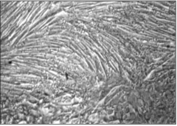
(3) (464 mCi/mmol, New England Nuclear) 또는 0.1 14. 포의 분화가 진행되어 평활근세포의 특징인 hills and. μCi/ml의 [ C]phenylalanine(86 mCi/mmol, New. valleys형태를 나타내었다(Fig. 2). 그리고 면역세포화. England Nuclear)을 함유하는 신선한 DMEM으로 교. 학적 방법으로 평활근 α-actin에 대한 특이 단일클론. 체하여 24시간 동안 세포배양기에서 혈관평활근세포를. 성 항체를 결합시킨후 염색한 결과 본 세포의 세포질에. 성장시킨 후 배양액을 흡인하였다. 혈관평활근세포를. 서 풍부한 α-actin을 확인할 수 있었다(Fig. 3).. 4℃ 세척용액(10 mM Tris, 150 mM NaCl, pH 7.4). 안지오텐신 Ⅱ로 3일간 자극한 후에 혈관평활근세포. 으로 세척한후 10% trichloroacetic acid로 고정하였다.. 들의 상태를 inverted microscope로 관찰한 결과 형. 95% ethanol로 다시 세척하고 건조시킨 후 0.2 N. 태학적으로는 대조군과 비교하여 유의한 차이가 발견. NaOH를 사용하여 단백질과 DNA를 추출하였으며,. 되지 않았다.. Liquid scintillation spectrometry로 incorporated. [3H]thymidine incorporation study. radioactivity를 측정하였다.. 혈관평활근세포를 quiescent 상태에서 1.0 μM 안지. 통. 오텐신 Ⅱ로 자극한지 1일째와 3일째에 [3H]thymidine. 계 측정된 성적은 평균과 표준편차로 표시하였으며, 검. incorporation의 radioactivity를 측정한 결과, 대조군. 정은 Student’s t-test에 의하여 P value <0.05인 경. 은 1352522±46155 cpm, 1340756±49868 cpm. 우 통계적으로 유의하다고 판정하였다.. 이었으며, 안지오텐신 Ⅱ 투여군은 1일째에 1334370 ±121104 cpm, 3일째에 1321885±30763 cpm로서. 결. 과. 안지오텐신 Ⅱ 단독으로 자극한 혈관평활근세포는 대 조군과 비교하여 [3H]thymidine incorporation 면에서. 혈관평활근세포의 배양. 유의한 차이가 없었다(Fig. 4).. 배양된 혈관평활근세포를 0.25% trypsin-EDTA 용 액으로 처리하고나서 1:4 비율로 계대배양을 시행하. [14C]phenylalanine incorporation study. 였을때 하루가 지나면 혈관평활근세포들이 culture dish. 혈관평활근세포에 섭취된 [14C]phenylalanine의 ra-. 에 붙어있는 상태가 확인되었다. 별모양으로 붙고 나서. dioactivity 측정 결과에서는, 대조군이 1일째 10703. 혈관평활근세포의 증식이 진행되어 70~80% confluence. ±518 cpm, 3일째 9752±1268 cpm인데 반하여 1.0. 상태에 이르게 되었고(Fig. 1) 이 상태의 혈관평활근세. μM 안지오텐신 Ⅱ로 자극한 군은 1일째 12910±. 포들을 본 연구에 사용하였다. 한편 이 세포가 순수한. 1094 cpm, 3일째 12671±708 cpm로서 대조군보다. 혈관평활근세포임을 확인하기 위하여 일부 세포를 계. 통계적으로 유의하게 높은 섭취율을 나타냈다(p<0.05).. 속 증식시켜 완전한 confluence상태에 이르게하면 세. Fig. 1. 70-80% confluent state of vascular smooth muscle cells. Cells assumed a flattened, well spread morphology.. Fig. 2. Characteristic“hill and valley morphology” . Vascular smooth muscle cells (VSMC) formed dense multilayers having the typical appearance of cultured vascular smooth muscle cells.. 211.
(4) A. B. Fig. 4. [3H]-thymidine incorporation after administration of angiotensin Ⅱ.. Fig. 3. A:The fixed VSMC were exposed to a monoclonal antibody reactive against smooth muscle-specific alpha actin. VS MC were clearly distinguished by brownish staining of actin fibers running the entire length of each cell. B:Negative control showed no cytoplasmic staining.. Fig. 6. Effects of angiotensin Ⅱ on VSMC growth.. 안지오텐신 Ⅱ와 혈관평활근세포의 성장 안지오텐신 Ⅱ 단독으로 혈관평활근세포의 성장에 미치는 직접적인 영향을 종합하여 [3H]thymidine과 [14C]phenylalanine의 섭취율을 비교한 결과, 대조군 을 100%로하여 각각의 백분율로 나타내보면, 안지오 텐신 Ⅱ 단독으로 자극한 군에서 [3H]thymidine의 섭 취율은 대조군에 비해 1일째에 98.7±7.9%, 3일째에 98.6±4.2%로 대조군과 비슷한 수준을 보였고, [14C] Fig. 5. [14C]-phenylalanine incorporation after administration of angiotensin Ⅱ.. phenylalanine의 섭취율은 대조군에 비하여 1일째에. 반면 saralasin으로 처치한후 안지오텐신 Ⅱ로 자극한. 20%, 3일에서는 약 30%의 증가를 보였다. Saralasin. 군에서는 섭취율이 증가하지 않고 대조군과 비슷한 수. 으로 전처치후 안지오텐신 Ⅱ로 자극한 군에서는 3일. 준을 나타내었다(Fig. 5).. 째에서 [3H]thymidine과 [14C]phenylalanine의 섭취. 212. 120.6±8.8%, 3일째에 129.9±22.2%로 1일에서 약. Korean Circulation J 1999;29(2):209-215.
(5) 율이 대조군에 비해 각각 102.2±3.1%, 102.2±7.4%. 조건에서 시행되었는데, 앞의 연구결과들을 보면, 안지. 로서, 모두 대조군과 비교하여 유의한 차이가 없었다. 오텐신 Ⅱ가 혈관평활근세포의 증식을 일으킨 연구들. (Fig. 6).. 은 비후만을 일으킨 연구들에 비해 다른 요소가 첨가되 었거나 비교적 높은 농도의 혈청조건하에서 이루어진. 고. 찰. 경향이 있다. 그러므로 안지오텐신 Ⅱ가 혈관평활근세 포의 증식을 일으킨 결과들은 그의 독립적인 효과이기. 본 연구에서, 안지오텐신 Ⅱ로 자극한 혈관평활근세 14. 보다는 혈청내의 다른 요소들과의 복합적인 작용에 의. 포에서 [ C]phenylalanine의 섭취율은 대조군과 비교. 해 나타났을 가능성이 있다. 또한 Black 등은 동물의. 하여 통계적으로 유의한 증가를 보였으나, [3H]thymidine. 생체내 실험에서 안지오텐신 Ⅱ가 대동맥의 혈관평활. 의 섭취율은 대조군과 비슷하게 나타났다. 즉 본 연구. 근세포에서는 비후만을 일으키지만 장간세동맥에서는. 의 조건에서 1.0 μM농도의 안지오텐신 Ⅱ 자극은 흰. 증식을 일으켰다고 보고하였고,13) 일부 보고에서는 안. 쥐 혈관평활근세포의 단백질합성을 20~30% 증가시. 지오텐신 Ⅱ가 혈관평활근에 미치는 영향이 동물과 사. 켰으나 DNA의 합성에는 유의한 영향을 미치지 않았다.. 람에서 서로 다르다고 나타나 있어서,11)14) 결국 안지. 또한 안지오텐신 Ⅱ 수용체 길항제인 saralasin으로 전. 오텐신 Ⅱ가 혈관평활근세포의 성장에 미치는 영향이. 처치후 안지오텐신 Ⅱ로 자극한 혈관평활근세포에서는. 종이나 해부학적 위치의 차이에 따라 다르게 나타날 수. 14. [ C]phenylalanine의 섭취율이 증가하지 않았던 것은,. 있는 가능성이 있다. 최근 Freeman 등은 혈청이 없는. 안지오텐신 Ⅱ가 혈관평활근세포의 단백질 합성의 증. 배지조건하에서 안지오텐신 Ⅱ가 흰쥐 대동맥 혈관평. 가를 일으킨 것이 안지오텐신 Ⅱ의 특이 수용체에 작용. 활근세포의 [3H]thymidine 섭취율을 증가시킨다는 것. 하여 발생한 독립적인 효과임을 나타낸다.. 을 보고하였으며, 7개의 아미노산으로 구성된 안지오텐. 안지오텐신 Ⅱ가 혈관평활근세포의 성장에 미치는. 신-(1-7)은 오히려 [3H]thymidine 섭취율을 감소시. 영향에 대해서는 현재까지도 여러 상반된 결과들이 보. 킨다고 주장하였는데, 그로 보아 안지오텐신 Ⅱ는 혈관. 17). 고되고 있다. Berk,. 18). 그리고 Scott-. 평활근세포의 성장을 촉진하기도 하지만 특정 조건에. Burden 등의 연구19)에서는 혈청이 없는 배지조건하의. 서는 성장을 억제할 수도 있다고 제안하였다.15) 혈관평. 동물실험에서 안지오텐신 Ⅱ가 혈관평활근세포의 비후. 활근세포는 내분비자극에 대한 정상적인 반응으로서 수. 만을 일으켰다. 그러나 Campbell-Boswell 등은 높은. 축과 이완을 하게된다. 그러나 특정한 병적 상황에서는. 농도의 혈청을 포함하는 조건에서 안지오텐신 Ⅱ에 의. 성장(growth), 비후(hypertrophy) 또는 내막으로의. Geisterfer,. 20). Makitta 등. 이주(migration)등이 보다 더 주요한 반응으로 나타. 은 2%의 혈청을 함유하는 배지조건에서 사람의 혈관. 한 혈관평활근세포의 증식을 보고했으며,. 난다. 혈관에 활성적으로 작용하는 몇가지 물질들로부. 평활근세포에 대해 안지오텐신 Ⅱ에 의한 증식효과를. 터 발생되는 생화학적 신호들이 이와 유사한 반응을. 14). Gibbons 등은 혈청이 없는 조건에서 안. 일으킬수 있으며, 최종적인 생리적 반응은 세포의 환. 지오텐신 Ⅱ만으로 자극하였을때는 혈관평활근세포의. 경과 유전자형에 의해 결정되어 실질적인 생화학적 경. 확인하였다.. 1. 증식이 없었으나, 안지오텐신Ⅱ와 TGF-β 항체를 같. 로가 활성화된다. 즉 정상세포에서는 성장인자(growth. 이 투여하였을때는 증식이 나타나는 결과를 보였다.21). factor)들이 혈관수축인자(vasoconstrictor)로서 작용. Bunkenberg 등은 배양된 동물의 혈관평활근세포를 안. 하는 반면 변형된 평활근세포에서는 혈관수축인자들이. 지오텐신 Ⅱ로 자극한 결과, 혈청이 없는 조건하에서는. 증식(hyperplasia)과 비후(hypertrophy)를 일으킬수. [3H]thymidine의 섭취율은 증가하였으나 세포의 수에는. 있다.23)24). 변화가 없었고, 혈청이 있는 조건하에서는 [3H]thymidine. 혈관평활근세포의 성장은 증식과 비후의 두가지 형. 의 섭취율과 함께 세포의 수도 증가되었으며 1%의 혈. 식으로 이루어진다. 통상적으로 비후는 혈관수축인자에. 청에서보다 5%와 10%의 혈청조건에서 더욱 현저한. 의한 장기적인 자극에 의해 발생되는 것에 비해, 증식. 22). 본 연구. 은 전형적인 성장인자들에 대한 반응으로서 나타난다.. 는 0.4%의 비교적 낮은 농도의 혈청을 함유하는 배지. 고혈압하의 큰 혈관에서 볼수 있듯이 비후는 단백질합. 세포수의 증가가 관찰되었다고 보고하였다.. 213.
(6) 성의 증가로 인한 혈관평활근세포체의 증가이며 안지. 요. 오텐신 Ⅱ와 thrombin에 대한 반응으로 일어난다.17). 약. 증식은 세포의 복제로 특징지어지며 혈관손상후에 나 타나는 platelet-derived growth factor(PDGF)나. 연구목적:. fibroblast growth factor(FGF)같은 성장인자들에 의. 최근 고혈압을 비롯하여 심근경색, 그리고 심부전등. 25-27). 해 자극된다.. 한편 위와같은 성장(growth)의 두. 각종 심혈관계 질환에서 레닌-안지오텐신계의 역할이. 가지 형태를 일으키는 생화학적신호에 대해서는 아직. 재조명되고 있는 상태이다. 특히 안지오텐신 Ⅱ는 강력. 명확히 밝혀지지 않았다. 이전의 연구들에서 안지오텐. 한 혈관수축작용을 갖고 있으며, 혈관평활근세포의 성. 신 Ⅱ가 c-fos, c-myc, c-jun같은 proto-oncogene. 장과 분화에도 관여하는 것으로 알려져 있다. 본 연구. 28)29). 이러. 에서는 인지오텐신 Ⅱ가 단독으로 혈관평활근세포의 증. 한 증가는 새로운 단백질합성과는 무관하게 전사(trans-. 식과 단백질생성에 어떠한 영향을 미치는지 알아보고. cription)의 증가에 기인한다는 것이 밝혀졌다.30) 또한. 자 하였다.. 안지오텐신 Ⅱ는 여러 유전자의 표현을 증가시키며 가. 방 법:. 의 mRNA 농도를 증가시킨다고 보고되었고,. 장 특징적인 것은 platelet-derived growth factor. 혈관평활근세포는 휜쥐 대동맥의 중막에서 효소소화. (PDGF)의 표현증가이다. Naftilan등은 쥐의 대동맥평. 방법으로 분리하여 1차 배양에 성공한 후 계대배양을. 활근세포를 배양하여 안지오텐신 Ⅱ를 투여한후 4~6. 계속하여 4~8대의 세포들을 사용하였고, 배양된 혈관. 시간에 PDGF-A chain의 증가를 관찰하였다.31) 위의. 평활근세포를 안지오텐신 Ⅱ와 saralasin으로 3일간. 결과들은 안지오텐신 Ⅱ가 proto-oncogene을 초기에. 매일 처치한 후, [3H]thymidine과 [14C]phenylala-. 유도하여 PDGF-A chain의 유전자를 유도하고 결과. nine의 섭취율을 liquid scintillation spectrometry로. 적으로 PDGF의 증가를 가져온다는 가능성을 시사하고. 측정하였다.. 있다. 그 후의 연구결과들이 이러한 가능성을 뒷받침하. 결 과:. 고 있다. Rainer등은 antisense RNA로 특이유전자의. 안지오텐신 Ⅱ로 3일간 자극한 후에 혈관평활근세포. 유도를 차단한 결과 안지오텐신 Ⅱ에 의한 c-fos의 증. 들의 상태를 inverted microscope로 관찰한 결과 형. 가효과가 나타나지 않았다고 보고하였고,32) Itoh등은. 태학적으로는 대조군과 비교하여 유의한 차이가 발견. 안지오텐신 Ⅱ에 의한 PDGF의 증가를 차단한 결과 안. 되지 않았다. 세포를 quiescent상태에서 안지오텐신. 지오텐신 Ⅱ에 의한 단백질합성의 증가가 현저히 저하. Ⅱ로 자극하고 1일과 3일후에 24시간 [3H]thymidine. 되었다고 보고하였다.33). 섭취율을 측정한 결과 유의한 증가가 없었다. 반면에 상. 결론적으로 본 연구의 조건에서는, 안지오텐신 Ⅱ가 혈관평활근세포의 비후를 일으키고 그 효과는 안지오. 기 조건에서 [14C]phenylalanine의 섭취는 약 30%의 증가소견이 증명되었다.. 텐신 Ⅱ의 독립적인 작용에 의해 나타나는 것이며, 안. 결 론:. 지오텐신 Ⅱ 그 자체만으로는 혈관평활근세포의 증식. 이상의 결과로 본 연구의 조건에서 안지오텐신 Ⅱ 단. 에는 아무 영향도 미치지 않음을 알 수 있었다. 또한. 독으로는 주로 혈관평활근세포의 비후효과를 가지고 있. 이전의 다른 연구들을 종합하여 보면 안지오텐신 Ⅱ의. 으며, 적어도 혈관평활근세포의 증식에는 영향을 미치. 역할에는 복합성이 있다고 생각되어진다. 즉 다른 성장. 지 않음을 알 수 있었다.. 요소들과 복합적으로 작용하여 혈관평활근세포의 성장. 중심 단어:안지오텐신 Ⅱ・혈관평활근세포・비후.. 에 영향을 미치기도 하며, 유사분열물질(mitogen)에 의해 안지오텐신 Ⅱ의 역할이 강화되기도 하고, 안지오 텐신 Ⅱ가 독립적으로 성장인자들의 생성을 자극하기. 본 연구는 1997년도 중앙대학교 학술연구비의 지원으로 이 루어졌음.. 도 한다. 그 정확한 기전을 밝히기 위해서는 여러 다른. REFERENCES. 조건 하에서 계속적인 연구가 이루어져야 하겠다.. 214. 1) Zimmerman BG, Dunham EW. Tissue renin-angiotensin. Korean Circulation J 1999;29(2):209-215.
(7) 2). 3) 4) 5) 6) 7) 8). 9) 10). 11) 12) 13) 14). 15) 16) 17) 18). system: A site of drug action?. Annu Rev Pharmacol Toxicol 1997;37:53-69. Touyz R, Schiffrin EL. Angiotensin Ⅱ regulates vascular smooth muscle cell pH, contraction and growth via tyrosine kinase-dependent signaling pathways. Hypertension 1997; 30:222-9. Cody RJ. The integrated effects of angiotensin Ⅱ. Am J Cardiol 1997;79(5A):9-11. Matsusaka T, Ichikawa I. Biological functions of angiotensin and its receptors. Annu Rev Physiol 1997;59:395412. Folkow B. Cardiovascular structural adaptations: Its role in the initiation and maintenance of primary hypertension. Clin Sci Mol Med 1978;55:3S-22S. Owens GK. Influence of blood pressure on development of aortic medial smooth muscle hypertrophy in spontaneously hypertensive rats. Hypertension 1987;9:178-87. Goodfriend TL, Elliot ME, Catt KJ. Angiotensin receptors and their antagonists. N Engl J Med 1996;334:1649-54. Daeman MJ, Lombardi DM, Bosman FT, Schwarz SM. Angiotensin Ⅱ induces smooth muscle cell proliferation in the normal and injured rat arterial wall. Circ Res 1991;68:450-6. Powell JS, Closel JP, Muller RKM, Kuhn H, Hefti F, Hosang M, et al. Inhibition of ACE prevent myointimal proliferation after vascular injury. Science 1989;245:186-8. Clozel JP, Hess P, Michael C, Scheitinger K, Baumgartner RH. Inhibition of converting enzyme and neointima formation after vascular injury in rabbits and guinea pigs. Hypertension 1991;18(Suppl):Ⅱ55-9. Mii S, Ware JA, Mallette SA, Kent KC. Effect of angiotensin Ⅱ on human vascular smmoth musle cell growth. J Surgical Research 1994;57:174-8. Weber H, Taylor DS, Molloy CJ. Angiotensin Ⅱ induces delayed mitogenesis and cellular proliferation in rat aortic smooth muscle cells. J Clin Invest 1994;93:788-98. Black MJ, Bertram JF, Campbell JH, Campbell GR. Angiotensin Ⅱ induces cardiovascular hypertrophy in perindopriltreated rats. J Hypertension 1995;13:683-92. Makita S, Nakamura M, Yoshida H, Hiramori K. Effect of angiotensin Ⅱ receptor blocker on angiotensin Ⅱ stimulated DNA synthesis of cultured human aortic smooth muscl cells. Life Sci 1995;56:383-8. Freeman EJ, Chisolm GM, Ferrario CM, Tallant E. Angiotensin-(1-7) inhibits vascular smooth muscle cell growth. Hypertension 1996;28:104-8. Smith JB, Brock TA. Analysis of angiotensin-stimulated sodium transport in cultured smooth muscle cells from rat aorta. J Cell Physiol 1983;114:284-90. Berk BC, Vekshtein V, Gordon HM, Terukata T. Angiotensin Ⅱ-stimulated protein synthesis in cultured vascular smooth muscle cells. Hypertension 1989;13:305-14. Geisterfer AA, Peach MJ, Owens GK. Angiotensin Ⅱ induces hypertrophy, not hyperplasia, of cultured rat aortic smooth muscle cells. Circ Res 1988;62:749-56.. 19) Scott-Burden T, Resink TJ, Baur U, Bwegin M, Buhler. 20) 21). 22). 23) 24) 25). 26). 27). 28) 29) 30) 31). 32). 33). FR. Amiloride sensitive activation of S6 kinase by angiotensin Ⅱ in cultured vascular smooth muscle cells. Biochem Biophys Res Commun 1988;151:583-9. Campbell-Bowsell M, Robertson AL. Effects of angiotensin Ⅱ and vasopressin on human smooth muscle cells in vitro. Exp Mol Pathol 1981;35:265-76. Gibbons GH, Pratt RE, Dzau VJ. Vascular smooth muscle cell hypertrophy vs. hyperplasia. Autocrine transforming growth factor-beta1 expression determines growth response to angiotensin Ⅱ. J Clin Invest 1992;90:456-61. Bukenburg B, van Amelsvoort T, Rogg H, Wood JM. Receptor mediated effects of angiotensin Ⅱ on growth of vascular smooth muscle cells from spontaneously hypertensive rats. Hypertension 1992;20:746-54. Berk BC, Alexander RW, Brok TA, Gimbrone MA Jr, Webb RC. Vasoconstriction; A new activity for PDGF. Science 1986;232:87-90. Owens GK. Control of hypertrophic vs. hyperplastic growth of vascular smooth muscle cells. Am J Physiol 1989; 257:H1755-65. Mangiarua EI, Palmer VL, Lloyd LL, McCumbee WD. Platelet-derived growth factor mediates angiotensin Ⅱinduced DNA synthesis in vascular smooth muscle cells. Arch Physiol Biochem 1997;105:151-7. Golden MA, Au YPT, Kirkman Tr, Wilkox JN, Raines EW, Ross R, et al. PDGF activity and mRNA expression in healing vascular grafts in baboons. J Clin Invest 1991;87:406-14. Myers PR, Minor RL, Guerra R Jr, Bates JN, Harrison DG. The vasorelaxant properties of the endothelium derived relaxing factor more closely resemble S-nitrocysteine than nitric oxide. Nature 1990;345:161-3. Naftilan AJ, Pratt RE, Eldridge CS, Lin HL, Dzau VJ. Angiotensin Ⅱ induces c-fos expression in smooth muscle via transcriptional control. Hypertension 1989;13:706-11. Berk BC, Corson MA. Angiotensin Ⅱ signal transduction in vascular smooth muscle: role of tyrosine kinases. Circ Res 1997;80:607-16. Naftilan AJ, Gilliland GK, Eldridge CS, Kraft AS. Induction of the proto-oncogene c-jun by angiotensin Ⅱ. Mol Cell Biol 1990;10:536-40. Naftilan AJ, Pratt RE, Dzau VJ. Induction of plateletderived growth factor A chain and c-myc gene expresions by angiotensin Ⅱ in cultured rat vascular smooth muscl cells. J Clin Invest 1989;83:1419-24. Rainer RS, Eldridge CS, Gilliland GK, Naftilan AJ. Antisense oligonucleotide to c-fos blocks the angiotensin Ⅱ-induced stimulation of protein synthesis in rat aortic smooth muscle cells. Hypertension 1990;16:326. Itoh H, Mukoyama M, Pratt RE, Gibbons GH, Dzau VJ. Multiple autocrine factors modulate vascular smooth muscle cell growth response to angiotensin Ⅱ . J Clin Invest 1993;91:2268-74.. 215.
(8)
수치
![Fig. 5. [14C]-phenylalanine incorporation after admi- admi-nistration of angiotensin Ⅱ](https://thumb-ap.123doks.com/thumbv2/123dokinfo/5456549.655640/4.892.126.532.179.701/fig-c-phenylalanine-incorporation-admi-admi-nistration-angiotensin.webp)
관련 문서
Diabetes (HR 1.811), elevated e/e’ (HR 2.479), elevated pulmonary artery systolic pressure (HR 2.833), anemia (HR 1.860), beta blocker non-use (HR 2.148) and
입원하여 환자가 최근 복용하던 약물을 검토한 결과 sulfonylurea, metformin, angiotensin II receptor blocker, thiazide 이 뇨제였고, 그 중 thiazide 이뇨제인
Lastly, statin, angiotensin II receptor blocker, chymase inhibitor, bilirubin and biliverdin, PKC β isoform inhibitor, and glucagon-like peptide-1 analog, are shown to serve
STAT pathway by the angiotensin II AT1 receptor. Angiotensin II induces c-fos mRNA in aor- tic smooth muscle: role of Ca 2+ mobilization and protein kinase C
분자 생물학적 관점에서 관동맥질환과 관련이 있는 angiotensin converting enzyme insertion/deletion polymorphism, angiotensin Ⅱ type 1 receptor gene
ABBREVIATIONS: Ang II, angiotensin II; AT1, Angiotensin II type 1 receptor; IGFBP-5, insulin-like growth factor; VSMC, vascular smooth muscle cell; ROS, reactive oxygen species;
Effects of ranitidine (H 2 receptor blocker) on histamine- induced tonic relaxation of circular smooth muscle in human gastric corpus.. To identify specific receptor type
Effects of Prunus on renal Angiotensin Ⅱ type 1 receptor (AT1) expression in streptozotocin- induced diabetic nephropathy rats.. Original
