The Treatment of Acute Shoulder Instability in Double Disruption of Superior Shoulder Suspensory Complex
5
0
0
전체 글
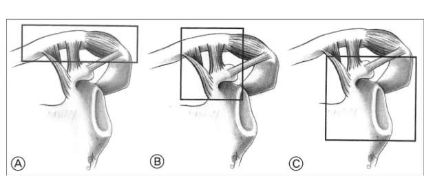
(2) HS Song, et al. Shoulder suspensory complex, Shoulder instability, Bony Bankart lesion, Shoulder reduction. Fig. 1.The superior shoulder suspensory complex comprises three component: A; the clavicular- acromioclavicular joint-acromial strut; B; the clavicular-coracoclavicular ligamentous-coracoid linkage; C; the three-process-scapular body.. 유지와 기능 회복에 심각한 장애를 가져오며, 반드시 수술적 1). 치료로 구조물의 회복이 필요하다 . 저자들은 이러한 상부 견갑 현수 복합체 이중 분리의 3예에 대하여 수술을 시행하고 보고하고자 한다.. 증. 례. 증례 1 27세 남자 환자가 교통사고로 우측 견관절의 골절 및 탈구와 경추 손상, 비장 및 간 파열 등을 주소로 본원에 전원되었다. 견관절의 정복 후 10일간의 경추부와 복부 손상의 치료 중. Fig. 2. A; Anteroposterior radiograph of 27 years old male shows dislocation in right shoulder with fracture of coracoid and acromion. B; This computed tomograph of the patient shows displaced acromion, coracoid fracture and large bony Bankart lesion which are unstable.. 견관절의 전방 탈구가 지속되었으며, Ogawa 분류상4) 제 1형에 해당하는 약 20 mm 전위된 오구돌기와 Kuhn 분류상5) 제 3형에 해당하는 약 5 mm 전위된 후외측부 견봉 골절이 관찰되었으며, 견관절 관절와의 전하방에 약 30% 정도의 골 결손이 발생되어 골성 방카트의 골편은 약 10 mm 하방으로 전위되어 있었다(Fig. 2A, B). 수술은 오구돌기의 외하방으로 약 10 cm의 절개선을 가하여 골절된 오구돌기의 끝에 부착된 상완 이두건 단두와 오구 완 근의 연합 건을 박리하였다(Fig. 3). 견갑하건을 소결절 로부터 약 1 cm 내측에서 세로로 상방 3/4만 절개한 후 내측으로 젖힌 다음 전방 관절낭을 관절와에 연하여 종절개를 가한 후 전방 관절와를 노출시켰다. 이때 상완골 이두건구에 파열된. Fig. 3. We detached the coracoid process (white arrow) with conjoined tendon (dark arrow).. 이두근 장두가 관찰되어 이두건구에 고정술을 시행하였다. 관절와의 하방에 전위된 약 2×1.5 cm 크기의 관절와 골을 골성. levo®, Linvatec, Largo, Florida)를 이용하여 골에 봉합사의 네. 방카트 병변 하방에 위치시킨 후 흡수성 봉합나사(Biomini-. 가닥을 통과시켜 고정하였다. 전위된 오구돌기를 Bristow 술식6). 232. 대한스포츠의학회지.
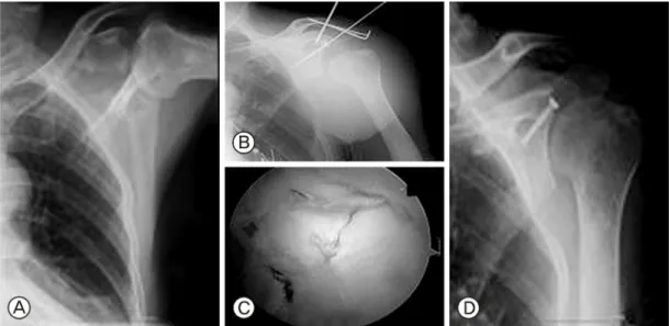
(3) 송호섭 외. 상부 견갑 현수 복합체 이중 분리에서의 급성 견관절 불안정성의 치료. 을 응용하여 연합 건이 부착된 상태로 방카트 병변의 전하방. 정복을 시행하였고, 단순 방사선 검사 및 컴퓨터단층촬영 결과. 부위에 봉합 나사 두 개와 고정 나사 한 개를 이용하여 고정시키. Ideberg 분류상 제 2형에 해당하는 견갑골 관절와 골절과. 고 상완 골두를 정복시켰다(Fig. 4). 이후 별도의 절개를 가하여. 함께 Kuhn 분류 제 3형에 해당하는 견봉 골절이 관찰되었고. 견봉 골절에 긴장대 강선고정으로 고정하였으며, 견관절의. (Fig. 6A-C), 관절와의 골절은 전상방 골편이 약 10 mm 전위를. 안정성을 확인한 후 절개부를 봉합하였다(Fig. 5A, B). 수술. 보였고 방카트나, SLAP병변은 관찰되지 않았다. 후방 삽입구. 후 약 3주간 보조기 착용 하에 안정을 취하였으며, 이후 수동적. 를 통한 관절경으로 관절내를 관찰한 다음 탐색자로 함몰된. 관절운동을 3주간 시행하고, 수술 후 6주 후에는 능동적 관절. 관절와를 들어 올려 관절면을 맞추고 전방에서 후방으로 두. 운동 및 근력 강화운동을 병행하였다.. 개의 유관나사를 이용하여 정복된 관절와를 고정하였고, 관절. 7). 5). 운동 시 상완골두에 충돌되지 않는 것을 확인한 후 견봉 위에. 증례 2. 사선으로 약 3 cm의 절개를 가한 후 유관나사를 이용하여 전위. 26세 남자 환자가 2 m 높이에서 낙상하여 좌측 견관절의. 된 견봉을 정복하였다. 견봉 골절과 견봉 쇄골 관절은 별도의. 골절 및 탈구를 주소로 내원하였으며, 내원 즉시 견관절의. Fig. 4. This figure demonstrates reattaching fractured coracoid with conjoined tendon (white arrow) and bony Bankart fragment. These were well fixed and restored the contact surface of the glenoid cavity (white star).. 절개를 통하여 고정하였다(Fig. 7A, B). 수술 후 약 2주 후 수동적 관절 운동을 시행하였으며, 수술 후 4주 후부터 근력. Fig. 5. A; The anteroposterior radiograph shows well reduced humeral head on the center of the glenoid cavity. B; The three dimentional computed tomograph shows well fixed large bony Bankart and transferred coronoid process on the anteroinferior of the glenoid.. Fig. 6. A; Plain radiograph of shoulder suggests fracture of glenoid cavity and acromion. B; Axial computed tomograph shows about 10mm displaced glenoid cavity and no bony Bankart lesion in anterior or posterior glenoid. C; The three dimensional computed tomograph shows a displaced glenoid cavity fracture and acromion fracture. 제27권 제2호 2009. 233.
(4) HS Song, et al. Shoulder suspensory complex, Shoulder instability, Bony Bankart lesion, Shoulder reduction. 강화 운동을 병행하였다.. 고. 찰. 증례 3 53세 남자가 쇠 파이프로 어깨의 후방에 직접 손상을 받은 후 발생한 좌측 견관절의 통증과 운동 제한으로 내원하였다. 7). 수술 전 방사선 사진 상 Ideberg 분류상 제2형의 관절와 손상과 견봉의 손상이 관찰되었으며, 수술 시 관절경 관찰 하에 전위 된 관절와의 외상방에서 내하방으로 스테인만 강선을 삽입한 후 이것을 Joystick으로 활용하여 관절면을 정복한 후 유관나사 를 이용하여 고정하였다(Fig. 8A-D).. 견갑골의 골절을 동반한 견갑 현수 복합체의 손상은 단일 분리의 경우 대부분 보존적 치료에 좋은 결과를 보인다고 1,2). 3). 보고되고 있다 . Goss 는 이러한 상부 견갑 현수 복합체의 손상을 원형 단일체의 개념으로 설명하고 비교적 흔하게 발생 하는 단일 분리의 경우 구조적으로 안정적이며, 보존적 치료에 잘 반응하며, 좋은 결과를 얻을 수 있다고 하였다. 그러나 상부 견갑 현수의 이중 분리의 경우 두 개의 골 연골 조직의 고리 골절, 두 개의 고리 인대 파열, 양 지주골의 골절, 고리 분리를 동반한 지주골의 골절 등 다양한 형태로 나타나며, 불안정하고 수술적 치료가 필요하며, 수술 후에도 견봉하 충돌 증후군, 근력약화, 신경 혈관계 합병증, 퇴행성 관절염의 유발 8). 등의 후유증이 남을 수 있다고 하였다. Oh 등 도 이중 분리 손상의 경우 손상된 구조물의 고정으로 좋은 결과를 보고하였 고, 이는 견관절의 안정성에 중요한 요소라 주장하였다. 이에 따라 저자들은 전례에서 이중 분리의 손상으로 손상된 모든 Fig. 7. A; The arthroscopic findings show well reduced joint surface of the displaced glenoid cavity. B; The anteroposterior radiograph shows well reduced glenoid and acromion fracture. Acromiocalvicular injury was treated with modified Phemister method.. 2). 구조물에 대하여 고정을 시행하였다. Mayo 등 은 Ideberg의 견갑골 관절와 골절의 분류를 기초로 변형된 Ideberg 분류를 제시하였으며, 증례 1은 이러한 분류상 제1형에 해당하며,. Fig. 8. A; Anteroposterior radiograph of 53 years old male shows acromioclvicular injury and glenoid fracture of Ideberg’s type 2. B; We used joystick technique for the reduction of the displaced glenoid cavity C; The arthroscopic findings show well reduced glenoid cavity. D; This 6 months follow radiograph shows good maintenance of the fracture reduction and well healed fracture.. 234. 대한스포츠의학회지.
(5) 송호섭 외. 상부 견갑 현수 복합체 이중 분리에서의 급성 견관절 불안정성의 치료. 관절와 전하방의 골성 방카트 병변을 동반한 예이다. Gries 9). 2. Mayo KA, Benirschke SK, Mast JW: Displaced fracture of. 등 은 관절와 전하방의 약 30%의 골 결손은 정상측에 비해. the glenoid fossa. Results of open reduction and internal. 약 53%의 접촉면의 상실을 가져오고, 약 300-400%의 평균. fixation. Clin Ortho Relat Res, 347:122-130, 1998.. 접촉 압력의 상승을 가져온다고 보고하였다. 이러한 골 결손을 10). 해결하는 수술적 요법으로 Latarjet. 6). 술식과 Bristow 술식. 등이 이용되고 있으며, 본 저자의 예에서는 골절된 오구돌기와. 3. Goss TP: Fractures of the scapula. In: Rockwood CA, et al ed. The shoulder. 3rd ed. Philadelphia, Saunders: 432-437, 2004.. 연합 건을 이용하여 Latarjet 술식을 응용하여 전방 골 결손부위. 4. Ogawa K, Yoshida A, Takahashi M, Ui M: Fracture of the. 에 이식하였다. 본 연구의 증례 2, 3과 같이 Ideberg 제2형의. coracoid process, J Bone Joint Surg Br, 79:17-19, 1997.. 관절와 골절과 동반된 견갑 현수 복합체의 손상은 약 5 mm. 5. Kuhn JE, Blasier RB, Carpenter JE: Fracture of the acromion. 이상의 관절와의 함몰이 있는 경우나, 견관절 불안정성으로. process: a proposed classification system. J Orthop Trauma,. 견관절의 정복을 유지할 수 없을 경우 수술이 반드시 필요하다.. 8:6-13, 1994.. 본 증례의 경우 관절와의 정복에 관절경만을 이용하여 정복한. 6. Helfet AJ: Coracoid transplantation for recurring dislocation. 후 경피적 나사 고정을 통해 충분한 고정력을 얻을 수 있었고,. of the shoulder. J Bone Joint Surg Br, 40:198-202, 1958.. 견봉 쇄골 관절과 견봉 골절에 대해서는 별도의 고정을 시행해. 7. Ideberg R: Fractures of the scapula involving the glenoid. 관절의 안정성을 얻을 수 있었다. 견관절의 상부 견갑 현수. fossa. In: Bateman JE, Welsh RP ed. Surgery of the shoulder.. 복합체의 손상에 대한 이해가 최근 관절경의 발달로 더욱 높아. 1st ed. Philadelphia, Decker: 63-66, 1984.. 졌다. 저자들은 이러한 급성 손상으로 인하여 견관절의 불안정. 8. Oh W, Jeon IH, Kyung S, Park C, Kim T, Ihn C: The treatment. 성을 야기한 다른 형태의 세가지 증례 치료에서 모든 손상된. of double disruption of the superior shoulder suspensory. 구조를 수술적으로 고정하여 견관절의 안정성을 얻었으며,. complex. Int Orthop, 26:145-149, 2002.. 효과적인 치료 방법이라고 사료된다. 그러나 장기적인 추시가 필요하며, 더 많은 증례의 결과를 참고하여야 할 것이다.. 9. Gries PE, Scuderi MG, Mohr RA, Bachus KN, Burks RT: Glenohumeral articular contact areas and pressures following labral and osseous injury to the anterior-inferior quadrant. 참 고 문 헌 1. Goss TP: Double disruption of the superior shoulder suspensory complex. J Orthop Trauma, 7:99-106, 1993.. of the glenoid. J Shoulder Elbow Surg, 11:442-451, 2002. 10. Latarjet M: Technic of coracoid preglenoid arthrodesis in the treatment of recurrent dislocation of the shoulder. Lyon Chir, 54:604-607, 1958.. 제27권 제2호 2009. 235.
(6)
수치

관련 문서
It considers the energy use of the different components that are involved in the distribution and viewing of video content: data centres and content delivery networks
After first field tests, we expect electric passenger drones or eVTOL aircraft (short for electric vertical take-off and landing) to start providing commercial mobility
1 John Owen, Justification by Faith Alone, in The Works of John Owen, ed. John Bolt, trans. Scott Clark, "Do This and Live: Christ's Active Obedience as the
The greater tubercle is palpable on the line from the lateral epicondyle of the distal humerus in the direction of the humeral longitudianl axis and just below the acromion
Evaluations of the potential for the unpredictable disruption of nuclear fuel markets and the possibility of mutual assistance in the event of such disruption;.
Based on the above results, the 12-week complex type correction program had a positive effect on the shoulder angle, trunk rotation angle, and body shape
In contrast, Chiropractic has shown better result than sports massage in the range of motion of the shoulder joint, Thoracic Scapular rhythm test, and the
Using this leap motion, a surgeon can perform the virtual shoulder arthroscopic surgery skillfully and reduce mistakes in surgery.. The simulation using leap motion shows
