377
서 론
Cyclooxygenase-2 (Cox-2)는 arachidonic acid에서 prostagl- andins (PG)이 합성되는 과정에 관여하는 효소로 체내에서 주로 염증반응에 관여를 한다.(1,21,22) Cox-2를 억제하는 약제들은 항염증작용 뿐만 아니라 위장관암, 폐암, 유방암 등의 발생 및 증식을 억제하는 것으로 알려져 있는데, 이는 종양의 혈관생성 억제와 세포사멸의 조장에 의한 기전으로 보고되고 있다.(1) 유방암 조직에서도 Cox-2의 발현이 보고 된 이후 Cox-2 및 Cox-2억제제의 역할에 대해 세포주 및 동 물 모델을 이용한 여러 연구결과들이 보고되고 있으며,(1) Cox-2 발현의 예후인자적 가치에 대한 임상연구를 비롯하 여 Cox-2 억제제를 이용한 유방암의 예방 및 치료약제로서 의 가능성이 연구되고 있다.(2) Cox-2 발현은 종양의 크기가 클수록, 조직학적 등급이 높을수록, 호르몬 수용체가 음성 일 때 증가하며, 또한 분화도가 높을수록, p53 발현이 높을 수록 증가한다고 보고되고 있다.(8)
저자들은 유방암에서 Cox-2 발현율을 조사하고 기존의 병리조직학적 인자들과의 상관관계를 분석하고자 본 연구 를 진행하였다.
방 법
1990년 1월부터 1991년 6월까지 한림대학교 의과대학 강 동성심병원에서 유방암 진단하에 수술 받은 환자 중 파라 핀포매 조직의 상태가 양호한 40예 환자의 파라핀포매 조 직을 이용하여 Cox-2의 종양 내 발현율을 특이항인 단일클 론성항체(specific antihuman monoclonal antibody)를 사용한 면역조직화학염색법을 이용하여 측정하였다.
면역조직화학염색은 파라핀포매 조직을 4μm로 절단하
유방암에서 Cyclooxygenase-2 발현의 예후인자적 가치
한림대학교 의과대학 외과학교실, 1연세대학교 의과대학 병리학교실
설진원․김승일․박찬흔․박철재․양우익1
Prognostic Value of Elevated Cyclooxygenase-2 Expression in Breast Cancer
Jin Won Seol, M.D., Seung Il Kim, M.D., Chan Heun Park, M.D., Ph.D., Chul Jae Park, M.D. and Woo Ick Yang, M.D., Ph.D.1
Purpose: Cyclooxygenase-2 (Cox-2) is the rate-limiting en- zyme in the conversion of arachidonic acid to prostaglandins and can be induced by various agents such as growth factors and tumor promoters. Cox-2 contributes to carcino- genesis and tumor growth. This study was performed to demonstrate the correlation between elevated expression of Cox-2 and pathologic factors in breast cancer.
Methods: Cox-2 expression was analyzed immunohistoche- mically in paraffin-embedded tumor samples from 40 patients withbreast cancer. Cox-2 expression was defined as nega- tive or positive. The correlation between Cox-2 expression and pathologic factors (tumor size, axillary lymph node metastasis, TNM stage and histologic grade) was analyzed.
Results: Cox-2 was highly expressed in proportion to tumor size, but the difference was not significant (P>0.05). High Cox-2 expression was observed in the presence of axillary lymph node metastasis and TNM stage III, but was not significant (P>0.05). The Cox-2 expression rate was signi- ficantly associated with high histologic grade (I: 42.9%, II:
50.0%, III: 80.0%) (P=0.046).
Conclusion: Elevated levels of Cox-2 expression were associated with large tumor size, presence of axillary lymph node metastasis, high TNM stage and high histologic grade, and can therefore be a possible marker for poor prognosis.
Due to the small number of cases, we couldn't confirm the statistical significance except in terms of histologic grade.
Further prospective studies with a large number of cases are required. (J Korean Surg Soc 2003;65:377-381)
책임저자:박찬흔, 서울시 강동구 길동 445 ꂕ 134-010, 강동성심병원 외과 Tel: 02-2224-2226, Fax: 02-2224-2570 E-mail: [email protected]
접수일:2003년 7월 3일, 게재승인일:2003년 9월 5일
Key Words: Breast cancer, Cyclooxygenase-2, Prognostic factors
중심 단어: 유방암, Cyclooxygenase-2, 예후인자 ꠏꠏꠏꠏꠏꠏꠏꠏꠏꠏꠏꠏꠏꠏꠏꠏꠏꠏꠏꠏꠏꠏꠏꠏꠏꠏꠏꠏꠏꠏꠏꠏꠏꠏꠏꠏꠏꠏꠏꠏꠏꠏꠏꠏꠏ Department of Surgery, Hallym University College of Medi- cine, 1Department of Pathology, Yonsei University College of Medicine, Seoul, Korea
ꠏꠏꠏꠏꠏꠏꠏꠏꠏꠏꠏꠏꠏꠏꠏꠏꠏꠏꠏꠏꠏꠏꠏꠏꠏꠏꠏꠏꠏꠏꠏꠏꠏꠏꠏꠏꠏꠏꠏꠏꠏꠏꠏꠏꠏꠏꠏꠏꠏꠏꠏꠏꠏꠏꠏꠏꠏꠏꠏꠏꠏꠏꠏꠏꠏꠏꠏꠏꠏꠏꠏꠏꠏꠏꠏꠏꠏꠏꠏꠏꠏꠏꠏꠏꠏꠏꠏꠏꠏꠏꠏꠏꠏꠏꠏꠏꠏꠏꠏꠏꠏꠏꠏꠏꠏꠏꠏꠏꠏꠏꠏꠏꠏꠏꠏ 여, xylene으로 탈파라핀한 후 알콜로 재수화(rehydrated)하
였다. 절단면은 항원복구를 위해 0.01 M Na-citrate 완충액 (pH 6.0)을 이용해 820 wtts 극초단파 오븐에서 20분 동안 전처치를 하였다. 20분 동안 냉각 후 슬라이드를 3% 수소과 산화물에 10분 동안 담그어 내인성 과산화 효소의 활동을 억제하였다. Cox-2 특이항인 단일클론성항체(Cayman Che- mical Co., Ann Arbor, MI)는 실온에서 0.1% sodium azide와 0.5% 우형 혈청 알부민이 함유된 말초혈액에서 1:200으로 희석하여 사용하였다. 그 후, 절단면은 biotinylated horse antimouse immunoglobulin (1:200; Vector Laboratories, Inc., Burlingame, CA)으로 처치 후 avidin-biotin peroxidase com- plex (Vectastain ABC complex; Vector Laboratories Inc.)와 diaminobezidine (Vector Laboratories Inc.)로 발색하였다.
Harris hematoxylin으로 대비염색을 시행하였다.
Cox-2의 발현정도는 각각 음성 및 양성으로 분류하였으 며 암세포의 세포질 염색이 10% 이상인 경우를 양성으로 하였다. Cox-2의 발현정도와 기타 병리학적 인자들(원발종 양의 크기, 액와림프절 전이여부, TNM 병기 및 종양의 조 직학적 분류)과의 상관관계를 분석하였다. 자료의 분석은 chi-sqaure test를 이용하였다.
결 과 1) 대상환자의 일반적 특성
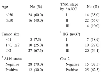
유방암 환자 40예의 연령분포는 27세에서 73세까지였으며, 평균연령은 45.2세였고, 원발종양의 크기는 1 cm 이하가 3예 (7.5%), 1 cm에서 2 cm 사이가 10예(25.0%), 2 cm 이상이 27 예(67.5%)였다(Table 1). 액와림프절 전이여부는 음성이 28예 (70.0%), 양성이 12예(30.0%)로 각각 조사되었다. TNM 병기 는 I이 14예(35.0%), II가 22예(55.0%), III이 4예(10.0%)였다.
종양의 조직학적 등급은 Bloom & Richardson 분류법을 이용 하였으며 I이 7예(18.9%), II가 10예(27.0%), III이 20예(54.1%) 의 분포를 보였다(Table 1). Cox-2의 발현은 15예(37.5%)에서 음성, 25예(62.5%)에서 양성을 보였다(Table 1, Fig 1).
2) 원발종양의 크기에 따른 Cox-2 발현율
원발종양의 크기에 따른 Cox-2 발현율의 비교 결과, 1cm 이하에서 Cox-2 발현율은 33.3%, 2 cm 이상에서 Cox-2 발현 율은 66.7%로 원발종양의 크기가 클수록 Cox-2의 발현이 증가하는 경향을 보였으나 통계적 의의는 없었다(P>0.05) (Table 2).
Fig. 1. Immunohistochemistry in the breast tumor tissue using Cox-2 specific monoclonal antibody: A) Positive: pigmentation (arrow) in cytoplasm (H-E stain, ×400), B) Negative.
Table. 1. General characteristics of patients (n=40) ꠚꠚꠚꠚꠚꠚꠚꠚꠚꠚꠚꠚꠚꠚꠚꠚꠚꠚꠚꠚꠚꠚꠚꠚꠚꠚꠚꠚꠚꠚꠚꠚꠚꠚꠚꠚꠚꠚꠚꠚꠚꠚꠚꠚꠚꠚꠚꠚꠚꠚꠚꠚꠚꠚꠚ
TNM stage
Age No (%) No (%)
by *AJCC
<50 24 (60.0) I 14 (35.0)
≥50 16 (40.0) II 22 (55.0)
III 4 (10.0)
Tumor size †HG (n=37)
≤1 3 (7.5) I 7 (18.9)
1<, ≤2 10 (25.0) II 10 (27.0)
>2 27 (67.5) III 20 (54.1)
‡ALN status Cox-2
Negative 28 (70.0) Negative 15 (37.5)
Positive 12 (30.0) Positive 25 (62.5)
ꠏꠏꠏꠏꠏꠏꠏꠏꠏꠏꠏꠏꠏꠏꠏꠏꠏꠏꠏꠏꠏꠏꠏꠏꠏꠏꠏꠏꠏꠏꠏꠏꠏꠏꠏꠏꠏꠏꠏꠏꠏꠏꠏꠏꠏꠏꠏꠏꠏꠏꠏꠏꠏꠏꠏ
*AJCC = American Joint Committee on Cancer; †HG = Histologic grade; ‡ALN = Axillary lymph node.
ꠏꠏꠏꠏꠏꠏꠏꠏꠏꠏꠏꠏꠏꠏꠏꠏꠏꠏꠏꠏꠏꠏꠏꠏꠏꠏꠏꠏꠏꠏꠏꠏꠏꠏꠏꠏꠏꠏꠏꠏꠏꠏꠏꠏꠏꠏꠏꠏꠏꠏꠏꠏꠏꠏꠏꠏꠏꠏꠏꠏꠏꠏꠏꠏꠏꠏꠏꠏꠏꠏꠏꠏꠏꠏꠏꠏꠏꠏꠏꠏꠏꠏꠏꠏꠏꠏꠏꠏꠏꠏꠏꠏꠏꠏꠏꠏꠏꠏꠏꠏꠏꠏꠏꠏꠏꠏꠏꠏꠏꠏꠏꠏꠏꠏꠏ
3) 액와림프절 전이여부에 따른 Cox-2 발현율 액와림프절 전이여부에 따른 Cox-2 발현율의 비교 결과, 전이가 되지 않은 경우 Cox-2 발현율은 57.1%, 전이가 된 경우 Cox-2 발현율은 75.0%로 액와림프절 전이가 있을수록 발현이 증가하는 경향을 보였으나 통계적 의의는 없었다(P>
0.05)(Table 2).
4) TNM 병기에 따른 Cox-2 발현율
TNM 병기에 따른 Cox-2의 발현율의 비교 결과, 병기 I에 서 Cox-2 발현율은 50.0%, 병기 II에서 Cox-2 발현율은 68.2%, 병기III에서 Cox-2 발현율은 75.0%로 TNM 병기가 높을수 록 Cox-2의 발현율도 높아짐을 볼 수 있으나 통계적 의의는 없었다(P>0.05)(Table 2).
5) 조직학적 등급에 따른 Cox-2 발현율
조직학적 등급에 따른 Cox-2 발현율의 비교 결과, 등급 I에서 Cox-2 발현율은 42.9%, II에서 Cox-2 발현율은 50.0%, III에서 Cox-2 발현율은 80.0%로 조직학적 등급이 높을수록 유의하게 높은 발현율을 보였다(P=0.046)(Table 2).
6) Cox-2 발현에 따른 생존율분석
전체 40예 중 35예에서 추적 관찰이 가능하였으며 평균 추적기간은 67.3개월, 중간 추적기간은 73.0개월이었다(범 위 1∼151개월). Kaplan-Meier 생존율 분석을 이용한 결과 Cox-2 발현에 따른 무병생존율은 차이가 없는 것으로 분석
되었다(P>0.05)(data not shown).
고 찰
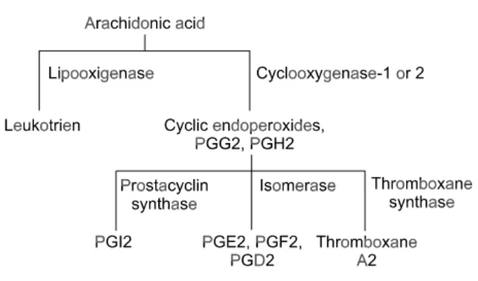
Cyclooxygenase-2 (Cox-2)는 arachidonic acid로부터 prosta- glandin (PG)과 thromboxane (TX)의 합성과정에 관여하는 효 소이며, 이는 성장인자(growth factor), cytokines, 그리고 내 독소(endotoxin)와 같은 여러 요소들에 의해 유발된다.(1,3) Fig. 2는 arachidonic acid 대사과정을 보여주며 Cox enzyme 은 PG 생산의 속도제한 단계(rate-limiting step)에 관여하고 있다. Cox-1은 주로 항상성 유지(homeostasis)와 세포조절 기능(cell regulatory function)에 관여하고, Cox-2는 염증반응 기능(proinflammatory function)에 관여한다고 알려져 있 다.(1) 그 외 Cox-2의 역할로는 신장 조직에서 발현되어 prostanoids를 형성하고, 이온 조절에 관여하여 신체의 항상 성을 유지하게 된다. 뇌 조직에서도 발현되어 체온 및 수면 조절에 관여한다. 또한 췌도세포(pancreatic islet cells)에서 발현되어 당의존적 인슐린 분비를 억제하게 되며, 혈관 평 활근 세포에서는 염증반응과 세포증식을 억제하는 기능을 하며, 호흡기에서는 기도세포의 과증식을 억제하여 천식과 같이 기도세포가 과증식되는 질환을 예방하는 역할을 한 다.(2) 한편 Cox-2는 다양한 암과 전암 병변에서 과발현되 있는데, 그 기전은 PG생성을 증가시키고, 전발암물질(pro- carcinogen)을 발암물질로 전환시키며, 세포사멸(apoptosis) 을 억제하고, 혈관형성을 조장하고, 암세포 침습을 증가시 키는 기능 등을 통해 발암현상에 관여하는 것으로 생각되 고 있다.(3)
Cox-2는 위장관암, 유방암, 폐암 및 방광암 등에서도 과 표현된다고 보고되고 있다.(4,5) 위암에서는 83%, 대장암에 서는 85%,(6) 유방암에서는 56%, 폐암에서는 90%,(7) 방광 암에서는 65%(4) 정도까지 Cox-2 단백질의 상향조정(up regulation)이 관찰된다고 보고되고 있으나 그 기전은 아직 명확하지 않다.
연구보고에 의하면 Cox-2의 과발현이 PGE2의 생합성을 증가시키고 혈관형성을 야기하여 암을 유발시키는 것으로 설 Table 2. Association of Cox-2 immunopositivity with clinico-
pathological parameters
ꠚꠚꠚꠚꠚꠚꠚꠚꠚꠚꠚꠚꠚꠚꠚꠚꠚꠚꠚꠚꠚꠚꠚꠚꠚꠚꠚꠚꠚꠚꠚꠚꠚꠚꠚꠚꠚꠚꠚꠚꠚꠚꠚꠚꠚꠚꠚꠚꠚꠚꠚꠚꠚꠚ Clinicopathological COX-2 positive
P-value parameters no (%)
ꠏꠏꠏꠏꠏꠏꠏꠏꠏꠏꠏꠏꠏꠏꠏꠏꠏꠏꠏꠏꠏꠏꠏꠏꠏꠏꠏꠏꠏꠏꠏꠏꠏꠏꠏꠏꠏꠏꠏꠏꠏꠏꠏꠏꠏꠏꠏꠏꠏꠏꠏꠏꠏꠏꠏ Tumor size
≤1 1 (33.3) >0.05
1<, ≤2 6 (60.0)
>2 18 (66.7)
*ALN status
(-) 16 (57.1) >0.05
(+) 9 (75.0)
TNM stage
I 7 (50.0) >0.05
II 15 (68.2)
III 3 (75.0)
HG
I 3 (42.9) =0.046
II 5 (50.0)
III 16 (80.0)
ꠏꠏꠏꠏꠏꠏꠏꠏꠏꠏꠏꠏꠏꠏꠏꠏꠏꠏꠏꠏꠏꠏꠏꠏꠏꠏꠏꠏꠏꠏꠏꠏꠏꠏꠏꠏꠏꠏꠏꠏꠏꠏꠏꠏꠏꠏꠏꠏꠏꠏꠏꠏꠏꠏꠏ
*ALN = Axillary lymph node; HG = Histologic grade.
Fig. 2. Arachidonic acid pathway.
ꠏꠏꠏꠏꠏꠏꠏꠏꠏꠏꠏꠏꠏꠏꠏꠏꠏꠏꠏꠏꠏꠏꠏꠏꠏꠏꠏꠏꠏꠏꠏꠏꠏꠏꠏꠏꠏꠏꠏꠏꠏꠏꠏꠏꠏꠏꠏꠏꠏꠏꠏꠏꠏꠏꠏꠏꠏꠏꠏꠏꠏꠏꠏꠏꠏꠏꠏꠏꠏꠏꠏꠏꠏꠏꠏꠏꠏꠏꠏꠏꠏꠏꠏꠏꠏꠏꠏꠏꠏꠏꠏꠏꠏꠏꠏꠏꠏꠏꠏꠏꠏꠏꠏꠏꠏꠏꠏꠏꠏꠏꠏꠏꠏꠏꠏ 명하고 있다. PGE2는 장기에서 특히 더욱 강한 효과를 보이
는데 이는 암세표 성장 및 암세포의 침습을 증가시킨다.(3) Cox-2 발현의 증가는 위장관 종양에만 특이한 것이 아니라 유방암에서도 과발현이 보고되고 있다.(8,9) 유방암 환자에서 얻은 파라핀포매 조직을 Cox-2 특이항인 단일클론성 항체를 이용하여 면역조직화학염색법으로 측정하였을 때 중등도 또 는 강한 Cox-2 면역반응이 암세포의 세포질에만 국한되어 나 타나며, 간질에서는 음성 또는 약한 양성 소견을 보인다.(8) Cox-2의 발현은 유방암의 예후에 영향을 미치는 여러 병 리학적 인자와도 연관이 있는 것으로 보고되고 있다. 본 연 구에서도 나타난 바와 같이 Cox-2 발현의 증가는 나쁜 예후 를 나타내는 요소들과 상관관계를 가지는데, 본 연구에서 는 조직학적 등급만 유의성이 확인되었으나 다른 연구 결 과에 의하면 원발 종양의 크기, 액와림프절 전이여부, 조직 학적 등급 등과 유의한 상관관계를 가진다고 보고하고 있 다.(8) 이 연구들에 의하면 큰 원발종양의 크기, 액와림프절 전이의 양성, 높은 조직학적 등급, 호르몬 수용체 음성, p53 발현의 증가, HER-2의 증폭, 진행된 분류병기 등에서 Cox-2 과발현이 관찰된다고 보고하고 있다.(8) 또한 Cox-2가 Dis- tant Disease-free survival (DDFS)와도 연관을 보이는데, 본 연구에서는 연구 대상의 수가 적어 Cox-2 발현에 따른 무병 생존율이 차이가 없는 것으로 나타났으나 다른 연구에 의 하면 Cox-2 발현이 낮은 군에서는 5년 DDFS가 83%, Cox-2 발현이 높은 군에서는 73%로 유의하게 DDFS가 낮다는 보 고가 있다.(8)
유방암에서 Cox-2의 상향조정에 대한 기전은 명확히 알 려지지 않았지만, Cox-2발현이 비암세포보다 암세포에서 활발하며, p53과 같은 암억제 유전자의 비활성화와 HER-2 와 같은 암유발 유전자의 활성화가 Cox-2 발현과 관련이 있 다고 추정된다.(8)
한편 Cox-2가 인체에서 암의 발생과 증식에 관여함에 따 라 Cox-2를 억제하여 암의 예방 및 치료에 이용하려는 연구 들이 진행되고 있다. 최근 들어 실험적 임상연구에서 아스 피린 및 비스테로이드성 항염증제(NSAIDs) 등이 식도암, 위암, 대장암, 유방암, 폐암 및 방광암 등의 발생률을 감소 시킨다고 보고하였다.(3) NSAIDs는 Cox 의존적, Cox 비의 존적인 기전으로 암발생을 억제하게 되는데, 아직 명확한 기전이 밝혀져 있지는 않다. 현재까지 알려진 바에 의하면 Cox 의존적 기전으로는 Cox-2에 의해 생성되는 PG의 생성 억제를 통해 DNA 합성과 세포분화에 관여하는 PG의 기능 을 방해하는 것이다. 이로써 암조직의 혈관 생성을 억제하 고, 암세포 성장을 막게 된다. 또한 Cox 효소는 PG의 생성 뿐만 아니라 다른 세포들간의 반응에도 관여하는데, 예를 들어 Cox 효소는 발암물질-DNA 부가물(carcinogen-DNA adducts)을 생성하는데 이 부가물의 생성이 NSAIDs에 의해 억제된다.(1) Cox 비의존적 기전으로는 Cox-2를 억제하지 않고 세포사멸을 조장시켜 암세포 성장을 억제하는 것이
다. 현재까지 여러 연구에서 규칙적인 NSAIDs의 사용, 특 히 Cox-2 선택적 억제제가 대장암을 비롯한 일부 위장관암 의 예방에 효과가 있음을 보고하고 있는데,(10) 아직까지 유방암에서 Cox-2 억제제의 예방적 효과가 입증되지 않았 으나 그 가능성은 제시되고 있다.
결 론
유방암에서 Cox-2는 원발종양의 크기가 클수록, 액와림 프절 전이가 많을수록, TNM 병기 및 종양의 조직학적 등급 이 높을수록 발현율이 높은 경향을 보여 Cox-2은 높은 발현 율 자체가 나쁜 예후의 지표로 판단되며 따라서 Cox-2의 임 상적 유용성의 가능성을 보고하며 향후 좀더 많은 환자를 대상으로 한 전향적 연구가 필요하리라 생각된다.
REFERENCES
1) Shaheen NJ, Straus WL, Sandler RS. Chemoprevention of gastrointestinal malignancies with nonsteroidal antiinflamma- tory drugs. Am Can Sci 2002;94:950-61.
2) Mechell JA, Warner TD. Cyclo-oxygenase-2: pharmacology, physiology, biochemistry and relevance to NSAID therapy. Br J Pharmacol 1999;128:1121-32.
3) Xu X-C. Cox-2 inhibitors in cancer treatment and prevention, a recent development. Anti-cancer Drugs 2002;13:127-37.
4) Ristimaki, A, Nieminen O, Saukkonen K, Hotakainen K, Nordling S, Haglund C. Expression of cyclooxygenase-2 in human transitional cell carcinoma of the urinary bladder. Am J Pathol 2001;158:849-53.
5) Marrogi AJ, Travis WK, Welsh JA, Khan MA, Rahim H, Tazelaar H, et al. Nitric oxide synthase, cyclooxygenase-2, and vascular endothelial growth factor in the angiogenesis of non- small cell lung carcinoma. Clin Cancer Res 2000;6:4739-44.
6) Tsujii M, Kawano S, DuBois RN. Cyclooxygenase-2 expre- ssion in human colon cancer cells increases metastatic pote- ntial. Proc Natl Acad Sci USA 1997;94:3336-40.
7) Soslow RA, Dannenberg AJ, Rush D, Woennier BM, Khan KN, Masferrer J, et al. COX-2 is expressed in human pulmo- nary, colonic, and mammary tumors. Cancer (Phila) 2000;89:
2637-45.
8) Ristimaki A, Sivula A, Lundin J, Lundin M, Salminen T, Haglund C, et al. Prognostic significance of elevated cycloyge nase-2 expression in breast cancer. Cancer Res 2002;62:632-5.
9) Hwang D, Scollard D, Byme J, Levine E. Expression of cyclooxygenase-1 and cyclooxygenase-2 in human breast can- cer. J Natl Cancer Inst (Bethesda) 1998;90:455-60.
10) Kawamon T, Rao CV, Seibert K, Reddy BS. Chemopreventive activity of Colocoxib, a specific cyclooxygenase-2 inhibitor, against colon carcinogenesis. Cancer Res 1998;58:409-12.
11) Uefuji K, Ichikura T, Mochezuki H, Shinomiya N. Expression
ꠏꠏꠏꠏꠏꠏꠏꠏꠏꠏꠏꠏꠏꠏꠏꠏꠏꠏꠏꠏꠏꠏꠏꠏꠏꠏꠏꠏꠏꠏꠏꠏꠏꠏꠏꠏꠏꠏꠏꠏꠏꠏꠏꠏꠏꠏꠏꠏꠏꠏꠏꠏꠏꠏꠏꠏꠏꠏꠏꠏꠏꠏꠏꠏꠏꠏꠏꠏꠏꠏꠏꠏꠏꠏꠏꠏꠏꠏꠏꠏꠏꠏꠏꠏꠏꠏꠏꠏꠏꠏꠏꠏꠏꠏꠏꠏꠏꠏꠏꠏꠏꠏꠏꠏꠏꠏꠏꠏꠏꠏꠏꠏꠏꠏꠏ of cyclooxygenase-2 protein in gastric adenocarcinoma. J Surg
Oncol 1998;69:168-72.
12) Uefuji K, Ichikura T, Mochizuki H. Cyclooxygenase-2 to tumor angiogenesis in human gastric cancer. Clin Cancer Res 2000;6:135-8.
13) Khuder SA, Mutugi AB. Breast cancer and NSAID use: a meta-analysis. Br J Cancer 2001;84:1188-92.
14) Harris RE, Alshafie GA, Scibert K, Abou-Issa A. Chemo- prevention of breast cancer in rats by celecoxib, a cycloo- xygenase 2 inhibitor. Cancer Res 2000;60:2101-3.
15) Coogan FP, Rao RS, Rosenberg L, Palmer JR, Strom BL, Zauber AG, et al. The relationship of nonsteroidal antiinfla- mmatory drug use to the risk of breast cancer. Prev Med 1999;29:72-6.
16) Eberhart CE, Coffey RJ, Radhika A, Giardiello FM, Ferre- nbach S, DuBots RN, et al. Up-regulation of cyclooxygenase 2 gene expression in human colorectal adenomas and adeno- carcinomas. Gastroenterology 1994;197:1183-8.
17) Tsujii M, Kawano S, Tsuji S, Sawaoka H, Hori M, DuBois RN. Cyclooxygenase regulates angiogenesis induced by colon cancer cells. Cell 1998;93:705-16.
18) Rosenberg L, Palmer JR, Zouber AG, warshuer ME, Stolley PD, Shaprio S, et al. A hypothesis: nonsteroidal anti-infla- mmatory drugs reduce the incidence of the large-bowel cancer.
J Natl Cancer Inst 1991;83:355-8.
19) Marks F, Furstenberger G, Muller-Decker K. Metabolic targets of cancer chemoprevention: interruption of tumor development by inhibitors of arachidonic acid metabolism. Recent Results Cancer Res 1999;151:45-67.
20) Van Rees BP, Ristimaki A. Cyclooxygenase-2 in carcino- genesis of the gastrointestinal tract. Scand J Gastroenterol 2001;36:897-903.
21) Gately S. The contributions of cyclooxygenase-2 to tumor angiogenesis. Cancer Metastasis Rev 2000;19:19-27.
22) Sawaoka H, Tsuji S, Tsuji M, Gunawan ES, Sasaki Y, Kawano S, et al. Inhibition of angiogenesis and reduce tumor growth in vivo. Lab Invest 1999;79:1469-77.
23) Chan TA, Morin PJ, Vogelstein B, Kinzler KW. Mechanism underlying nonsteroidal antiinflammatory drug-mediated apo- ptosis. Proc Natl Acad Sci USA 1998;95:681-6.
24) Masferrer JL, Zweifel BS, Manning PT, Hauser SD, Leany KM, Smith WG, et al. Selective inhibition of inducible cycloo- xygenase-2 in vivo is antiinflammatory and nonulcerogenic.
Proc Natl Acad Sci USA 1994;91:3228-32.
25) Egan KM, Stampfer MJ, Giovannucci E, Rosner BA, Coditz GA. Prospective study of regular aspirin use and the risk of breast cancer. J Natl Cancer Inst 1996;88:988-93.