INTRODUCTION
One of the most important issues for treating patients with early stage breast cancer is determining those who could bene- fit from adjuvant chemotherapy and which chemotherapy regimen would provide the best results. Prognostic factors can be used to predict the natural history of breast cancer, so they are included in the decision to apply adjuvant systemic che- motherapy for patients with breast cancer. Currently, the pres- ence of axillary lymph node involvement, tumor size, nuclear grade, hormone receptor status, and patient age are well-known prognostic factors for patients with operable invasive breast
cancer [1]. These prognostic factors are widely used to deter- mine whether to apply adjuvant chemotherapy in patients with breast cancer. Among the prognostic factors, axillary lymph node involvement is the most significant [1,2]. Because the presence of lymph node involvement shows that the cancer has already developed the ability for distant metastasis [2-4].
Thus, the presence of axillary lymph node involvement pre- dicts the choice of adjuvant chemotherapy and radiation ther- apy after surgery for primary breast cancer [5].
Lymphovascular invasion (LVI) is defined as tumor emboli present within a definite endothelial-lined space in the breast surrounding invasive carcinoma [6,7]. The existence of LVI may help identify who is at increased risk for axillary lymph node and distant metastasis [2,3] Although LVI has been ac- cepted as an important prognostic factor in patients with lymph node-negative invasive breast cancer [8], LVI is controversial regarding prognostic value in patients with lymph node-posi- tive breast cancer [3,9-11]. The aim of this study was to ana-
The Role of Lymphovascular Invasion as a Prognostic Factor in Patients with Lymph Node-Positive Operable Invasive Breast Cancer
Young Ju Song, Sun Hyoung Shin, Jin Seong Cho, Min Ho Park, Jung Han Yoon, Young Jong Jegal
Department of Surgery, Chonnam National University Medical School, Gwangju, Korea ORIGINAL ARTICLE
Purpose: Lymphovascular invasion (LVI) is an important prognostic factor in patients with lymph node-negative patients with inva- sive breast cancer. However, the prognostic value of LVI it is un- clear and controversial about its prognostic value in patients with lymph node-positive breast cancer patients. So, we report the an analysis of the prognostic significance of LVI in a large cohort study of patients with lymph node-positive patients with invasive breast cancer. Methods: We retrospectively reviewed 967 patients with invasive breast cancer that had undergone surgical treatment at our hospital, from January 2004 to December 2007. Among these thempatients, 349 patients with lymph node-positive breast cancer patients are were included in this study. We evaluated clinical and pathological data in these patients, we compared with 5-year overall survival and disease-free survival between an LVI-present group and an LVI-absent group. Results: The median follow-up was 48 months (range, 12-78 months), and the mean age of the patients was 48 years (range, 23-78 years). LVI was present in 192 patients (55%) of with tumors and was associated with age ≤40 years (p=0.009), high histologichistological grade
(p=0.007), estrogen receptor status (p=0.001), tumor size ≥2 cm (p<0.001), and number of involved lymph nodes (p<0.001), but not with progesterone receptor status, HER2 status, p53 status, or tumor multiplicity. LVI was a significant independent prognos- tic factor for disease-free survival (p<0.001) and overall survival (p=0.006). By multivariate analysis revealed that LVI (p=0.003), number of involved lymph nodes (≥4; p=0.005), and high histo- logical grade (II and III; p=0.02) was were an independent signif- icant predictors of disease-free survival and overall survival in the whole group of patients. Conclusion: In this case, we demonstrated that LVI is a significant predictor of poor prognosis in patients with lymph node-positive patients with primary invasive breast cancer, LVI is a significant predictive predictor value of poor prognosis. So, LVI should be considered in the therapeutic strategy as a decision making tool in the adjuvant chemotherapy setting.
Key Words: Breast neoplasms, Lymph node metastasis, Lymphovascular invasion, Prognostic factor
Correspondence: Min Ho Park
Department of Surgery, Chonnam National University Medical School, 671 Jebongno, Dong-gu, Gwangju 501-757, Korea
Tel: +82-62-220-6456, Fax: +82-62-227-1635 E-mail: [email protected]
Received: April 4, 2011 Accepted: August 12, 2011
Cancer
lyze the prognostic significance of LVI in a large cohort study of patients with lymph node-positive invasive breast cancer.
METHODS
Patients and methods
From January 2004 to December 2007, we retrospectively reviewed 989 patients with invasive breast cancer that had un- dergone surgical treatment at the Department of Surgery, Chonnam National University Hospital. In total, 596 lymph node-negative and 371 lymph-node positive patients were en- rolled. Among the 371 lymph-node positive patients, 22 (six patients with a previous or a concurrent contralateral breast cancer, two males with breast cancer, and 14 patients who un- derwent neoadjuvant chemotherapy) were excluded. Thus, 349 patients with lymph node-positive breast cancer were in- cluded. Among the 349 patients, 11 were treated with sentinel lymph node biopsy only because the frozen biopsy was nega- tive, whereas permanent sections revealed positive findings.
We evaluated the 349 patients with lymph-node positive in- vasive breast cancer during a mean follow-up of 48 months (range, 12-78 months). These patients had been surgically treated with either a modified radical mastectomy or wide lo- cal tumor resection, with sentinel lymph node biopsy or axil- lary lymph node dissection, followed by postoperative radia- tion therapy. The need for adjuvant systemic therapy (chemo- therapy and hormonal therapy) was determined according to axillary lymph node status, hormone receptor status, and meno- pausal status. All patients visited the hospital every 6 months for 5 years, then once per year.
Pathologic examination
We analyzed the patient’s clinicopathological data including age, tumor size, hormone receptor status, axillary lymph node status, nuclear grade, and presence of LVI. Diameter of the tu- mor was measured on pathological specimens that were he- matoxylin and eosin (H&E) stained. Nuclear grade was iden- tified by a modified Scarff–Bloom–Richardson grading sys- tem. Lymph nodes were stained with H&E and examined for tumor cell metastasis. Hormone receptor (estrogen [ER] and progesterone [PR] receptors) and HER2 status was determined by immunohistochemical (IHC) analysis using a tissue micro- array [13]. The immunohistochemical analyses used an ER antibody (1D5; DAKO, Carpinteria, USA), a PR antibody (PgR636; DAKO), and a HER2 antibody (4B5; DAKO). Hor- mone receptors were considered positive if expression was
≥10%. The HER2 expression results by IHC analysis were scored as negative, 1+, 2+, or 3+, according to the manufac- turer’s recommendations. Patients with a score of 3+ were con-
sidered HER2 positive. HER2/chromosome 17 fluorescence in situ hybridization was performed on patients in which the IHC analysis score was 2+.
LVI was assessed on H&E-stained slides, as defined by Rosen and Oberman [6]. LVI was defined as carcinoma cells present within a definite endothelial-lined space, at a distance from the tumors, in the breast surrounding the invasive carcinoma.
[6-8] That is, LVI must be assessed outside the boundaries of the main breast tumor (at least more than one high power microscopic field away) to avoid misinterpretation [9]. When interpretation results were vague, the immunostaining mark- ers D2-40 and CD34 were used to confirm that LVI by H&E staining was truly present.
The clinicopathological data and systemic or locoregional Table 1. Clinicopathological data of patients
Patient characteristic LVI negative No. (%) (n=157)
LVI positive No. (%) (n=192)
Total
No. (%) p-value
Age (yr) 0.009
≤40 21 (30.9) 47 (69.1) 68 (19.5)
>40 136 (48.4) 145 (51.6) 281 (80.5)
Tumor size (mm) <0.001
≤10 13 (54.2) 11 (45.8) 24 (6.9)
10>, ≤20 69 (65.1) 37 (34.9) 106 (30.4) >20 75 (34.2) 144 (65.8) 219 (62.8)
Multiplicity NS
Single 149 (44.6) 185 (55.4) 334 (95.7)
multiple 8 (53.3) 7 (46.7) 15 (4.3)
Grade 0.007
I 29 (58.0) 21 (42.0) 50 (14.3)
II 86 (48.6) 91 (51.4) 177 (50.7)
III 42 (34.4) 80 (65.6) 122 (35.0)
No. of positive LN <0.001
1 64 (64.3) 37 (36.6) 101 (28.9)
2 31 (50.8) 30 (49.2) 61 (17.5)
3 14 (45.2) 17 (54.8) 31 (8.9)
≥4 48 (30.8) 108 (69.2) 156 (44.7)
ER 0.001
Negative 54 (35.3) 99 (64.7) 153 (43.8) Positive 103 (52.6) 93 (48.4) 196 (56.2)
PR NS
Negative 89 (43.2) 117 (56.8) 206 (59) Positive 68 (47.6) 75 (52.4) 143 (41)
Hormonal receptors 0.005
ER and PR negative 46 (35.4) 84 (64.6) 130 (37.2) ER or PR positive 111 (50.7) 108 (49.3) 219 (62.8)
HER2 status NS
Negative 124 (46.8) 141 (53.2) 265 (76.1) Positive 32 (38.6) 51 (61.4) 83 (23.9)
p53 NS
Negative 66 (44.6) 82 (55.4) 148 (45.7) Positive 76 (42.9) 100 (57.1) 176 (54.3)
LVI=lymphovascular invasion; NS=not significant; LN=lymph node; ER=
estrogen receptor; PR=progesterone receptor.
treatment of the 349 patients are summarized in Tables 1 and 2. Clinicopathological data were analyzed according to the presence of LVI to establish the relationship between LVI and other factors.
Statistical analysis
Correlations between LVI and patient clinicopathological data were analyzed with the chi-square test. Overall survival (OS), disease-free survival (DFS), and metastasis-free survival (MFS) rates were evaluated using the Kaplan–Meier method from the date of surgery to death, recurrence, or distant meta- stasis. Univariate analyses were performed with the log-rank test. Multivariate analyses were performed using the Cox re- gression model. A p-value<0.05 was considered significant.
RESULTS
The mean age of the patients at the time of surgery was 48 years (range, 23-78 years), and the median follow-up period
was 48 months (range, 12-78 months). The number of distant metastases with or without locoregional recurrence was 61 (17.5%). The number of locoregional recurrences or distant metastases was 72 (20.9%). Thirty-six of 343 (10.3%) patients died because of breast cancer at the time of the study. Five-year OS, MFS, and DFS were 93%, 89.1%, and 87%, respectively.
LVI was present in 192 of 349 (55%) patients and was sig- nificantly associated with patient age ≤40 years (p=0.009), high histological grade (grades II and III, p=0.007), tumor diameter >20 mm (p=0.001), number of involved lymph nodes ≥4 (p=0.001), and negative ER (p=0.001) tumors.
But it was not related to tumor multiplicity, PR status, HER2 receptor status, or p53 status (Table 1). Patients with negative LVI tumors were more frequently treated with adjuvant endo- crine therapy (52.5% vs. 47.5, p=0.001). Surgical treatment and adjuvant chemotherapy were not significantly associated with the presence of LVI (Table 2).
In a univariate survival analysis, both 5-year OS and 5 year DFS were significantly different in patients with or without LVI: 88.8% vs. 94.1% (p=0.007) for OS (Figure 1A) and 76.4%
vs. 90.9% (p<0.001) for DFS (Figure 1B). Furthermore, 5-year MFS was shorter in patients with LVI: 80.1% vs. 91.5% than those without LVI (p<0.001) (Figure 2).
In the univariate analysis, presence of LVI, tumor size (>20 mm), high histological grade (grades II and III), number of involved lymph nodes (≥4), hormone receptor status (nega- tive ER and PR), and HER2 receptor status (positive) were as- sociated with poor DFS. But age, tumor multiplicity, and p53 status did not influence DFS (Table 3).
LVI was an independent prognostic factor for DFS in all patients (relative risk [RR], 2.59; 95% confidence interval [CI], 1.35-5.53; p=0.005). Furthermore, histological grade II (RR, Table 2. Locoregional and systemic adjuvant therapy
Therapy LVI negative
No. (%) (n=157)
LVI positive No. (%) (n=192)
Total
No. (%) p-value
Surgery NS
MRM only 38 (45.8) 45 (54.2) 83 (23.8)
MRM+RT 45 (36.9) 77 (63.1) 122 (35.0)
BCS+RT 74 (51.4) 70 (48.6) 144 (41.3)
Systemic treatment
Chemotherapy 152 (44.4) 190 (55.6) 342/349 (98) NS Hormone therapy 106 (52.5) 96 (47.5) 202/349 (58.2) 0.001 LVI =lymphovascular invasion; MRM =modified radical mastectomy; RT = radiotherapy; BCS=breast conserving surgery; NS=not significant.
Cumulative overall survival Cumulative disease-free survival
Time to death Time to recurrence
0.00 20.00 40.00 60.00 80.00 0.00 20.00 40.00 60.00 80.00
1.0
0.8
0.6
0.4
0.2
0.0
1.0
0.8
0.6
0.4
0.2
0.0
A B
Figure 1. Overall survival and disease-free survival of whole patients. (A) Overall survival curves are shown according to presence or absence of lym- phovascular invasion (LVI). (B) Disease-free survival curves according to presence or absence of LVI are shown.
3.20; 95% CI, 1.00-10.65; p=0.049), histological grade III (RR, 4.12; 95% CI, 1.25-13.57; p=0.02), and a high number of in- volved lymph nodes (≥4; RR, 2.73; 95% CI, 1.35-5.53; p=
0.003) were independent predictors in all patients. However, age, tumor size, hormone receptor status, and HER2 status were not significant independent factors (Table 4).
In the hormone receptor-positive group (n=218), positive LVI (p=0.021), number of involved lymph nodes (p=0.037) and histological grade III (p=0.023) were also independent factors for a poor prognosis (Table 5).
DISCUSSION
Previous well-designed studies have analyzed the prognos- tic factors in patients with invasive breast cancer. Axillary lymph node involvement, younger age, high histological grade, large tumor size, and negative hormone receptor status were signifi- cantly associated with a poor prognosis (DFS and OS) [1]. Pre- vious studies have also shown that LVI is an independent poor prognostic factor in patients with invasive breast cancer [2,3,14].
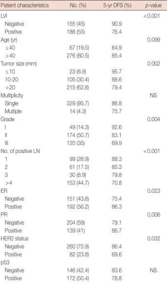
Nevertheless, although LVI is an important prognostic factor in patients with lymph node-negative invasive breast cancer, its prognostic value in patients with lymph node-positive breast cancer is unclear and controversial [3,9-11]. So, we report the analysis of the prognostic significance of LVI in a large cohort Table 3. Univariate analysis of disease-free survival and clinicopatholog- ical data
Patient characteristics No. (%) 5-yr DFS (%) p-value
LVI <0.001
Negative 155 (45) 90.9
Positive 188 (55) 76.4
Age (yr) 0.099
≤40 67 (19.5) 64.9
>40 276 (80.5) 85.4
Tumor size (mm) 0.002
≤10 23 (6.9) 95.7
10-20 105 (30.4) 88.6
>20 215 (62.8) 79.4
Multiplicity NS
Single 329 (95.7) 86.8
Multiple 14 (4.3) 75.7
Grade 0.004
I 49 (14.3) 92.6
II 174 (50.7) 83.1
III 120 (35) 69.9
No. of positive LN <0.001
1 99 (28.9) 88.3
2 61 (17.5) 85.3
3 30 (8.9) 79.8
>4 153 (44.7) 70.8
ER 0.023
Negative 151 (43.8) 75.4
Positive 192 (56.2) 86.3
PR 0.006
Negative 204 (59) 79.1
Positive 139 (41) 86.7
HER2 status 0.032
Negative 260 (75.9) 86.4
Positive 82 (23.8) 69.6
p53
Negative 146 (42.4) 83.6 NS
Positive 172 (50.4) 78.8
DFS=disease-free survival; LVI=lymphovascular invasion; NS=not significant;
LN=lymph node; ER=estrogen receptor; PR=progesterone receptor.
Cumulative metastasis-free survival
Time to metastasis
0.00 20.00 40.00 60.00 80.00
1.0
0.8
0.6
0.4
0.2
0.0
Figure 2. Metastasis-free survival of whole patients. Metastasis-free survival curves are shown according to presence or absence of lym- phovascular invasion (LVI).
Table 4. Disease-free survival: multivariate analysis in the whole group of patients
Patient characteristic RR 95% CI p-value
≥4 involved LN 2.737 1.352-5.539 0.005
Grade II 3.207 1.004-10.650 0.049
Grade III 4.127 1.253-13.587 0.020
LVI 2.594 1.384-4.862 0.003
RR=relative risk; CI=confidence interval; LN=lymph node; LVI=lymphovas- cular invasion.
Table 5. Disease-free survival: multivariate analysis in the hormone re- ceptor-positive group of patients
Patient characteristic RR 95% CI p-value
≥4 involved LN 2.688 1.064-6.792 0.037
Grade III 5.712 1.277-25.544 0.023
LVI 2.489 1.147-5.398 0.021
RR=relative risk; CI=confidence interval; LN=lymph node; LVI=lymphovas- cular invasion.
study of lymph node-positive patients with invasive breast cancer.
In our retrospective study, LVI was observed in 55% (n=192) of all patients (n=349), which was similar to other series of lymph node-positive cancer. LVI added prognostic informa- tion for patients with lymph node-positive breast cancer with a RR of 2.59 and a p-value of 0.003. These results contrast with those reported by Colleoni et al. [10] This discrepancy may be explained by the short-term follow-up period and analysis limitations in patients with 1 to 3 metastatic lymph nodes in their study.
In addition to LVI, our study demonstrated that high histol- ogical grade, number of lymph nodes involved, and both fac- tors constituting the Nottingham Prognostic Index were other significant independent clinicopathological factors. Because LVI appears to be independent from these pathological factors, our results propose that LVI should be considered in the prog- nostic index for patients with lymph node-positive breast can- cer as well as in the group of patients with lymph node-nega- tive breast cancer [15]. However, tumor size and patient age were not significant independent prognostic factors in our study. This result may be explained by the shorter follow-up period compared to that in previous studies.
We also assessed whether LVI had an effect on the hormone receptor-positive subgroup of patients as an independent prog- nostic factor. This analysis was restricted to the large subgroup of 218 patients who were hormone receptor-positive. In this subgroup, with a RR of 2.48 and a p-value of 0.021, LVI was an significant prognostic factor in patients who were hormone receptor-positive. These results suggest that LVI and other pathological factors may be useful to determine the need for adjuvant treatment in some patients for whom adjuvant che- motherapy could be dangerous despite axillary lymph node in- volvement. Eventually this may be applicable to the elderly or fragile patients (patients with severe underlying disease) with other treatment options such as endocrine and trastuzumab therapy.
Our analysis had three limitations. First, our analysis had a short follow-up period, and it was retrospective in nature. Sec- ond, not all patients with HER2-positive breast cancer were treated with a trastuzumab-based regimen. Third, LVI was as- sessed by H&E staining, and the selective endothelial cell mark- ers such as D2-40, CD34 were not used in the routine patho- logical evaluation, but use of such markers could potentially have improved the accuracy of detecting LVI [16].
In conclusion, this study emphasized the role of pathologic- almetastasis analysis in the prognostic evaluation of breast cancer, particularly those factors that cannot be subjected to molecular analyses, such as size, lymph node status, histologi-
cal grade, and LVI. These pathological factors are useful to evaluate prognostic and predictive factors for more effective and convenient clinical decision-making tools during adju- vant treatment.
The presence of LVI was an independent significant prog- nostic factor in patients with lymph node-positive breast can- cer as well as patients with lymph node-negative breast cancer.
The existence of LVI alone cannot be used to decide to omit adjuvant chemotherapy in all patients with lymph-node posi- tive breast cancer but, it may be considered in the adjuvant treatment decision in a specific subgroup of patients for whom chemotherapy is contraindicated. We suggest that patients with LVI-positive breast cancer require a shorter follow up and ad- ditional management.
CONFLICTS OF INTEREST
All authors declare no conflicts of interest.
REFERENCES
1. Clark GM. Prognostic and predictive factors. In: Harris JR, Lippman ME, Morrow M, Osborne CK, editors. Diseases of the Breast. 2nd ed.
Philadelphia: Lippincott Williams & Wilkins; 2000. p.489-514.
2. Woo CS, Silberman H, Nakamura SK, Ye W, Sposto R, Colburn W, et al.
Lymph node status combined with lymphovascular invasion creates a more powerful tool for predicting outcome in patients with invasive breast cancer. Am J Surg 2002;184:337-40.
3. Davis BW, Gelber R, Goldhirsch A, Hartmann WH, Hollaway L, Russell I, et al. Prognostic significance of peritumoral vessel invasion in clinical trials of adjuvant therapy for breast cancer with axillary lymph node metastasis. Hum Pathol 1985;16:1212-8.
4. Page DL, Anderson TJ, Connelly JL, Schnitt SF. Miscellaneous features of carcinoma. In: Page DL, Anderson TJ, editors. Diagnostic Histopa- thology of the Breast. Edinburgh: Churchill Livingstone; 1987. p.283-4.
5. Fisher B, Bauer M, Wickerham DL, Redmond CK, Fisher ER, Cruz AB, et al. Relation of number of positive axillary nodes to the prognosis of patients with primary breast cancer. An NSABP update. Cancer 1983;
52:1551-7.
6. Rosai J, Sobin LH, editors. Tumors of the Mammary Gland. Washing- ton, DC: Armed Forces Institute of Pathology; 1993.
7. de Mascarel I, MacGrogan G, Debled M, Sierankowski G, Brouste V, Mathoulin-Pthoulin- S, et al. D2-40 in breast cancer: should we detect more vascular emboli? Mod Pathol 2009;22:216-22.
8. de Mascarel I, Bonichon F, Durand M, Mauriac L, MacGrogan G, Soubeyran I, et al. Obvious peritumoral emboli: an elusive prognostic factor reappraised. Multivariate analysis of 1320 node-negative breast cancers. Eur J Cancer 1998;34:58-65.
9. Yildirim E, Berberoglu U. Lymph node ratio is more valuable than level III involvement for prediction of outcome in node-positive breast carcin- oma patients. World J Surg 2007;31:276-89.
10. Colleoni M, Rotmensz N, Maisonneuve P, Sonzogni A, Pruneri G, Casa-
dio C, et al. Prognostic role of the extent of peritumoral vascular invasion in operable breast cancer. Ann Oncol 2007;18:1632-40.
11. MacGrogan G, Desrousseaux M, de Mascarel I. Prognostic value of Mib1 in a tissue microarray of 855 invasive breast carcinomas. 5th European Breast Cancer Conference. 2006;4. Abstract #261.
12. Truong PT, Berthelet E, Lee J, Kader HA, Olivotto IA. The prognostic significance of the percentage of positive/dissected axillary lymph nodes in breast cancer recurrence and survival in patients with one to three positive axillary lymph nodes. Cancer 2005;103:2006-14.
13. Rosen PP. Tumor emboli in intramammary lymphatics in breast carci- noma: pathologic criteria for diagnosis and clinical significance. Pathol Annu 1983;18 Pt 2:215-32.
14. McCready DR, Chapman JA, Hanna WM, Kahn HJ, Murray D, Fish EB, et al. Factors affecting distant disease-free survival for primary inva- sive breast cancer: use of a log-normal survival model. Ann Surg Oncol 2000;7:416-26.
15. Mohammed RA, Martin SG, Gill MS, Green AR, Paish EC, Ellis IO.
Improved methods of detection of lymphovascular invasion demon- strate that it is the predominant method of vascular invasion in breast cancer and has important clinical consequences. Am J Surg Pathol 2007;
31:1825-33.
16. Sun Y, Goodison S, Li J, Liu L, Farmerie W. Improved breast cancer prog- nosis through the combination of clinical and genetic markers. Bioin- formatics 2007;23:30-7.