© 2015 Korean Breast Cancer Society. All rights reserved. http://ejbc.kr | pISSN 1738-6756
INTRODUCTION
Axillary lymph node (ALN) metastasis is one of the most significant prognostic factors for survival in breast cancer pa- tients. Sentinel lymph node dissection (SLND) has become the standard practice for nodal staging in patients with early- stage breast cancer. Several studies revealed that SLND accu- rately evaluated the status of ALN and was associated with de- creased morbidity than axillary lymph node dissection (ALND) in clinically node-negative patients [1-3].
Neoadjuvant chemotherapy (NAC) has become the stan- dard treatment in patients with inoperable locally advanced and large operable breast cancers. It is also widely used in pa-
tients with early invasive breast cancer. Rates of pathologic complete remission (pCR) vary from 16% to 20% according to the histologic subtypes of tumor and treatment modalities [4-6]. Achievement of pCR has been correlated with better disease-free survival and overall survival [7-9].
ALN metastases can be eradicated by NAC and the initial nodal stage will change substantially after NAC [10,11]. Re- sidual metastatic ALNs after NAC are important prognostic factor for disease-free survival [12,13], and lymph nodes in- volvement at the time of surgery, not the initial axillary node stage, is significantly associated with distant disease-free sur- vival [14].
Dynamic contrast-enhanced magnetic resonance imaging (MRI) is known to be the most accurate diagnostic tool for the assessment of tumor response after NAC [15,16]. On maximum intensity projection (MIP) images of MRI, it is eas- ier and more accurate to measure the longest dimension of primary tumor than mammography or ultrasound (US).
The purpose of our study was to find out any histologic fac-
Histologic Grade and Decrease in Tumor Dimensions Affect Axillary Lymph Node Status after Neoadjuvant Chemotherapy in Breast Cancer Patients
Tae Hee Kim, Doo Kyoung Kang, Ji Young Kim1, Sehwan Han1, Yongsik Jung1
Departments of Radiology and 1Surgery, Ajou University School of Medicine, Suwon, Korea ORIGINAL ARTICLE
Purpose: The purposes our study was to find out any histologic factors associated with negative conversion of axillary lymph node (ALN) after neoadjuvant chemotherapy (NAC). We also evaluated the association between the decrease in size of pri- mary breast tumor and negative conversion of ALN. Methods:
From January 2012 to November 2014, we included 133 breast cancer patients who underwent NAC and who had ALN metas- tases which were confirmed on fine-needle aspiration or core needle biopsy at initial diagnosis. All 133 patients underwent ini- tial magnetic resonance imaging (MRI) at the time of diagnosis and preoperative MRI after completion of NAC. We measured the longest dimension of primary breast cancer on MRI. Results:
Of 133 patients, 39 patients (29%) showed negative conversion of ALN and of these 39 patients, 25 patients (64%) showed pathologic complete remission of primary breast. On univariate analysis, mean percent decrease in longest dimension, estrogen receptor, progesterone receptor, human epidermal growth factor
receptor 2 status and histologic grade were significantly associ- ated with the ALN status after NAC (p<0.001, p=0.001, p<
0.001, p=0.001, p=0.002, respectively). On multivariate logistic regression analysis, percent decrease in longest dimension (odds ratio, 1.026; 95% confidence interval [CI], 1.009–1.044) and his- tologic grade (odds ratio, 3.964; 95% CI, 1.151–13.657) were identified as being independently associated with the ALN status after NAC. The area under the receiver operating characteristic curve was 0.835 with the best cutoff value of 80% decrease in longest dimension. Combination of high histologic grade and more than 80% decrease in longest dimension showed 64%
sensitivity and 92% specificity. Conclusion: High histologic grade and more than 80% decrease in primary tumor dimension were associated with negative conversion of ALN after NAC.
Key Words: Axillary lymph node, Breast neoplasms, Magnetic resonance imaging, Neoadjuvant chemotherapy
Correspondence to: Yongsik Jung
Department of Surgery, Ajou University School of Medicine, 164 Worldcup- ro, Yeongtong-gu, Suwon 16499, Korea
Tel: +82-31-219-5200, Fax: +82-31-219-5755 E-mail: [email protected]
Received: July 22, 2015 Accepted: November 4, 2015
Cancer
tors associated with negative conversion of ALN after NAC.
We also evaluated the association between the decrease of pri- mary breast tumor and negative conversion of ALN.
METHODS
Patients
Our Institutional Review Board approved this retrospective study (MED-MDB-14-476). From January 2012 to November 2014, 191 consecutive patients with breast cancer underwent NAC. Of 191 patients, 139 patients had ALN metastases which were confirmed on fine-needle aspiration or core nee- dle biopsy at initial diagnosis. We excluded three patients who did not undergo follow-up MRI, one patient who underwent vacuum assisted biopsy in the outside hospital before initial MRI, and two patients who had human epidermal growth factor receptor 2 (HER2)-positive cancer but did not receive trastuzumab treatment. Finally 133 patients were included for the analysis. Patient characteristics are summarized in Table 1.
ALND was performed in 127 patients regardless of SLN sta- tus. ALND was omitted in six patients who had converted to clinically negative ALN, near complete remission of primary
breast tumor on imaging after NAC and had confirmed nega- tive SLN on pathology.
All patients were scheduled for four cycles of adriamycin and cyclophosphamide (AC) regimen (AC, 50 mg/m2 and 500 mg/m2, respectively) followed by four cycles of paclitaxel 175 mg/m2 at a 3-week interval. Of 133 patients, 12 patients underwent the operation after four cycles of NAC because of complete remission of primary breast tumor on MRI (n=3), disease progression (n=2) or little response to NAC (n=7). All 30 patients who had HER2-positive cancer received trastu- zumab treatment.
MRI technique and interpretation
Patients underwent MR examinations three times, at the time of initial diagnosis, after four cycles of adriamycin and cyclophosphamide and after four cycles of paclitaxel. We used a 1.5-T MR system (Signa HDxt; General Electric Medical Systems, Milwaukee, USA) with a dedicated breast coil (8-channel HD breast array; General Electric Medical Sys- tems). An unenhanced coronal fast low-angle shot three-di- mensional T1-weighted image was acquired. Gadobutrol (Gadovist; Bayer Schering Pharma, Berlin, Germany) was in- jected into an antecubital vein at a dose of 0.1 mmol/kg of body weight and at a rate of 3 mL/sec, followed by a 20-mL saline flush for all patients. Subsequently, five consecutive contrast-enhanced series were acquired. The imaging param- eters were repetition time (msec)/echo time (msec) of 5.1/2.4, flip angle of 10°, field of view of 300×300 mm, image matrix of 300×300 pixels, section thickness of 1.5 mm, and section gap of 0 mm.
Subtraction images were reconstructed by subtracting the precontrast images from the early peak postcontrast image obtained at 60 seconds after contrast injection. MIP recon- structions were applied to the subtraction images. Two radiol- ogists with 11 and 7 years’ experiences interpreted MRI before and after NAC in consensus. The lesion size was measured as the longest diameter of the lesion on MIP image. In patients with multiple cancers, we recorded the sum of the longest dia- meter of each lesion.
Pathologic examination
All patients underwent core needle biopsy before surgery and surgical resection for breast cancer with sentinel lymph node biopsy (SLNB) and/or axillary lymph node dissection after NAC. The localization of primary tumor was performed by charcoal injection after four cycles of NAC. The routinely formalin-fixed, paraffin-embedded tissue blocks of tumors and ALNs were sectioned to 4 μm thickness and stained with hematoxylin and eosin.
Table 1. Patient and tumor characteristics (n=133)
Characteristic No. (%)
Age (yr)* 46.4±9.9
Tumor size (cm)* 3.8±2.2
T stage
T1 23 (17.3)
T2 76 (57.1)
T3 13 (9.8)
T4 21 (15.8)
Tumor histology
Invasive ductal 126 (94.7)
Invasive lobular 3 (2.3)
Others 4 (3.0)
Nuclear grade
Low 53 (49.5)
High 54 (50.5)
Histologic grade
Low 70 (64.8)
High 38 (35.2)
Estrogen receptor
Negative 40 (30.1)
Positive 93 (69.9)
Progesterone receptor
Negative 64 (48.1)
Positive 69 (51.9)
HER2
Negative 103 (77.4)
Positive 30 (22.6)
HER2=human epidermal growth factor receptor 2.
*Mean±SD.
The specimens of core needle biopsy were evaluated accord- ing to the following histopathologic features: histological type of carcinoma, Black nuclear grade (nuclear grade 1, poorly differentiated; grade 2, moderately differentiated; and grade 3, well differentiated), and modified Bloom-Richardson histol- ogical grade (histological grade 1, well differentiated; grade 2, moderately differentiated; and grade 3, poorly differentiated).
For dichotomous-dependent variables, nuclear grade was clas- sified as high (grade 1) versus low (grades 2 and 3) and histologic grade as low (grade 1 and 2) versus high (grade 3). Expression of estrogen receptor (ER), progesterone receptor (PR), and HER2 was evaluated using standard avidin-biotin complex immunohistochemical staining methods. The ER and PR sta- tus were assessed using the Allred score, which was expressed as the sum of the proportion score and the intensity score of positively stained tumor cells. Tumors with an Allred score of at least 3 were regarded as positive. The intensity of HER2 staining was scored as 0, 1+, 2+, or 3+. Tumors with a 3+ score were classified as HER2 positive, and tumors with a 0 or 1+
score were classified as negative. In tumors with a 2+ score, gene amplification by using fluorescence in situ hybridization was used to determine HER2 status.
After completion of NAC, the size and extent of residual cancer were measured. The pCR was defined as the complete disappearance of invasive carcinoma in the breast. Residual ductal carcinoma in situ (DCIS) was included in the pCR cat- egory. All specimens were reviewed by an experienced pa- thologist with 16 years of experience.
Statistical analysis
The mean values of initial tumor size, percent decrease in longest dimension and duration of NAC were compared us- ing two-sample t-test. Univariate analysis was performed by using chi-square and Fisher exact test for the evaluation of re- lationships between ALN status with clinical and histopatho- logic factors. Multivariate analysis was performed using logis- tical regression of the variables that were found to be statisti- cally significant on univariate analyses. Analyses were per- formed using the SPSS version 19.0 statistical software pack- age (IBM Corp., Armonk, USA), with a value of p<0.05 con- sidered to be significant.
For the evaluation of diagnostic performance of percent de- crease in longest dimension, we used receiver operating char- acteristic (ROC) analysis. Diagnostic accuracy was calculated from the area under the ROC curve (AUC). The best cutoff was determined from the ROC analysis and we used the best cutoff values to calculate the sensitivity, specificity, positive predictive value (PPV), and negative predictive value (NPV) for predicting negative conversion of ALN. We used MedCalc
software (version 10.4.8; MedCalc Software, Ostend, Belgium) for ROC analysis.
RESULTS
Of 133 patients, 39 patients (29%) showed negative conver- sion of ALN. Of these 39 patients, 25 patients (64%) showed pCR of primary breast tumor and five patients (13%) had re- sidual tumor less than 5 mm.
Univariate analysis was performed for the evaluation of re- lationships between the negative conversion of ALN and clini- cal and histological factors (Table 2). Mean percent decrease in longest dimension was significantly higher in patients with negative conversion compared to patients with residual meta- static ALN (82% vs. 37%, p<0.001). There were no significant differences in mean initial tumor size, mean duration of NAC treatment and cycles of NAC (p=0.145, p=0.510, and p=
0.508, respectively). ER, PR, HER2 status and histologic grade were significantly associated with the ALN status after NAC (p=0.001, p<0.001, p=0.001, p=0.002, respectively). Nucle- ar grade was not significantly different between two groups (p=0.222).
Table 2. Association of axillary lymph node status after neoadjuvant chemotherapy with clinical and histopathologic prognostic factors
Pathologic factor
Negative ALN (n=39) No. (%)
Positive ALN (n=96)
No. (%) p-value Initial tumor size (cm)* 4.2±2.22 3.62±2.22 0.145 Decrease in longest
dimension (%)* 81.79±35.57 36.66±31.11 <0.001 Duration of NAC (mo)* 5.95±1.52 5.71±2.00 0.510
NAC cycles 0.508
4 cycles 2 (5.1) 10 (10.6)
8 cycles 37 (94.9) 84 (89.4)
Estrogen receptor 0.001
Negative 20 (51.3) 20 (21.3)
Positive 19 (48.7) 74 (78.7)
Progesterone receptor <0.001
Negative 29 (74.4) 35 (37.2)
Positive 10 (25.6) 59 (62.8)
HER2 0.001
Negative 23 (59.0) 80 (85.1)
Positive 16 (41.0) 14 (14.9)
Nuclear grade 0.222
Low 7 (36.8) 46 (52.3)
High 12 (63.2) 42 (47.7)
Histologic grade 0.002
Low 7 (35.0) 63 (71.6)
High 13 (65.0) 25 (28.4)
ALN=axillary lymph node; NAC=neoadjuvant chemotherapy; HER2=human epidermal growth factor receptor 2.
*Mean±SD.
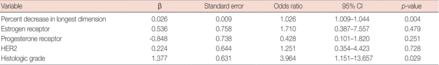
Multivariate logistic regression analysis was performed with the variables associated with axillary lymph node status through univariate analysis (Table 3). Percent decrease in lon- gest dimension (odds ratio, 1.026; 95% confidence interval [CI], 1.009–1.044) and histologic grade (odds ratio, 3.964;
95% CI, 1.151–13.657) were identified as being independently associated with the ALN status after NAC. ER, PR, and HER2 were not significant independent factor for ALN status after NAC.
A ROC analysis was performed to differentiate negative conversion group from the residual metastasis group by using percent decrease in longest dimension of primary breast tu- mor (Figure 1). The AUC was 0.835. The best cutoff for differ- entiating negative conversion group from residual metastatic ALN group was 80% decrease in longest dimension. With this cutoff, percent diameter decrease showed 89.4% sensitivity, 79.5% specificity, 91.2% PPV, and 73.8% NPV.
Diagnostic performance of combination of high histologic
grade or more than 80% decrease in longest dimension was assessed for the diagnosis of metastatic ALN after NAC. The sensitivity, specificity, PPV and NPV were 64% (60/94), 92%
(36/39), 95% (60/63), and 100% (36/36), respectively.
DISCUSSION
Lymph node status after NAC have known to be an impor- tant prognostic factor for disease-free survival [12-14]. In a recent prospective multicenter study [17], detection rate of SLNB was 80.1% and the false-negative rate was 14.2% in pa- tients with lymph node conversion after NAC. The false-nega- tive rate of SLNB was 12.6%, if two or more sentinel lymph nodes were removed and dual agents were used, in patients with breast cancer with clinical N1 stage receiving NAC in ACOSOG Z1071 trial [18].
However, the detection rate of SLNB after NAC is limited by the effects of NAC, including anatomical alterations or dis- ruptions of lymphatic vessels by tumors, inflammation or fibrosis, blockage by necrotic and/or apoptotic cells or induc- tion of nonuniform tumor regression among ALNs [19,20].
In the study of Park et al. [21], patients with poor tumor or nodal response to NAC had higher SLN detection failure rates. They suggested that disruption or blockage of lymphatic pathways by residual tumors might affect the detection rate of radioisotope-based SLNB. Thus, accurate prediction of ALN status before surgery is important because it could help sur- geons to proceed directly to ALND or to do SLNB without ALND.
In a recent study analyzing the diagnostic performance of US, MRI, and F-18 fluorodeoxyglucose positron emission tomo- graphy/computed tomography (FDG PET/CT) for metastatic lymph node after NAC, the sensitivity was 70%, 61%, and 63% and the specificity was 58%, 59%, and 85%, respectively [22]. In a recent review article, the sensitivity of US, MRI, and PET/CT was 58%–86%, 59%, and 48–85%, respectively for the detection of pathologic complete remission of ALN [23].
Previous result of our institution revealed that the sensitivity of US, MRI, and PET/CT was 50%, 72%, and 22% and the specificity of US, MRI, and PET/CT was 77%, 54%, and 85%, Table 3. Logistic regression analysis for variables associated with negative conversion of axillary lymph node
Variable β Standard error Odds ratio 95% CI p-value
Percent decrease in longest dimension 0.026 0.009 1.026 1.009–1.044 0.004
Estrogen receptor 0.536 0.758 1.710 0.387–7.557 0.479
Progesterone receptor -0.848 0.738 0.428 0.101–1.820 0.251
HER2 0.224 0.644 1.251 0.354–4.423 0.728
Histologic grade 1.377 0.631 3.964 1.151–13.657 0.029
CI=confidence interval; HER2=human epidermal growth factor receptor 2.
Figure 1. Receiver operating characteristic (ROC) analysis in the differ- entiation of negative conversion group and residual metastasis group.
Area under the ROC curve of percent decrease in longest dimension is 0.835. The best cutoff value is 80% decrease in longest dimension.
100
80
60
40
20
0
0 20 40 60 80 100
100-Specificity (%) Percent decrease in longest dimension
Sensitivity (%)
respectively [24].
Add of axillary ultrasound (AUS) to SLNB could reduce the false-negative rate from 12.6% to 9.8% in ACOSOG Z1071 trial [25]. In this study, patients with suspicious LNs on AUS had a greater number of positive LNs and larger size of metas- tasis in ALN and the negative predictive value of AUS was 43% (145/334). In our study, by using the combination of high histologic grade or more than 80% decrease in longest dimen- sion, the PPV and NPV were very high, 95% and 100% for the diagnosis of metastatic ALN. Thus, high PPV could be useful in routine practice for surgeons to proceed to ALND without SLNB and high NPV for surgeons to do only SLNB without ALND.
There are several studies reporting the association of ALN status and the pathologic primary tumor response to NAC. In the study of Nagashima et al. [26], response rate of primary breast tumor was correlated with that of lymph node and these were well correlated with disease-free survival. However, tumor size, histological grade and HER2 were not correlated with patient outcome. In the study of Rouzier et al. [14], high histologic grade and more than 50% response to chemother- apy were associated with negative conversion of ALN after NAC. Our results also showed that high histologic grade and better response of primary breast tumor were associated with negative conversion of ALN.
In the study of Kuerer et al. [27], of 30 patients with pCR of primary breast tumor, 19 patients (63%) had negative ALN at dissection. Thirteen patients (33%) of 40 who had near pCR (residual cancer ≤1 cm3) and 15 patients (17%) of 86 who had residual cancer larger than 1 cm3 showed negative ALN at dissection. Our results also revealed that mean percent de- crease in longest dimension was significantly higher in pa- tients with negative conversion compared to patients with re- sidual metastatic ALN (82% vs. 37%, p<0.001). With 80%
cutoff value, percent diameter decrease showed 89.4% sensi- tivity, 79.5% specificity, 91.2% PPV, and 73.8% NPV for the diagnosis of metastatic ALN after NAC.
There are several limitations in our study. First, this was a retrospective study from a single center and the total number of patients was relatively small. Larger multicenter study with more patients is needed to validate our results. Second, we did not evaluate the recurrence of ALN after surgery because of relatively short duration of follow-up. The follow-up study of ALN recurrence is needed for better understanding of cancer biology after NAC.
In conclusion, high histologic grade and more than 80%
decrease in primary tumor dimension were associated with negative conversion of ALN after NAC.
CONFLICT OF INTEREST
The authors declare that they have no competing interests.
REFERENCES
1. Mansel RE, Fallowfield L, Kissin M, Goyal A, Newcombe RG, Dixon JM, et al. Randomized multicenter trial of sentinel node biopsy versus standard axillary treatment in operable breast cancer: the ALMANAC Trial. J Natl Cancer Inst 2006;98:599-609.
2. Cox CE, Bass SS, Ku NN, Berman C, Shons AR, Yeatman TJ, et al.
Sentinel lymphadenectomy: a safe answer to less axillary surgery?
Recent Results Cancer Res 1998;152:170-9.
3. Lucci A, McCall LM, Beitsch PD, Whitworth PW, Reintgen DS, Blumencranz PW, et al. Surgical complications associated with sentinel lymph node dissection (SLND) plus axillary lymph node dissection compared with SLND alone in the American College of Surgeons On- cology Group Trial Z0011. J Clin Oncol 2007;25:3657-63.
4. Ju NR, Jeffe DB, Keune J, Aft R. Patient and tumor characteristics asso- ciated with breast cancer recurrence after complete pathological re- sponse to neoadjuvant chemotherapy. Breast Cancer Res Treat 2013;
137:195-201.
5. Gonzalez-Angulo AM, McGuire SE, Buchholz TA, Tucker SL, Kuerer HM, Rouzier R, et al. Factors predictive of distant metastases in patients with breast cancer who have a pathologic complete response after neo- adjuvant chemotherapy. J Clin Oncol 2005;23:7098-104.
6. Tanioka M, Shimizu C, Yonemori K, Yoshimura K, Tamura K, Kouno T, et al. Predictors of recurrence in breast cancer patients with a pathologic complete response after neoadjuvant chemotherapy. Br J Cancer 2010;
103:297-302.
7. Bear HD, Anderson S, Brown A, Smith R, Mamounas EP, Fisher B, et al. The effect on tumor response of adding sequential preoperative docetaxel to preoperative doxorubicin and cyclophosphamide: prelimi- nary results from National Surgical Adjuvant Breast and Bowel Project Protocol B-27. J Clin Oncol 2003;21:4165-74.
8. Kaufmann M, von Minckwitz G, Smith R, Valero V, Gianni L, Eiermann W, et al. International expert panel on the use of primary (preoperative) systemic treatment of operable breast cancer: review and recommenda- tions. J Clin Oncol 2003;21:2600-8.
9. Smith IC, Heys SD, Hutcheon AW, Miller ID, Payne S, Gilbert FJ, et al.
Neoadjuvant chemotherapy in breast cancer: significantly enhanced re- sponse with docetaxel. J Clin Oncol 2002;20:1456-66.
10. Bear HD, Anderson S, Smith RE, Geyer CE Jr, Mamounas EP, Fisher B, et al. Sequential preoperative or postoperative docetaxel added to pre- operative doxorubicin plus cyclophosphamide for operable breast can- cer: National Surgical Adjuvant Breast and Bowel Project Protocol B-27. J Clin Oncol 2006;24:2019-27.
11. Zhang GC, Liao N, Guo ZB, Qian XK, Ren CY, Yao M, et al. Accuracy and axilla sparing potentials of sentinel lymph node biopsy with methy- lene blue alone performed before versus after neoadjuvant chemother- apy in breast cancer: a single institution experience. Clin Transl Oncol 2013;15:79-84.
12. Kuerer HM, Newman LA, Buzdar AU, Hunt KK, Dhingra K, Buchholz TA, et al. Residual metastatic axillary lymph nodes following neoadju-
vant chemotherapy predict disease-free survival in patients with locally advanced breast cancer. Am J Surg 1998;176:502-9.
13. von Minckwitz G, Untch M, Blohmer JU, Costa SD, Eidtmann H, Fasching PA, et al. Definition and impact of pathologic complete response on prognosis after neoadjuvant chemotherapy in various intrinsic breast cancer subtypes. J Clin Oncol 2012;30:1796-804.
14. Rouzier R, Extra JM, Klijanienko J, Falcou MC, Asselain B, Vincent- Salomon A, et al. Incidence and prognostic significance of complete axillary downstaging after primary chemotherapy in breast cancer patients with T1 to T3 tumors and cytologically proven axillary met- astatic lymph nodes. J Clin Oncol 2002;20:1304-10.
15. Balu-Maestro C, Chapellier C, Bleuse A, Chanalet I, Chauvel C, Largil- lier R. Imaging in evaluation of response to neoadjuvant breast cancer treatment benefits of MRI. Breast Cancer Res Treat 2002;72:145-52.
16. Rosen EL, Blackwell KL, Baker JA, Soo MS, Bentley RC, Yu D, et al. Ac- curacy of MRI in the detection of residual breast cancer after neoadju- vant chemotherapy. AJR Am J Roentgenol 2003;181:1275-82.
17. Kuehn T, Bauerfeind I, Fehm T, Fleige B, Hausschild M, Helms G, et al.
Sentinel-lymph-node biopsy in patients with breast cancer before and after neoadjuvant chemotherapy (SENTINA): a prospective, multicen- tre cohort study. Lancet Oncol 2013;14:609-18.
18. Boughey JC, Suman VJ, Mittendorf EA, Ahrendt GM, Wilke LG, Taback B, et al. Sentinel lymph node surgery after neoadjuvant chemo- therapy in patients with node-positive breast cancer: the ACOSOG Z1071 (Alliance) clinical trial. JAMA 2013;310:1455-61.
19. Charfare H, Limongelli S, Purushotham AD. Neoadjuvant chemother- apy in breast cancer. Br J Surg 2005;92:14-23.
20. Brown AS, Hunt KK, Shen J, Huo L, Babiera GV, Ross MI, et al. Histo- logic changes associated with false-negative sentinel lymph nodes after preoperative chemotherapy in patients with confirmed lymph node-
positive breast cancer before treatment. Cancer 2010;116:2878-83.
21. Park S, Park JM, Cho JH, Park HS, Kim SI, Park BW. Sentinel lymph node biopsy after neoadjuvant chemotherapy in patients with cytologi- cally proven node-positive breast cancer at diagnosis. Ann Surg Oncol 2013;20:2858-65.
22. Hieken TJ, Boughey JC, Jones KN, Shah SS, Glazebrook KN. Imaging response and residual metastatic axillary lymph node disease after neo- adjuvant chemotherapy for primary breast cancer. Ann Surg Oncol 2013;20:3199-204.
23. Schipper RJ, Moossdorff M, Beets-Tan RG, Smidt ML, Lobbes MB.
Noninvasive nodal restaging in clinically node positive breast cancer patients after neoadjuvant systemic therapy: a systematic review. Eur J Radiol 2015;84:41-7.
24. You S, Kang DK, Jung YS, An YS, Jeon GS, Kim TH. Evaluation of lymph node status after neoadjuvant chemotherapy in breast cancer patients: comparison of diagnostic performance of ultrasound, MRI and (18)F-FDG PET/CT. Br J Radiol 2015;88:20150143.
25. Boughey JC, Ballman KV, Hunt KK, McCall LM, Mittendorf EA, Ahrendt GM, et al. Axillary ultrasound after neoadjuvant chemothera- py and its impact on sentinel lymph node surgery: results from the American College of Surgeons Oncology Group Z1071 Trial (Alliance).
J Clin Oncol 2015;33:3386-93.
26. Nagashima T, Sakakibara M, Kadowaki M, Suzuki TH, Yokomizo J, Ohki Y, et al. Response rate to neoadjuvant chemotherapy measured on imaging predicts early recurrence and death in breast cancer patients with lymph node involvements. Acta Radiol 2011;52:241-6.
27. Kuerer HM, Newman LA, Buzdar AU, Dhingra K, Hunt KK, Buchholz TA, et al. Pathologic tumor response in the breast following neoadju- vant chemotherapy predicts axillary lymph node status. Cancer J Sci Am 1998;4:230-6.