© 2013 Korean Breast Cancer Society. All rights reserved. http://ejbc.kr | pISSN 1738-6756
INTRODUCTION
Breast cancer is the second most common cancer among women in Korea. Approximately 14,000 new cases of breast cancer occur each year in Korea and an estimated 240,000 oc- cur each year in the United States. Most breast cancers are in- vasive ductal carcinomas (IDC), ‘not otherwise specified’
(NOS), and medullary breast carcinoma (MBC) is a rare his- tological type of breast cancer, accounting for 1% to 7% of all
breast cancers [1-4].
In 1945, Moore and Foote described MBC as a unique his- tological subtype, characterized by high-grade structure with heavy lymphocytic infiltration, sharp circumscription, hem- orrhagic necrosis, and a relatively favorable prognosis [5,6].
Some researchers, however, have questioned the prognosis and morphologic definition of MBC. Ridolfi et al. [7] reas- sessed MBC and documented strict criteria for diagnosing typical and atypical MBC. Diagnostic criteria for typical MBC include a predominantly syncytial growth pattern in more than 75% of the tumor area, circumscription with a pushing margin, moderate to marked lymphoplasmacytic infiltration, poorly differentiated nuclear grade with a high mitotic rate, scant stroma, and absence of an intraductal component.
Atypical MBC differs from typical MBC in terms of patholog- ic features with an infiltrative margin, mild mononuclear infil- tration, a low nuclear grade, and presence of an intraductal component [8].
Comparison of the Characteristics of Medullary Breast Carcinoma and Invasive Ductal Carcinoma
Inhye Park*, Jiyoung Kim*, Minkuk Kim, Soo Youn Bae, Se Kyung Lee, Won Ho Kil, Jeong Eon Lee, Seok Jin Nam
Division of Breast and Endocrine Surgery, Department of Surgery, Samsung Medical Center, Sungkyunkwan University School of Medicine, Seoul, Korea ORIGINAL ARTICLE
Purpose: Medullary breast carcinomas (MBC) have been known to represent a rare breast cancer subtype associated with a more favorable prognosis than invasive ductal carcinomas (IDC). The purpose of this study was to compare the clinicopathologic char- acteristics and outcomes of MBC with those of IDC. Methods: We retrospectively reviewed medical records of patients with invasive breast cancer who were managed surgically from August 1995 to June 2010. Results: Fifty-two patients were identified with MBC and 5,716 patients were identified with IDC. The clinicopathologic features, disease-free survival (DFS), and overall survival (OS) of patients with MBC were compared with those of patients with IDC. The MBC group presented at a younger age (p=0.005) and had a significant association with a higher histological grade (p=
0.003) and nuclear grade (p<0.001) as well as negative estrogen receptor (p<0.001) and progesterone receptor (p<0.001) status.
Lymphatic invasion was absent (p<0.001) and lymph node me- tastasis was rare (p<0.001). The DFS and OS did not differ sig- nificantly between the two groups (5-year DFS: 88.0% vs. 89.2%,
p=0.920; 5-year OS: 93.4% vs. 94.4%, p=0.503). In multivariate analysis, the factors associated with DFS and OS were nuclear grade, histological grade, tumor size, lymph node metastasis, es- trogen receptor status, progesterone receptor status, and human epidermal growth factor receptor 2 status, chemotherapy, and hormone therapy. However, DFS and OS were not significantly different between IDC and MBC according to histological type it- self (DFS: hazard ratio 0.85, 95% confidence interval 0.12-6.05, p=0.866; OS: hazard ratio 1.49, 95% confidence interval 0.21- 10.77, p=0.692). Conclusion: Although MBC has specific clinico- pathologic features, its prognosis does not differ from IDC and is determined by prognostic factors such as tumor size and lymph node metastasis. Therefore, patients with MBC also require the same intensive treatment provided for IDC.
Key Words: Breast neoplasms, Lymphatic metastasis, Medullary carcinoma, Prognosis
Correspondence to: Jeong Eon Lee
Division of Breast and Endocrine Surgery, Department of Surgery, Samsung Medical Center, Sungkyunkwan University School of Medicine, 81 Irwon-ro, Gangnam-gu, Seoul 135-710, Korea
Tel: +82-2-3410-0926, Fax: +82-2-3410-6982 E-mail: [email protected]
*These authors contributed equally to this work.
Received: June 1, 2013 Accepted: October 10, 2013
Cancer
Many previous studies have indicated that this histological type is associated with a favorable prognosis despite the pres- ence of aggressive features such as large tumor size, a high nu- clear grade, and hormone receptor (HR) negativity, which are usually accepted as poor prognostic factors [3,9,10]. The prog- nosis of MBC is still under dispute. Some studies have report- ed that MBC survival rates do not differ from those of IDC [11-13]. The clinicopathologic features of MBC have various implications that dictate not only diagnosis but also therapeu- tic approaches. The aim of this study was to compare the clini- copathologic characteristics of MBC with those of IDC and to clarify the prognosis of patients with MBC.
METHODS
We retrospectively reviewed the medical records of patients with invasive breast cancer who were surgically managed at the Breast Cancer Center of Samsung Medical Center, Seoul, Korea from August 1995 to June 2010. Patients with distant metastasis at diagnosis, those with ductal carcinoma in situ, or patients who received neoadjuvant chemotherapy were ex- cluded. We also excluded patients with occult breast cancer presenting with axillary node metastasis, and patients with a history of ipsilateral or contralateral breast cancer.
In this study, MBC was defined according to the strict crite- ria of Ridolfi et al. [7], and only typical MBC was diagnosed as MBC. In our institution, atypical MBC is classified as IDC (NOS) in accordance with the literature indicating that the clinical course of atypical MBC is similar to that of IDC [14,15]. A total of 52 patients with MBC and 5,716 patients with IDC were included in this study. The authors retrospec- tively reviewed clinicopathologic features including tumor size; lymph node (LN) metastasis; pathologic stage; nuclear and histological grade with immunohistochemical findings, such as estrogen receptor (ER), progesterone receptor (PR), and human epidermal growth factor receptor 2 (HER2) sta- tus; treatment modalities (operation, adjuvant chemotherapy, radiotherapy, and hormone therapy); and recurrence or sur- vival status.
Tumor stage was assessed according to the American Joint Committee on Cancer (AJCC) 7th staging system [16]. The histological grade was determined according to the Bloom- Richardson classification [17]. The Allred score system (range from 0 to 8) was used to evaluate ER and PR status, and a total score of >2 was defined as positive. An HER2 score of 3+ or 2+ with gene amplification determined by fluorescence/silver in situ hybridization was considered positive. Disease-free sur- vival (DFS) was defined as no local or systemic recurrence and no newly developed contralateral breast cancer. The DFS
and overall survival (OS) of patients with MBC were com- pared with those of patients with IDC.
This study was approved by the Institutional Review Board of Samsung Medical Center (SMC 2013-07-090).
Statistical analysis
Statistical analyses were performed using PASW statistics 18 software (SPSS Inc., Chicago, USA). Student t-test and the chi-square test (or Fisher exact test when appropriate) were used to compare the clinicopathologic characteristics of the MBC and IDC groups. The Kaplan-Meier method was used to determine DFS and OS rates, and survival curves were compared using log-rank tests. Multivariate analysis was per- formed using Cox proportional hazards model to yield the relative hazards of multiple variables, including histological type, on breast cancer recurrence or death. All p-values are two-tailed, and a p-value of <0.05 was considered statistically significant.
RESULTS
A total of 7,848 patients with primary invasive breast cancer were treated surgically at our institution during the study peri- od. Among them, 6,828 patients (87.0%) were diagnosed with IDC-NOS, and 52 patients (0.7%) had MBC. A total of 5,716 IDC-NOS cases and 52 MBC cases met the eligibility criteria and were included in this study. The median follow-up periods were 53.5 months (range, 0.3-182.8 months) for IDC and 88.6 months (range, 0.9-194.6 months) for MBC. MBC patients were younger than IDC patients (mean age at diagnosis±
standard deviation 44±9 vs. 48±10, p=0.005) (Table 1). All surgeries were classified as mastectomy or breast-conserving surgery (BCS), regardless of axillary node dissection. There was no significant difference in the operative method (p=
0.341). More women in the MBC group were premenopausal (IDC 64.9% vs. MBC 74.5%, p=0.280).
The mean MBC tumor size was 2.25±1.47 cm and was not different from the mean IDC tumor size (2.37±1.21 cm, p=
0.536). All MBCs manifested as a single lesion, whereas 16.9%
of IDC had multiple foci (p<0.001). Interestingly, MBC had distinct features, such as no lymphovascular invasion (LVI) (IDC 27.8% vs. MBC 0.0%, p<0.001) and rare LN metastasis (N0 IDC 58.4% vs. MBC 86.5%, p<0.001). The average num- bers of metastatic LNs were 0.2 in the MBC group and 1.9 in the IDC group (p<0.001). The MBC group had a significantly higher proportion of patients with a poor histological grade (grade III IDC 38.3% vs. MBC 80.0%, p=0.003) and higher nuclear grade (high grade IDC 41.7% vs. MBC 82.8%, p<
0.001) as well as negative ER (IDC 31.0% vs. MBC 84.8%,
Table 1. Clinicopathologic characteristics of patients with medullary breast carcinoma and invasive ductal carcinoma Characteristic MBC (n=52)
No. of cases (%)
IDC (n=5,716)
No. of cases (%) p-value Characteristic MBC (n=52) No. of cases (%)
IDC (n=5,716)
No. of cases (%) p-value
Mean age (yr)* 44±9 48±10 0.005 N stage <0.001
Menopause 0.280 N0 45 (86.5) 3,341 (58.4)
Yes 13 (25.5) 1,981 (35.1) N1 7 (13.5) 1,560 (27.3)
No 38 (74.5) 3,656 (64.9) N2 0 512 (9.0)
FHx 0.084 N3 0 302 (5.3)
Yes 7 (13.5) 412 (7.2) Stage 0.027
No 45 (86.5) 5,304 (92.8) I 26 (50.0) 2,240 (39.2)
Surgery 0.341 IIa 19 (36.5) 1,815 (31.8)
Mastectomy 18 (34.6) 2,352 (41.1) IIb 7 (13.5) 774 (13.5)
BCS 34 (65.4) 3,364 (58.7) III 0 887 (15.6)
Tumor size (cm)* 2.25±1.47 2.37±1.21 0.536 ER <0.001
Mean no. of metastatic lymph node (range)
0.2 (0-2) 1.9 (0-49) <0.001 (+) (-)
7 (15.2) 39 (84.8)
3,896 (69.0) 1,754 (31.0)
Multiplicity 0.001 PR <0.001
Yes 0 956 (16.9) (+) 4 (8.7) 3,457 (61.2)
No 48 (100) 4,716 (83.1) (-) 42 (91.3) 2,188 (38.8)
LVI <0.001 HER2 0.293
Yes 0 1,579 (27.8) (+) 15 (37.5) 1,516 (28.9)
No 50 (100) 4,094 (72.2) (-) 25 (62.5) 3,721 (71.1)
NG <0.001 Molecular subtype <0.001
Low 0 701 (12.6) HR+/HER2- 4 (10.0) 2,839 (54.2)
Intermediate 5 (17.2) 2,549 (45.8) HR+/HER2+ 2 (5.0) 848 (16.2)
High 24 (82.8) 2,320 (41.7) HR-/HER2+ 13 (32.5) 668 (12.8)
HG 0.003 HR-/HER2- 21 (52.5) 882 (16.8)
I 0 1,088 (20.0) Radiotherapy 1.000
II 3 (20.0) 2,278 (41.8) Yes 34 (68.0) 3,807 (68.6)
III 12 (80.0) 2,087 (38.3) No 16 (32.0) 1,740 (31.4)
T stage 0.918 Chemotherapy 0.469
T1 28 (53.8) 3,200 (56.0) Yes 43 (86.0) 4,519 (81.0)
T2 22 (42.3) 2,276 (39.8) No 7 (14.0) 1,063 (19.0)
T3 2 (3.8) 226 (4.0) Hormone therapy <0.001
T4 0 13 (0.2) Yes 6 (12.0) 4,041 (72.8)
No 45 (88.0) 1,508 (27.2)
MBC=medullary breast carcinoma; IDC=invasive ductal carcinoma; FHx=family history; BCS=breast-conserving surgery; LVI=lymphovascular in- vasion; NG=nuclear grade; HG=histological grade; ER=estrogen receptor; PR=progesterone receptor; HER2=human epidermal growth factor re- ceptor 2; HR=hormone receptor.
*Mean±SD.
A B C
Disease-free survival
Months
0 12 24 36 48 60 72 84 96 108 120 Log rank p=0.920
Histology IDC (n=5,716) MBC (n=52)
Total 1.0
0.8
0.6
0.4
0.2
0.0
Disease-free survival
Months
0 12 24 36 48 60 72 84 96 108 120 Log rank p=0.193
Histology IDC (n=2,237) MBC (n=26)
Stage I 1.0
0.8
0.6
0.4
0.2
0.0
Disease-free survival
Months
0 12 24 36 48 60 72 84 96 108 120 Log rank p=0.853
Histology IDC (n=2,587) MBC (n=26)
Stage II 1.0
0.8
0.6
0.4
0.2
0.0
Figure 1. Disease-free survival curve for patients with medullary breast carcinoma (MBC) and invasive ductal carcinoma (IDC) according to the 7th American Joint Committee on Cancer stage system. (A) Total, (B) stage I, (C) stage II.
p<0.001) and PR (IDC 38.8% vs. MBC 91.3%, p<0.001) sta- tuses which are generally regarded as poor prognostic factors of invasive breast cancer. HER2 status did not differ signifi- cantly between the two histological groups (IDC 28.9% vs.
MBC 37.5%, p=0.293). When patients were categorized ac- cording to molecular subtype, the MBC group predominantly had HR-/HER2+ tumors or triple-negative breast cancer (p<
0.001).
There was no difference in the rates of chemotherapy be- tween groups. More patients with IDC received hormone therapy than did patients with MBC (IDC 72.8% vs. MBC 12.0%, p<0.001) owing to HR status.
Kaplan-Meier curves showed that IDC and MBC had simi- lar DFS and OS rates (5-year DFS 88.0% vs. 89.2%, p=0.920;
5-year OS 93.4% vs. 94.4%, p=0.503) (Figures 1 and 2). We compared DFS and OS between histological subtypes of same
A B C
Overall survival
Months
0 12 24 36 48 60 72 84 96 108 120 Log rank p=0.503
Histology IDC (n=5,716) MBC (n=52)
Total 1.0
0.8
0.6
0.4
0.2
0.0
Overall survival
Months
0 12 24 36 48 60 72 84 96 108 120 Log rank p=0.684
Histology IDC (n=2,237) MBC (n=26)
Stage I 1.0
0.8
0.6
0.4
0.2
0.0
Overall survival
Months
0 12 24 36 48 60 72 84 96 108 120 Log rank p=0.815
Histology IDC (n=2,587) MBC (n=26)
Stage II 1.0
0.8
0.6
0.4
0.2
0.0
Figure 2. Overall survival curve for patients with medullary breast carcinoma (MBC) and invasive ductal carcinoma (IDC) according to the 7th Ameri- can Joint Committee on Cancer stage system. (A) Total, (B) stage I, (C) stage II.
Table 2. Comparison of disease-free survival and overall survival between medullary breast carcinoma and invasive ductal carcinoma after subcate- gorization (univariate analysis)
Subcategory
DFS OS
Subcategory
DFS OS
Events No.
Hazard
ratio p-value Events No.
Hazard
ratio p-value Events
No.
Hazard
ratio p-value Events No.
Hazard ratio p-value
Nodal status* Chemotherapy§
Negative 0.909 0.258 Yes 0.600 0.465
IDC (n=3,339) 249 Ref 135 Ref IDC (n=4,517) 564 Ref 307 Ref
MBC (n=45) 5 1.05 1 0.32 MBC (n=43) 6 0.81 3 0.66
Positive 0.210 0.037 No 0.158 0.801
IDC (n=2,372) 394 Ref 232 Ref IDC (n=1,061) 73 Ref 50 Ref
MBC (n=7) 3 2.04 3 3.16 MBC (n=7) 2 2.66 1 1.29
HR status† RadiotherapyII
Positive 0.556 Yes 0.659 0.824
IDC (n=3,996) 376 Ref 203 IDC (n=3,804) 401 Ref 217 Ref
MBC (n=8) 2 1.52 0 MBC (n=34) 6 1.20 3 0.88
Negative 0.275 0.475 No 0.452 0.495
IDC (n=1,650) 247 Ref 141 Ref IDC (n=1,739) 233 Ref 135 Ref
MBC (n=38) 4 0.58 3 0.66 MBC (n=16) 2 0.59 1 0.51
HER2 status‡ Hormone therapy¶
Positive 0.416 0.315 Yes 0.026 0.176
IDC (n=1,515) 274 Ref 189 Ref IDC (n=4,039) 374 Ref 187 Ref
MBC (n=15) 2 0.57 1 0.38 MBC (n=6) 2 4.25 1 3.56
Negative 0.964 0.724 No 0.193 0.133
IDC (n=3,718) 311 Ref 143 Ref IDC (n=1,506) 262 Ref 165 Ref
MBC (n=25) 4 1.03 2 1.29 MBC (n=45) 6 0.59 3 0.43
DFS=disease-free survival; OS=overall survival; IDC=invasive ductal carcino ma; MBC=medullary breast carcinoma; HR=hormone receptor; Ref=reference;
HER2=human epidermal growth factor receptor 2.
*Missing data, IDC=1; †Missing data, IDC=66, MBC=6; ‡Missing data, IDC=479, MBC=12; §Missing data, IDC=134, MBC=2; IIMissing data, IDC=169, MBC=2;
¶Missing data, IDC=167, MBC=1.
the pathologic stage categorized according to the 7th AJCC staging system, and found that survival did not differ accord- ing to histological subtype (DFS: stage I, p=0.193, stage II,
p=0.853; OS: stage I, p=0.684, stage II, p=0.815). As shown in Table 2, when the prognosis of the two histological groups were compared, no differences in recurrence or death due to Table 3. Multivariate analysis of factors associated with disease-free survival and overall survival of patients with medullary breast carcinoma and inva- sive ductal carcinoma
DFS OS
Hazard ratio 95% CI p-value Hazard ratio 95% CI p-value
Histology (MBC vs. IDC) 0.85 0.12-6.05 0.866 1.49 0.21-10.77 0.692
Age 0.99 0.98-1.00 0.011 1.01 0.99-1.02 0.201
Tumor size 1.09 1.04-1.14 <0.001 1.13 1.07-1.21 <0.001
N stage <0.001 <0.001
0 1.00 Ref <0.001 1.00 Ref
1 1.68 1.33-2.12 <0.001 1.67 1.22-2.29 0.001
2 3.06 2.28-4.10 <0.001 3.56 2.45-5.20 <0.001
3 5.99 4.41-8.13 <0.001 5.12 3.43-7.64 <0.001
ER negativity 0.78 0.56-1.08 0.130 0.70 0.48-1.04 0.078
PR negativity 1.46 1.12-1.89 0.005 1.60 1.16-2.22 0.005
HER2 positivity 1.38 1.14-1.66 0.001 1.67 1.30-2.13 <0.001
NG 0.012 0.032
Low 1.00 Ref 1.00 Ref
Intermediate 1.83 1.19-2.81 0.006 2.34 1.22-4.49 0.011
High 1.95 1.25-3.03 0.003 2.42 1.24-4.72 0.010
HG 0.004 0.021
I 1.00 Ref 1.00 Ref
II 1.52 1.08-2.14 0.018 1.39 0.87-2.24 0.169
III 1.81 1.27-2.58 0.001 1.81 1.12-2.94 0.015
LVI 1.29 1.06-1.58 0.013 1.35 1.03-1.77 0.029
Chemotherapy 0.57 0.42-0.77 <0.001 0.42 0.29-0.61 <0.001
Radiotherapy 0.77 0.63-0.95 0.013 0.82 0.63-1.06 0.129
Hormone therapy 0.58 0.43-0.79 0.001 0.42 0.28-0.63 <0.001
Ref=reference; ER=estrogen receptor; PR=progesterone receptor; HER2=human epidermal growth factor receptor 2; NG=nuclear grade; HG=histological grade; LVI=lymphovascular invasion.
A B
Figure 3. Disease-free survival (DFS) (A) and overall survival (OS) (B) curve for patients with medullary breast carcinoma (MBC) molecular subgroups of invasive ductal carcinoma (IDC). DFS and OS of MBC were closest to those of Luminal A type of IDC, but it did not show significant difference.
HR=hormone receptor; HER2=human epidermal growth factor receptor 2.
Disease-free survival Overall survival
Months Months
0 12 24 36 48 60 72 84 96 108 120 0 12 24 36 48 60 72 84 96 108 120
Molecular subtype of
IDC and MBC Molecular subtype of
IDC and MBC
MBC MBC
HR+/HER2- 0.686 Log rank
p-value
HR+/HER2+ 0.368 HR-/HER2+ 0.360 HR-/HER2- 0.398
HR+/HER2- 0.820 Log rank
p-value
HR+/HER2+ 0.175 HR-/HER2+ 0.225 HR-/HER2- 0.229
DFS OS
1.0
0.8
0.6
0.4
0.2
0.0
1.0
0.8
0.6
0.4
0.2
0.0
HR+/HER2- (n=2,838) HR+/HER2- (n=2,838)
HR+/HER2+ (n=847) HR+/HER2+ (n=847)
MBC (n=52) MBC (n=52)
HR-/HER2+ (n=668) HR-/HER2+ (n=668)
HR-/HER2- (n=880) HR-/HER2- (n=880)
disease were observed in subgroup analysis of nodal status, HR status, HER2 status, and adjuvant treatment although this dif- fered on subgroup analysis for node-positive disease and use of hormone therapy. On survival analysis for each molecular sub- type of IDC and MBC, the DFS and OS of MBC were the clos- est to the outcome of Luminal A type (HR+/HER2-) of IDC;
however, this finding was not statistically significant (Figure 3).
We performed multivariate analysis of the entire population using a Cox proportional hazards model to identify the effect of histological subtype on recurrence or death, after adjusting for already known prognostic factors including age, tumor size, lymph node metastasis, HR/HER2 status, LVI, nuclear grade, histological grade, chemotherapy, and hormone thera- py. Radiotherapy (p=0.013) and relatively advanced age (p=
0.011) improved DFS but were not associated with OS. When adjusting for other factors, the DFS and OS associated with medullary histology itself was not significantly different from those associated with IDC (DFS: hazard ratio 0.85, 95% confi-
dence interval 0.12-6.05, p=0.866; OS: hazard ratio 1.49, 95%
confidence interval 0.21-10.77, p=0.692) (Table 3).
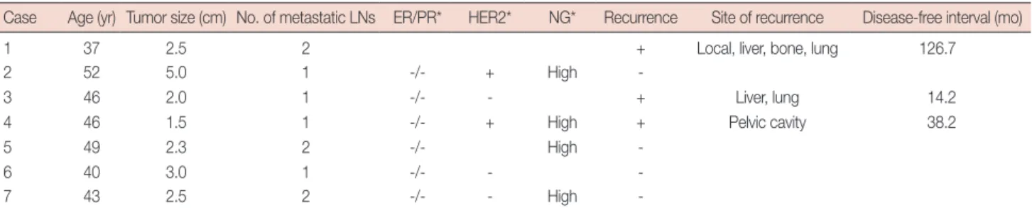
To identify the prognostic factors specific to MBC, a uni- variate survival analysis was performed for known prognostic factors using the Kaplan-Meier method with the log-rank test. We did not identify differences in DFS or OS in MBC patients according to tumor size, HR status, HER2 status, or adjuvant treatment. However, the survival curve for patients with LN metastasis differed significantly from that of patients without LN metastasis (DFS, p=0.010; OS, p<0.001) (Figure 4). The prognosis of patients with MBC seemed to be deter- mined by known prognostic factors, such as nodal status, rather than the histological type itself. As shown in Table 4, all MBCs associated with LN metastasis presented as HR- negative tumors with a high nuclear grade. Distant recur- rence with or without locoregional recurrence occurred in three of these patients.
Table 4. Clinicopathologic characteristics of medullary breast carcinoma with lymph node metastasis
Case Age (yr) Tumor size (cm) No. of metastatic LNs ER/PR* HER2* NG* Recurrence Site of recurrence Disease-free interval (mo)
1 37 2.5 2 + Local, liver, bone, lung 126.7
2 52 5.0 1 -/- + High -
3 46 2.0 1 -/- - + Liver, lung 14.2
4 46 1.5 1 -/- + High + Pelvic cavity 38.2
5 49 2.3 2 -/- High -
6 40 3.0 1 -/- - -
7 43 2.5 2 -/- - High -
LN=lymph node; ER=estrogen receptor; PR=progesterone receptor; HER2=human epidermal growth factor receptor 2; NG=nuclear grade.
*Unknown values are blank.
A B
Overall survival
Months
0 12 24 36 48 60 72 84 96 108 120 LN metastasis status
Log rank p=0.001 OS 1.0
0.8
0.6
0.4
0.2
0.0
Yes (n=7) No (n=45)
Figure 4. Disease-free survival (DFS) (A) and overall survival (OS) (B) curves for patients with medullary breast carcinoma according to lymph node (LN) metastasis status.
Disease-free survival
Months
0 12 24 36 48 60 72 84 96 108 120 LN metastasis status
Log rank p=0.010 DFS 1.0
0.8
0.6
0.4
0.2
0.0
Yes (n=7) No (n=45)
DISCUSSION
This retrospective review of invasive breast cancer at a sin- gle institution in Korea helped elucidate the clinicopathologic characteristics of MBC. In this study, MBC had several specif- ic features. However, prognosis of MBC did not differ from that of IDC and was determined by known prognostic factors such as tumor size, LN metastasis, and HR status, regardless of histologic type.
The MBC group presented at a younger age than did the IDC group. These results correspond with earlier studies [9- 11]. Vu-Nishino et al. [10] demonstrated that women with breast cancer with medullary histology presented at a younger age (mean, 47.5 years vs. 55.6 years), and a significantly higher proportion of patients were classified as very young age (≤35 years). Anderson et al. [18] explained this result using an age- specific incidence rate curve. MBC rates increased rapidly until age 50 and then plateaued, while IDC rates increased rapidly until age 50 and rose slowly thereafter. In our study, age at on- set of MBC was younger, and a higher proportion of patients in the MBC group was younger than 35 years, although this difference was not significant (13.5% vs. 9.2%, p=0.330). The age difference between the two groups was smaller than that reported in the Western literature. This difference may be at- tributable to the fact that Korean women present with breast cancer at younger ages than Western women. According to the Korean Breast Cancer Society, the peak of breast cancer incidence occurs at 40 to 49 years, and a total of 37.1% of women with newly developed breast cancer in 2010 belonged to this group [19]. Therefore, the age gap between histologic groups was smaller in this study.
In previous studies, the effect of tumor size has been con- troversial. Flucke et al. [20] found smaller tumor sizes in a MBC group than in an IDC group. In contrast, Vo et al. [11]
reported that a MBC group had larger tumors than an IDC group (T2 57.0% vs. 27.0%, p<0.001). In our study, tumor size did not differ significantly between groups.
The proportion of patients with LN metastasis was clearly lower in the MBC group without LVI, and patients who did show LN metastasis had no more than two metastases. This finding is consistent with other reports: Flucke et al. [20]
showed that patients with MBC had a higher node negative rate (75.0% vs. 47.9%, p=0.0014) and less frequent LVI (3.4%
vs. 14.4%, p=0.025) than those with IDC. This finding might be related to the histologic characteristics of MBC, which in- clude dense lymphocytic infiltration especially with CD3, CD8, TIA-1, and granzyme-B positive lymphocytes, and might also be affected by different immune responses in MBC, such as the absence of IgA antibody; the presence of
IgG; infiltration of plasma cells; and expression of tumor-spe- cific antigens such as β-actin, ganglioside D3, and HLA-DR [21,22]. These mechanisms could be involved in control of tu- mor spread and improvement of the prognosis of MBC.
Many previous studies have suggested that typical MBC is associated with a favorable prognosis [7,10,23,24]. Oh et al.
[14] reported a difference in the 10-year OS rate of typical MBC and IDC patients, which were 86.0% and 74.7%, respec- tively, in Korean women. Some researchers have suggested that the good prognosis of MBC is associated with the downregula- tion of genes involved in cell growth and proliferation, elevated levels of metastasis-inhibiting factors, and low levels of metas- tasis-promoting factors [4]. However, not all studies have con- firmed this finding or have reported similar survival rates for MBC and IDC, including the studies by Fisher et al. and Thur- man et al. [25,26]. These authors did not find significant differ- ences between the DFS and OS of patients with MBC and IDC. We also did not find any differences in death or recur- rence based on histological type. In addition, in subgroup analyses by pathologic stage, nodal involvement, HR status, HER2 status, and adjuvant treatment, MBC did not differ from IDC in either DFS or OS, as shown in Figures 1, 2 and Table 2.
However, in the subgroups with nodal metastasis, patients with IDC had better survival than did those with MBC. This differ- ence may be attributable to the small number of cases.
Another possible explanation for these results may be the implication of LN metastasis in patients with MBC. The MBC group had better nodal staging results (N1 86.5% vs. 58.4%, p<0.001) and microscopic LVI results than did the IDC group (negative 100.0% vs. 70.2%, p<0.001). Rare LN involvement is not only a typical feature of MBC, but also the most important prognostic factor of invasive carcinoma, including medullary type. The significance of the LN status in MBC has been re- ported in many previous studies [8,23,27]. Martinez et al. [27]
showed that 10-year survival rates in patients with MBC with and without LN metastasis were 67.5% and 81.9%, respective- ly, and Ridolfi et al. [7] reported that patients with axillary LN metastasis to less than three LNs did not die from the disease.
Our results were similar. Only 13.5% of MBC patients showed LN involvement, and these were limited to two LNs. Both DFS and OS in patients with MBC with LN metastasis were poorer than in patients without LN metastasis (p=0.010 and p<
0.001, respectively), even in patients with IDC. No prognostic factors other than LN metastasis affected survival in the MBC group (data not shown). We could not clearly determine the role of adjuvant treatment in MBC because of the small num- ber of cases. However, among patients treated with adjuvant chemotherapy, those with MBC had better DFS and OS than did those with IDC, unlike the case in patients without che-
motherapy, although the difference was not statistically signifi- cant. Whether less adjuvant treatment should be given and the prognostic value of adjuvant therapy in this population is still debatable, but on the basis of our results, we suggest that MBC should be treated as other invasive carcinoma according to the National Comprehensive Cancer Network guidelines. Further studies are needed to clarify the implications of adjuvant che- motherapy.
Our study has some limitations. The sample size was small.
The MBC group comprised 52 cases, less than 1% of IDC group. We could not perform multivariate analysis to identify prognostic factors in the MBC group because of the small sample size and rare deaths or recurrences. Some immunohis- tochemistry data, such as the Ki-67 index, CK 5/6 positivity, and epidermal growth factor receptor status, which are im- portant MBC characteristics and might be related to progno- sis, were missing. These missing data increase the difficulty in obtaining results of statistical significance.
In conclusion, MBC is a rare breast cancer subtype associat- ed with unique clinicopathologic features, such as rare LN me- tastasis, ER and PR negativity, advanced tumor grade, and nu- clear pleomorphism. The prognosis of MBC is not significant- ly different from that of IDC, and histologic type in itself was not an independent prognostic factor for invasive breast carci- noma in multivariate analysis. LN metastasis was rare in pa- tients with MBC; however, in patients with LN metastasis, re- currence and death were significantly more common than in patients without LN metastasis or patients with IDC histology.
Therefore, patients with MBC require the same intensive treatment as those with IDC, especially patients with LN me- tastasis, to improve survival.
CONFLICT OF INTEREST
The authors declare that they have no competing interests.
REFERENCES
1. Tavassoli FA. Pathology of the Breast. 2nd ed. New York: McGraw-Hill;
1999. p.333-9.
2. Abdul Rashid S, Rahmat K, Jayaprasagam K, Alli K, Moosa F. Medul- lary carcinoma of the breast: role of contrast-enhanced MRI in the di- agnosis of multiple breast lesions. Biomed Imaging Interv J 2009;5:e27.
3. Li CI. Risk of mortality by histologic type of breast cancer in the United States. Horm Cancer 2010;1:156-65.
4. Weigelt B, Horlings HM, Kreike B, Hayes MM, Hauptmann M, Wessels LF, et al. Refinement of breast cancer classification by molecular charac- terization of histological special types. J Pathol 2008;216:141-50.
5. Moore OS Jr, Foote FW Jr. The relatively favorable prognosis of medul- lary carcinoma of the breast. Cancer 1949;2:635-42.
6. Cotran RS, Kumar V, Collins T, Robbins SL. Robbins Pathologic Basis of Disease. 6th ed. Philadelphia: Saunders; 1999. p.1111.
7. Ridolfi RL, Rosen PP, Port A, Kinne D, Miké V. Medullary carcinoma of the breast: a clinicopathologic study with 10 year follow-up. Cancer 1977;40:1365-85.
8. Foschini MP, Eusebi V. Rare (new) entities of the breast and medullary carcinoma. Pathology 2009;41:48-56.
9. Li CI, Uribe DJ, Daling JR. Clinical characteristics of different histologic types of breast cancer. Br J Cancer 2005;93:1046-52.
10. Vu-Nishino H, Tavassoli FA, Ahrens WA, Haffty BG. Clinicopathologic features and long-term outcome of patients with medullary breast car- cinoma managed with breast-conserving therapy (BCT). Int J Radiat Oncol Biol Phys 2005;62:1040-7.
11. Vo T, Xing Y, Meric-Bernstam F, Mirza N, Vlastos G, Symmans WF, et al. Long-term outcomes in patients with mucinous, medullary, tubular, and invasive ductal carcinomas after lumpectomy. Am J Surg 2007;194:
527-31.
12. Fisher ER, Anderson S, Redmond C, Fisher B. Pathologic findings from the National Surgical Adjuvant Breast Project protocol B-06: 10-year pathologic and clinical prognostic discriminants. Cancer 1993;71:2507- 14.
13. Ellis IO, Galea M, Broughton N, Locker A, Blamey RW, Elston CW.
Pathological prognostic factors in breast cancer. II. Histological type.
Relationship with survival in a large study with long-term follow-up.
Histopathology 1992;20:479-89.
14. Oh JW, Park S, Kim JH, Koo JS, Hur H, Yang WI, et al. Clinical analysis of medullary carcinoma of the breast. J Breast Cancer 2009;12:47-53.
15. Rapin V, Contesso G, Mouriesse H, Bertin F, Lacombe MJ, Piekarski JD, et al. Medullary breast carcinoma. A reevaluation of 95 cases of breast cancer with inflammatory stroma. Cancer 1988;61:2503-10.
16. Edge SB; American Joint Committee on Cancer; American Cancer So- ciety. AJCC Cancer Staging Handbook: from the AJCC Cancer Staging Manual. 7th ed. New York: Springer; 2010.
17. Bloom HJ, Richardson WW. Histological grading and prognosis in breast cancer: a study of 1409 cases of which 359 have been followed for 15 years. Br J Cancer 1957;11:359-77.
18. Anderson WF, Chu KC, Chang S, Sherman ME. Comparison of age- specific incidence rate patterns for different histopathologic types of breast carcinoma. Cancer Epidemiol Biomarkers Prev 2004;13:1128-35.
19. Korean Breast Cancer Society. Breast Cancer Facts & Figures. Seoul:
Korean Breast Cancer Society; 2012.
20. Flucke U, Flucke MT, Hoy L, Breuer E, Goebbels R, Rhiem K, et al. Dis- tinguishing medullary carcinoma of the breast from high-grade hor- mone receptor-negative invasive ductal carcinoma: an immunohisto- chemical approach. Histopathology 2010;56:852-9.
21. Malyuchik SS, Kiyamova RG. Medullary breast carcinoma. Exp Oncol 2008;30:96-101.
22. Kuroda H, Tamaru J, Sakamoto G, Ohnisi K, Itoyama S. Immunophe- notype of lymphocytic infiltration in medullary carcinoma of the breast. Virchows Arch 2005;446:10-4.
23. Reinfuss M, Stelmach A, Mitus J, Rys J, Duda K. Typical medullary car- cinoma of the breast: a clinical and pathological analysis of 52 cases. J Surg Oncol 1995;60:89-94.
24. Huober J, Gelber S, Goldhirsch A, Coates AS, Viale G, Öhlschlegel C, et al. Prognosis of medullary breast cancer: analysis of 13 International
Breast Cancer Study Group (IBCSG) trials. Ann Oncol 2012;23:2843-51.
25. Fisher ER, Kenny JP, Sass R, Dimitrov NV, Siderits RH, Fisher B. Med- ullary cancer of the breast revisited. Breast Cancer Res Treat 1990;16:
215-29.
26. Thurman SA, Schnitt SJ, Connolly JL, Gelman R, Silver B, Harris JR, et al. Outcome after breast-conserving therapy for patients with stage I or
II mucinous, medullary, or tubular breast carcinoma. Int J Radiat Oncol Biol Phys 2004;59:152-9.
27. Martinez SR, Beal SH, Canter RJ, Chen SL, Khatri VP, Bold RJ. Medul- lary carcinoma of the breast: a population-based perspective. Med Oncol 2011;28:738-44.