84
INTRODUCTION
Breast carcinoma is the most common malignancy affecting women in Korea. Recent research for tumor biology has resulted in new insights in the regulation of cell kinetics, invasion and metastasis. However, the pathogenic mechanism underlying development and progression of breast carcinoma is still unknown. Recently, angiopoietins (Angs) have been identified as a group of the major physiological ligands for the tyrosine kinase receptor Tie-2 and are thought to be important factors in vascular maturation and stability during angiogenesis.(1-4) Angiopoietin-1 (Ang-1) binds to the Tie-2 receptor and activates it by inducing phosphorylation and dimerization of the known domains. Angiopoietin-2 (Ang-2) also binds to Tie-2 but dose not induce phosphorylation and antagonizes the action of Ang-1. Ang-1 helps to maintain and stabilize mature vessels by promoting interaction between endothelial cells and periendothelial supporting cells.(5-7) Ang-2 is expressed at sites of vascular remodeling and is thought to block the stabilizing action of Ang-1. Desta- bilization by Ang-2 in the presence of Vascular Endothelial Growth Factor (VEGF) has been hypothesized to induce an angiogenic response; however, in the absence of VEGF, Ang-2 leads to vessel regression.(2,5,8,9) Recently, the Ang-2 overexpression has been reported in brain tumor, gastric carcinoma and hepatocellular carcinomas.(10-13) However, there is little available information about the expression of
Expression and Clinical Significance of Angiopoietin-2 and its Receptor Tie-2 in Invasive Breast Cancer
Division of Breast․Endocrine Surgery, Department of Surgery, Chonbuk National University Medical School, Jeonju, Korea
Kui Seon Park and Sung Hoo Jung
Purpose: Breast carcinomas are highly malignant tumor that the angiogenesis factor, vascular endothelial growth factor and its receptors are overexpressed. To elucidate the role of Angiopoietin-2 (ANG2) and ANG2 receptor Tie-2 in in- vasive ductal carcinoma, we examined the expression of ANG2, and Tie-2 at the mRNA and protein levels in human breast cancer cell lines and samples.
Methods: Total RNA from 22 breast cancer patient biopsies were extracted. ANG2 and Tie-2 mRNA expression was measured by means of reverse transcription-PCR assay.
Results: RT-PCR indicated that the ANG2 and Tie-2 mRNA levels in carcinoma samples were significantly higher than those of the adjacent non-neoplastic breast tissues. For ANG2 and Tie-2, 41 of 71 invasive ductal carcinomass (58%) showed high expressions in Immunohistochemistry.
Immunohistochemical analysis demonstrated that ANG2 and Tie-2 were expressed by both tumor cells and endothelial elements. Expression in tumor cells were confirmed by studying a panel of human breast carcinoma cell lines cultured by RT-PCR. Our study showed that the ANG2 positivity was correlated with axillary lymph node metastasis among the clinicopathological parameter and confirmed that high expressions of ANG2 correlated highly with the axillary lymph node metastases, histological grade, positive PR status, and age, and Tie-2 expression correlated significantly with the p53 status. Moreover, ANG2 and Tie-2 co-expres- sion correlated significantly with the axillary lymph node metastases, compared with ANG2(-)/Tie-2 (-) and ANG2 (+)/Tie-2 (-) or ANG2 (-)/Tie-2 (+) cases.
Conclusion: These findings suggested that ANG2 and Tie-2 might be involved in the progression of invasive ductal carcinomas through autocrine and paracrine signaling and
that it may be clinically useful in selecting patients who could benefit from adjuvant treatment by further study. (Journal of Korean Breast Cancer Society 2004;7:84-91) ꠏꠏꠏꠏꠏꠏꠏꠏꠏꠏꠏꠏꠏꠏꠏꠏꠏꠏꠏꠏꠏꠏꠏꠏꠏꠏꠏꠏꠏꠏꠏꠏꠏꠏꠏꠏꠏꠏꠏꠏꠏꠏꠏꠏꠏ Key Words: Angiopoietin-2, Receptor Tie-2, Invasive Breast
Cancer
Correspondence: Sung Hoo Jung, Division of Breast․Endocrine Surgery, Department of Surgery, Chonbuk National University Medical School, Jeonju, 561-712, Korea. Tel: 063-250-1570, Fax:
063-271-6197, E-mail: [email protected] Received: 23 March, 2004 Accepted: 13 May, 2004
Ang-2 and Tie-2 in a series of human breast carcinoma.
Herein, we tried to reveal the correlations between the expression of Ang-2/its receptor Tie-2 and the various cli- nicopathologic prognostic factors such as age, lymph node metastases, estrogen receptor (ER), and progesterone receptor (PR), in the invasive ductal carcinoma of breast.
METHODS
1) Cell culture and Western blot analysis
Used cells were the human breast cancer cell lines; MDA231, MCF7 and colon adenocarcinoma cell lines; HCT116, Colo205, HT29, SW620 and KM12 (ATCC, Rockville, MD, USA).
The cells were cultured in appropriate media, as follows:
RPMI 1640 medium (Sigma Chemical Co., St. Louis, MO, USA); each was supplemented with 10% fetal bovine serum (Life Technologies, Inc.) and 1% antibiotic- antimycotic solution (Life Technologies, Inc.). The cells were kept at 37oC in a humidified incubator which was maintained with 5% CO2. Protein isolation and Western blotting analysis were performed as described.(14) Immunostaining and protein band visualization with the ECL system SuperSignalⓇ (Pierce) was carried out according to the manufacturer's protocol.
2) Patients and specimens
The breast carcinoma samples were obtained from patients who underwent routine surgery for breast cancer at the Department of Surgery, Chonbuk National University Hospital in 2002∼2003. Patients included in the study had an axillary dissection that sampled at least five lymph nodes. The can- cerous breast and paired normal breast tissues taken from a site distant from the tumorous lesion were snap frozen and stored in liquid nitrogen till further use. For the immuno- histochemical study, some of these tissue specimens were fixed in 10% neutralized buffered formalin solution for 24 hours. Clinical state of each patient was classified according to the pathological grade of the tumor size, lymph node state and metastasis (pTNM) classification system.(15)
3) Reverse transcription-polymerase chain reaction (RT-PCR) analysis
Total RNA was extracted from 22 breast carcinoma tissues and non-tumorous tissues of the patients using the AGPC method.(16) Reverse transcription reactions were done with a cDNA synthesis kit (Stratagene, La Jolla, CA, USA) follow- ing the instruction manual. cDNA was synthesized with 7μg of total RNA and oligo (dT) primer in 50μl of a solution
containing reverse transcriptase. The reverse-transcribed samples were used as templates for amplification of Ang-2, Tie-2 and G3APDH gene, which was used as an internal quantitative control. For the PCR, the following primers were used: sense primer of Ang2 5'-ACTGAAGAAAGAATGTGGCAGA-3' and antisense primer 5'-CACAGCCGTCTGGTTCTGTAC-3' sense primer of Tie-2 5'-AGCAGAAATGCATCGAACAA-3' and antisense primer 5'-CCTAACCAGATGAAGTTGCTGA-3'.
With respect to G3APDH, the primer sequences were as follows: sense primer 5'-CCCCTGGCCAAGGTCATCCATG ACAACTTT-3' and antisense primer 5'-GGCCATGAGGTCC ACCACCCTGTTGCTGTA-3'. The PCR reactions were per- formed following the cycling parameters on a MinicyclerTMPCR system (MJ Research, Inc.): 10 min at 94oC followed by 25 cycles of 1 min at 94oC, 1 min at 55oC, 1 min at 72oC, and a final cycle at 72oC for 10 min. Quantification of the PCR products was scanned and performed using a Quantity One program (Bio-Rad, Hercules, CA). Increased expression of the Ang-2 and Tie-2 mRNA in tumor tissue was defined as more than about 2∼3 times higher than that seen in normal breast tissue.
4) Immunohistochemical analysis
Commercially available goat polyclonal antibody against Ang-2 (1:50, Santa Cruz Biotechnology, Santa Cruz, CA., USA), Tie-2 (1:50, Santa Cruz Biotechnology, Santa Cruz, CA., USA), mouse monoclonal antibodies against p53 DO-7 (1:100, Dako, Glostrup, Denmark), Estrogen Receptor (1:
50, Dako), and Progesterone Receptor (1:10, Dako) were used as primary antibodies. A paraffin section of the breast carcinoma tissue was deparaffinized and dehydrated in graded alcohol. Antigenic enhancement was performed by submerg- ing in 1x citrate buffer (pH 6.0) and microwaving. The sections were then treated with 3% hydrogen peroxide in methanol to quench the endogenous peroxidase activity, fol- lowed by incubation with 1% BSA to block the non-specific binding, respectively. The primary polyclonal anti-Bmi-1 anti- body was incubated for 90 min at room temperature. After washing, the tissue section was then reacted with the biotiny- lated anti-goat secondary antibody, followed by incubation with streptavidin-horseradish-peroxidase complex. The tissue section was immersed in 3-amino-9-ethyl carbazole as a substrate. In negative controls, non-immune goat IgG of the same isotype or antibody dilution solution replaced the primary antibody.
Each section was evaluated by at least two independent observers, and moderate to strong nuclear staining was consi-
dered as a positive reaction. The distribution of Ang-2 and Tie-2 was scored on a semi-quantitative scale, as follows:
negative (<10% of tumor positive), focally positive (10∼
50% of tumor positive), and diffusely positive (>50% of tumor positive). For estrogen receptor, a nuclear staining in
>25% of the cells was considered estrogen receptor positive, and similarly for progesterone receptor. A nuclear staining in
>10% of the cells were considered positive for p53.
5) Statistical analysis
The relationship between the results of the immunohisto- chemical study and the clinicopathologic parameters was performed using the SASⓇ software package (version 8.01;
SAS Institute, Cary, NC, USA). Univariate and multivariate analysis was carried out using the proc logistic module. In all cases, the exact mid-P adjusted P values were reported and a P value<0.05 was considered to be statistically significant.
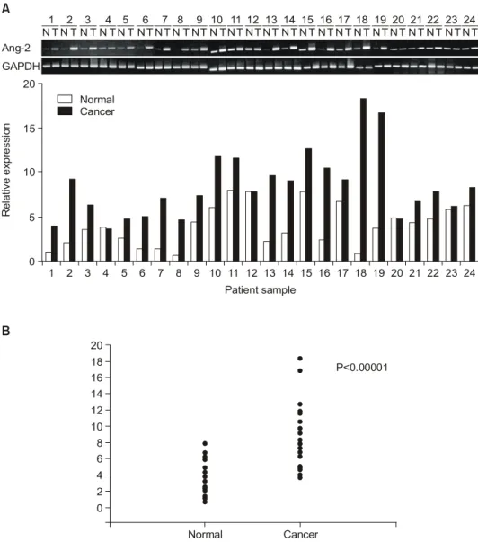
Fig. 1. The Ang-2 gene expression in human breast carcinoma. (A) Expression of the Ang-2 and G3APDHin 22 patients with invasive ductal breast cancer, determined by RT-PCR. Data were expressed relative to the expression of G3APDHgene in each breast carcinoma. The relative band density shows high expression level on breast carcinoma tissues. (B) The expression of Ang-2 gene was increased (P<0.0001) in tumor tissues when compared with corresponding non-tumorous tissues.
1 1
T T T T T T T T T T T T T T T T T T T T T T T T N N N N N N N N N N N N N N N N N N N N N N N N
2 2
3 3
4 4
5 5
6 6
7 7
8 8
9 9
10 10
11 11
12 12
13 13
14
P<0.00001 14
15 15
16 16
17 17
18 18
19 19
20 20
21 21
22 22
23 23
24 24
20 20
Relative expression
Patient sample 0
Ang-2 GAPDH
Normal 18
16 14 15
10 6 12 8 10
4 5
0 2
Cancer
B A
Cancer Normal
RESULTS
1) Ang-2 and Tie-2 expression in invasive ductal carcinomas
The relative levels of expression of the Ang-2 mRNA in 24 breast carcinoma tissues were compared with those of non-tumorous tissues by RT-PCR. The expression levels were determined as a ratio between the Ang-2 and the reference gene (G3APDH) to correct for the variation in the amounts of mRNA. The Ang-2 mRNA level was significantly increased (P<0.0001) in all the examined human breast car-
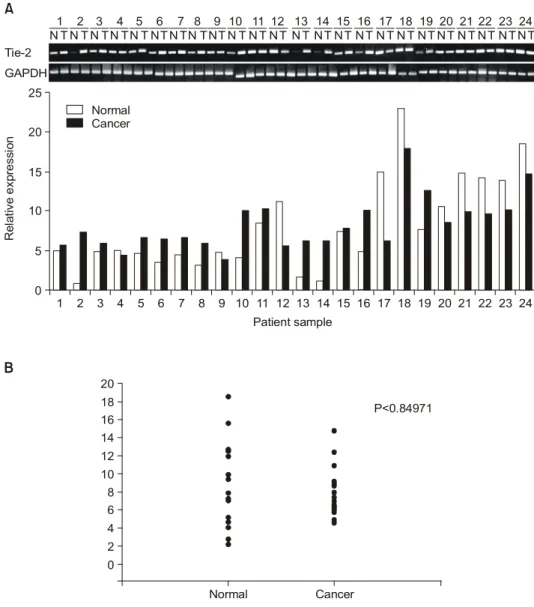
cinomas when compared with that of its corresponding normal breast tissue (Fig. 1A, B). Meanwhile, Tie-2 mRNA expression in breast cancer was not different from that in adjacent breast tissue (Fig. 2A, B). In the immunohistochemi- cal study, Ang-2 in tumor cells is positively expressed in 41(58%) cases of the 71 patients with breast cancer (Fig. 3).
Meanwhile, the normal breast tissue shows negative or weak Ang-2 staining in the breast epithelial cells. Ang-2 was largely distributed in the cytoplasmic areas of tumor cells(Fig.
3A, B). Of 31 (44%) cases of the 71 patients, Tie-2 is not only expressed as membranous pattern in the endothelial cells but also expressed in tumor cells (Fig. 3C, D). In all tested
Fig. 2. The Tie-2 gene expression in human breast carcinoma. (A) Expression of the Tie-2 and G3APDHin 22 patients with invasive ductal breast cancer, determined by RT-PCR. Data were expressed relative to the expression of G3APDHgene in each breast carcinoma. (B) The expression of Tie-2 gene was variablein tumor tissues when compared with corresponding non-tumorous tissues.
1 1
T T T T T T T T T T T T T T T T T T T T T T T T N N N N N N N N N N N N N N N N N N N N N N N N
2 2
3 3
4 4
5 5
6 6
7 7
8 8
9 9
10 10
11 11
12 12
13 13
14
P<0.84971 14
15 15
16 16
17 17
18 18
19 19
20 20
21 21
22 22
23 23
24 24
20 25
Relative expression
Patient sample 0
Tie-2 GAPDH
Normal 18
16 14 20
15 10
10 6 12 8 4 5
0 2
Cancer
B A
Cancer Normal
cancer tissues, Ang-2 were negative, focally and diffusely positive in 27 (38%), 21 (30%), and 23 (32%) cases, res- pectively. Ang-2 and Tie-2 expression in tumor cells was confirmed by western blot analyses using a panel of human two breast and five colon carcinoma cell lines (Fig. 4).
2) Relationship between Ang-2/its receptor Tie-2 ex- pression and clinicopathological parameters in invasive ductal carcinomas
We investigated the Ang-2/Tie-2 expression and various clinicopathological parameters to elucidate the roles of the Ang-2 and Tie-2 genes in tumoriogenesis of the breast cancer.
As the data summarized in Table 1 to 2, our study showed that the ANG2 positivity was correlated with axillary lymph node metastasis among the clinicopathological parameter and confirmed by multivariate analysis that high expression of Ang-2 was strongly correlated with axillary lymph node metastases, high histologic grade, positive PR status, and older age (Table 1, 2). Tie-2 expression was significantly
correlated with p53 positive status in univariate analysis (Table 3). Our multivariate analysis using data from the whole parameters also confirmed that high expression of Tie- 2 was strongly correlated with p53 positive status, axillary lymph node metastases and Age (Table 4). Also, Ang-2 and Tie-2 co-expression was significantly correlated with axillary lymph node metastases, compared with Ang-2 (-)/Tie-2 (-) and Ang-2 (+)/Tie-2 (-) or Ang-2 (-)/Tie-2 (+) cases.
These results suggested the Ang-2/its receptor Tie-2 co- Fig. 3. Immunohistochemical staining of human breastcarcinoma specimen using antibodies to Ang2 and Tie-2. (A) Ang-2 expression is
increased on the tumor cells of breast carcinoma (immunoperoxidase stain; original magnification ×200). (B) Note the distinct cytoplasmic staining in breast carcinoma cells (immunoperoxidase stain; original magnification ×400). (C) Tie- 2 expression is increased on the tumor cells of breast carcinoma (immunoperoxidase stain; original magnification ×200). (D) Note the distinct membranous staining pattern in breast carcinoma cells (immunoperoxidase stain; original magnification ×400).
A
C
B
D
Fig. 4. Western blot analysis for Ang-2 and Tie-2 expression in two human breast and five human colon carcinoma cell lines.
Ang-2
MCF7 MDA HCT
116 SW
620 KM 12 COLO
205 HUVEC HT29
Tie-2 Alpha- tubulin
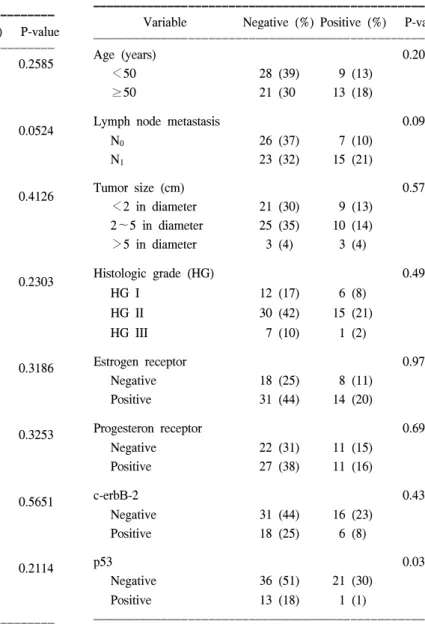
Table 3. Relationship between Tie-2 receptor expression and clinicopathological parameters
ꠚꠚꠚꠚꠚꠚꠚꠚꠚꠚꠚꠚꠚꠚꠚꠚꠚꠚꠚꠚꠚꠚꠚꠚꠚꠚꠚꠚꠚꠚꠚꠚꠚꠚꠚꠚꠚꠚꠚꠚꠚꠚꠚꠚꠚꠚꠚꠚꠚꠚꠚ Variable Negative (%) Positive (%) P-value ꠏꠏꠏꠏꠏꠏꠏꠏꠏꠏꠏꠏꠏꠏꠏꠏꠏꠏꠏꠏꠏꠏꠏꠏꠏꠏꠏꠏꠏꠏꠏꠏꠏꠏꠏꠏꠏꠏꠏꠏꠏꠏꠏꠏꠏꠏꠏꠏꠏꠏꠏ
Age (years) 0.2086
<50 28 (39) 9 (13)
≥50 21 (30 13 (18)
Lymph node metastasis 0.0994
N0 26 (37) 7 (10)
N1 23 (32) 15 (21)
Tumor size (cm) 0.5748
<2 in diameter 21 (30) 9 (13) 2∼5 in diameter 25 (35) 10 (14)
>5 in diameter 3 (4) 3 (4)
Histologic grade (HG) 0.4915
HG I 12 (17) 6 (8)
HG II 30 (42) 15 (21)
HG III 7 (10) 1 (2)
Estrogen receptor 0.9762
Negative 18 (25) 8 (11)
Positive 31 (44) 14 (20)
Progesteron receptor 0.6923
Negative 22 (31) 11 (15)
Positive 27 (38) 11 (16)
c-erbB-2 0.4390
Negative 31 (44) 16 (23)
Positive 18 (25) 6 (8)
p53 0.0325
Negative 36 (51) 21 (30)
Positive 13 (18) 1 (1)
ꠏꠏꠏꠏꠏꠏꠏꠏꠏꠏꠏꠏꠏꠏꠏꠏꠏꠏꠏꠏꠏꠏꠏꠏꠏꠏꠏꠏꠏꠏꠏꠏꠏꠏꠏꠏꠏꠏꠏꠏꠏꠏꠏꠏꠏꠏꠏꠏꠏꠏꠏ
Table 4. Results of multivariate logistic regression analysis with Tie-2 expression
ꠚꠚꠚꠚꠚꠚꠚꠚꠚꠚꠚꠚꠚꠚꠚꠚꠚꠚꠚꠚꠚꠚꠚꠚꠚꠚꠚꠚꠚꠚꠚꠚꠚꠚꠚꠚꠚꠚꠚꠚꠚꠚꠚꠚꠚꠚꠚꠚꠚꠚꠚ Odds 95% confidence
Categories P-value
ratio limits
ꠏꠏꠏꠏꠏꠏꠏꠏꠏꠏꠏꠏꠏꠏꠏꠏꠏꠏꠏꠏꠏꠏꠏꠏꠏꠏꠏꠏꠏꠏꠏꠏꠏꠏꠏꠏꠏꠏꠏꠏꠏꠏꠏꠏꠏꠏꠏꠏꠏꠏꠏ
p53 <0.0001 0.170 0.078∼0.368
Age 0.0007 1.994 1.337∼2.975
Nodal status 0.0009 2.098 1.375∼3.201
Histologic grade 0.2046 0.827 0.576∼1.189 ꠏꠏꠏꠏꠏꠏꠏꠏꠏꠏꠏꠏꠏꠏꠏꠏꠏꠏꠏꠏꠏꠏꠏꠏꠏꠏꠏꠏꠏꠏꠏꠏꠏꠏꠏꠏꠏꠏꠏꠏꠏꠏꠏꠏꠏꠏꠏꠏꠏꠏꠏ Table 2. Results of multivariate logistic regression analysis with
angiopoietin-II expression and clinicopathologic para- meters
ꠚꠚꠚꠚꠚꠚꠚꠚꠚꠚꠚꠚꠚꠚꠚꠚꠚꠚꠚꠚꠚꠚꠚꠚꠚꠚꠚꠚꠚꠚꠚꠚꠚꠚꠚꠚꠚꠚꠚꠚꠚꠚꠚꠚꠚꠚꠚꠚꠚꠚꠚ Odds 95% confidence
Categories P-value
ratio limits
ꠏꠏꠏꠏꠏꠏꠏꠏꠏꠏꠏꠏꠏꠏꠏꠏꠏꠏꠏꠏꠏꠏꠏꠏꠏꠏꠏꠏꠏꠏꠏꠏꠏꠏꠏꠏꠏꠏꠏꠏꠏꠏꠏꠏꠏꠏꠏꠏꠏꠏꠏ Nodal status <0.0001 3.341 2.237∼4.988 Histologic grade <0.0001 0.387 0.273∼0.548
PR 0.0003 2.596 1.706∼3.949
Age 0.0003 1.966 1.325∼2.919
c-erbB-2 0.0344 1.541 1.034∼2.299
Size 0.0388 1.425 1.017∼1.997
ꠏꠏꠏꠏꠏꠏꠏꠏꠏꠏꠏꠏꠏꠏꠏꠏꠏꠏꠏꠏꠏꠏꠏꠏꠏꠏꠏꠏꠏꠏꠏꠏꠏꠏꠏꠏꠏꠏꠏꠏꠏꠏꠏꠏꠏꠏꠏꠏꠏꠏꠏ Table 1. Relationship between angiopoietin-2 expression and
clinico-pathological parameters in invasive breast cancers
ꠚꠚꠚꠚꠚꠚꠚꠚꠚꠚꠚꠚꠚꠚꠚꠚꠚꠚꠚꠚꠚꠚꠚꠚꠚꠚꠚꠚꠚꠚꠚꠚꠚꠚꠚꠚꠚꠚꠚꠚꠚꠚꠚꠚꠚꠚꠚꠚꠚꠚꠚ Variable Negative (%) Positive (%) P-value ꠏꠏꠏꠏꠏꠏꠏꠏꠏꠏꠏꠏꠏꠏꠏꠏꠏꠏꠏꠏꠏꠏꠏꠏꠏꠏꠏꠏꠏꠏꠏꠏꠏꠏꠏꠏꠏꠏꠏꠏꠏꠏꠏꠏꠏꠏꠏꠏꠏꠏꠏ
Age (years) 0.2585
<50 18 (25) 19 (27)
≥50 12 (17) 22 (31)
Lymph node metastasis 0.0524
N0 18 (25) 15 (21)
N1 12 (17) 26 (37)
Tumor size (cm) 0.4126
<2 in diameter 15 (21) 15 (21) 2∼5 in diameter 12 (17) 23 (32)
>5 in diameter 3 (4) 3 (4)
Histologic grade (HG) 0.2303
HG I 5 (7) 13 (18)
HG II 20 (28) 25 (35)
HG III 5 (7) 3 (4)
Estrogen receptor 0.3186
Negative 13 (18) 13 (18)
Positive 17 (24) 28 (39)
Progesteron receptor 0.3253
Negative 16 (23) 17 (24)
Positive 14 (20) 24 (34)
c-erbB-2 0.5651
Negative 21 (30) 26 (37)
Positive 9 (13) 15 (21)
p53 0.2114
Negative 22 (31) 35 (49)
Positive 8 (11) 6 (8)
ꠏꠏꠏꠏꠏꠏꠏꠏꠏꠏꠏꠏꠏꠏꠏꠏꠏꠏꠏꠏꠏꠏꠏꠏꠏꠏꠏꠏꠏꠏꠏꠏꠏꠏꠏꠏꠏꠏꠏꠏꠏꠏꠏꠏꠏꠏꠏꠏꠏꠏꠏ
expression might play a role in the progression and metastases of invasive ductal carcinomass. The Ang-2/Tie-2 expression may be also used as one of the novel prognostic markers available for detecting invasive ductal carcinoma.
DISCUSSION
Human breast cancer has been increasing steadily and many patients with this malignancy have suffered for several decades. Available clinicopathologic prognostic indicators are not accurate despite axillary nodal status being the most important factor that determines the overall survival in pa- tients with breast cancer, approximately 25∼30% of node- negative cases eventually relapse.(17) Thus, it is important to identify the marker that is associated with pathophysiologic processes of human breast cancer.
Angiopoietins (Angs) has been identified as ligands for Tie-2 receptor and are thought to be important factors in vascular maturation and stability during angiogenic process.
Angiopoietin-1 (Ang-1) binds to the Tie-2 receptor and activates it by inducing phosphorylation and dimerization of the known domains. Angiopoietin-2 (Ang-2) also binds to Tie-2 but dose not induce phosphorylation and antagonizes the action of Ang-1. Ang-1 helps to maintain and stabilize mature vessels by promoting interaction between endothelial cells and periendothelial supporting cells.(5-7) Ang-2 is expressed at sites of vascular remodeling and is thought to block the stabilizing action of Ang-1. Recently, it has been reported that Ang-1 was expressed in some human tumors and in- volved in pathophysiologic processes of tumorigenesis and worse clinical prognosis.(18-21) However, there is little available information about the expression of Ang-2/its re- ceptor Tie-2 and clinicopathologic correlation in a series of
primary breast cancer.
Herein, we demonstrated that the both angiopoietin-2 and Tie-2 were expressed in the tumor cells of the invasive carcinomass and was strongly correlated with various clinicopathologic prognostic factors, such as axillary lymph node metastasis. It is remarkable, indeed, that given the small number of cases included in our study, Ang-2 and Tie-2 mRNA expression levels were higher in invasive ductal carcinomass than in paired adjacent normal breast tissue.
Immunohistochemical analysis also showed that Ang-2 and Tie-2 protein were expressed in 58% and 44% of breast carcinoma, respectively. A correlation between the Ang-2 positivity and clinicopathological parameter; axillary lymph node metastasis was detected and confirmed by multivariate analysis that high expression of Ang-2 was strongly correlated with axillary lymph node metastases, high histol- ogic grade, positive PR status, and age. Tie-2 expression was significantly correlated with p53positive status in univariate analysis and also confirmed that high expression of Tie-2 was strongly correlated with p53 positive status, axillary lymph node metastases and age in multivariate logistic analysis.
Interestingly, Ang-2 and Tie-2 co-expression was correlated with the clinical grade. Therefore, it appears that a high level of these two protein may contribute to the invasion and progression of breast cancer, presumedly via autocrine and paracrine mechanisms.
With the aid of univariate and multivariate analyses, we identified the fact that the Ang-2 and Tie-2 co-expression was positively correlated with axillary lymph node metastases, but not with the other histopathological parameters in the primary breast cancers. Indeed, axillary lymph node status is currently one of the most significant prognostic factors for patients with breast cancer.(22,23) Therefore, this finding suggests that Ang-2 and Tie-2 co-expression may be one of the novel independent prognostic markers available in invasive ductal carcinomas.
p53 is a tumor suppressor gene and p53 mutations are common in breast cancer.(24) The p53 protein normally has a short half-life and is therefore not usually detected using immunohistochemistry, except for cell cycle dependent expres- sion in a small proportion of proliferating cells. Mutations of the gene are generally associated with the accumulation of the protein in the nucleus. The Tie-2 expression was cor- related with positive p53 status, although the exact biological meaning of Tie-2 over-expression in positive p53 status was not known.
In conclusion, the present study described how the Ang-2 Table 5. Relationship between Angiopoietin-2/Tie-2 receptor co-
expression and clinicopathological parameter; lymph node metastasis
ꠚꠚꠚꠚꠚꠚꠚꠚꠚꠚꠚꠚꠚꠚꠚꠚꠚꠚꠚꠚꠚꠚꠚꠚꠚꠚꠚꠚꠚꠚꠚꠚꠚꠚꠚꠚꠚꠚꠚꠚꠚꠚꠚꠚꠚꠚꠚꠚꠚꠚꠚ Lymph node
metastasis Variable
ꠏꠏꠏꠏꠏꠏꠏꠏꠏꠏꠏꠏꠏꠏꠏ P-value N0 (%) N1 (%)
ꠏꠏꠏꠏꠏꠏꠏꠏꠏꠏꠏꠏꠏꠏꠏꠏꠏꠏꠏꠏꠏꠏꠏꠏꠏꠏꠏꠏꠏꠏꠏꠏꠏꠏꠏꠏꠏꠏꠏꠏꠏꠏꠏꠏꠏꠏꠏꠏꠏꠏꠏ All Angiopoietin-2 (+)
4 (6) 12 (17) <0.05 and Tie-2 (+)
Angiopoietin-2 (+)/Tie-2 (-)
14 (20) 17 (24) or Angiopoietin-2 (-)/Tie-2 (+)
All negative 15 (21) 9 (13)
ꠏꠏꠏꠏꠏꠏꠏꠏꠏꠏꠏꠏꠏꠏꠏꠏꠏꠏꠏꠏꠏꠏꠏꠏꠏꠏꠏꠏꠏꠏꠏꠏꠏꠏꠏꠏꠏꠏꠏꠏꠏꠏꠏꠏꠏꠏꠏꠏꠏꠏꠏ
and Tie-2 is expressed differentially in human breast car- cinomas. These results suggests that tumor growth deregula- tion by the Ang-2 and Tie-2 might play a role in the progression and lymph node metastases of breast carcinoma, probably through the modulation of both angiogenesis and tumor cell proliferation.
REFERENCES
1) Lauren J, Gunji Y, Alitalo K. Is angiopoietin-2 necessary for the initiation of tumor angiogenesis? Am J Pathol 1998;153:
1333-9.
2) Suri C, Jones PF, Patan S, Bartunkova S, Maisonpierre PC, Davis S, et al. Requisite role of angiopoietin-1, a ligand for the TIE2 receptor, during embryonic angiogenesis. Cell 27 1996;87:1171-80.
3) Schnurch H, Risau W. Expression of tie-2, a member of a novel family of receptor tyrosine kinases, in the endothelial cell lineage. Development 1993;119:957-68.
4) Batard P, Sansilvestri P, Scheinecker C, Knapp W, Debili N, Vainchenker W, et al. The Tie receptor tyrosine kinase is expressed by human hematopoietic progenitor cells and by a subset of megakaryocytic cells. Blood 15 1996;87:2212-20.
5) Maisonpierr PC, Suri C, Jones PF, Bartunkova S, Wiegand SJ, Radziejewski C, et al. Angiopoietin-2, a natural anta- gonist for Tie2 that disrupts in vivo angiogenesis. Science 4 1997; 277:55-60.
6) Suri C, McClain J, Thurston G, McDonald DM, Zhou H, Oldmixon EH, et al. Increased vascularization in mice over- expressing angiopoietin-1. Science 16 1998;282:468-71.
7) Papapetropoulos A, Garcia-Cardena G, Dengler TJ, Maison- pierre PC, Yancopoulos GD, Sessa WC. Direct actions of angiopoietin-1 on human endothelium: evidence for network stabilization, cell survival, and interaction with other angi- ogenic growth factors. Lab Invest 1999;79:213-23.
8) Sato TN, Tozawa Y, Deutsch U, Wolburg-Buchholz K, Fuji- wara Y, Gendron-Maguire M, et al. Distinct roles of the receptor tyrosine kinases Tie-1 and Tie-2 in blood vessel formation. Nature 6 1995;376:70-4.
9) Dumont DJ, Gradwohl G, Fong GH, Puri MC, Gertsenstein M, Auerbach A, et al. Dominant-negative and targeted null mutations in the endothelial receptor tyrosine kinase, tek, reveal a critical role in vasculogenesis of the embryo. Genes Dev 15 1994;8:1897-909.
10) Koga K, Todaka T, Morioka M, Hamada J, Kai Y, Yano S, et al. Expression of angiopoietin-2 in human glioma cells and its role for angiogenesis. Cancer Res 15 2001;61: 6248-54.
11) Hu B, Guo P, Fang Q, Tao HQ, Wang D, Nagane M, et al.
Angiopoietin-2 induces human glioma invasion through the activation of matrix metalloprotease-2. Proc Natl Acad Sci USA 22 2003;100:8904-9.
12) Etoh T, Inoue H, Tanaka S, Barnard GF, Kitano S, Mori M.
Angiopoietin-2 is related to tumor angiogenesis in gastric carcinoma: possible in vivo regulation via induction of proteases. Cancer Res 1 2001;61:2145-53.
13) Mitsuhashi N, Shimizu H, Ohtsuka M, Wakabayashi Y, Ito H, Kimura F, et al. Angiopoietins and Tie-2 expression in angiogenesis and proliferation of human hepatocellular car- cinoma. Hepatology 2003;37:1105-13.
14) Gershoni JM, Palade GE. Protein blotting: principles and applications. Anal Biochem 1983;131:1-15.
15) Wittekind C, Compton CC, Greene FL, Sobin LH. TNM residual tumor classification revisited. Cancer 2002;94:2511- 16.
16) Chomczynski P, Sacchi N. Single-step method of RNA isola- tion by acid guanidinium thiocyanate-phenol-chloroform ex- traction. Anal Biochem 1987;162:156-9.
17) McGuckin MA, Cummings MC, Walsh MD, Hohn BG, Bennett IC, Wright RG. Occult axillary node metastases in breast cancer: their detection and prognostic significance. Br J Cancer 1996;73:88-95.
18) Mitsuhashi N, Shimizu H, Ohtsuka M, Wakabayashi Y, Ito H, Kimura F, et al. Angiopoietins and Tie-2 expression in angiogenesis and proliferation of human hepatocellular carci- noma. Hepatology 2003;37:1105-13.
19) Moon WS, Rhyu KH, Kang MJ, Lee DG, Yu HC, Yeum JH, et al. Overexpression of VEGF and angiopoietin 2: a key to high vascularity of hepatocellular carcinoma? Mod Pathol 2003;16:552-7.
20) Tanaka F, Ishikawa S, Yanagihara K, Miyahara R, Kawano Y, Li M, et al. Expression of angiopoietins and its clinical significance in non-small cell lung cancer. Cancer Res 2002;
62:7124-9.
21) Hata K, Udagawa J, Fujiwaki R, Nakayama K, Otani H, Miyazaki K. Expression of angiopoietin-1, angiopoietin-2, and Tie2 genes in normal ovary with corpus luteum and in ovarian cancer. Oncology 2002;62:340-8.
22) Carter CL, Allen C, Henson DE. Relation of tumor size, lymph node status, and survival in 24,740 breast cancer cases. Cancer 1989;63:181-7.
23) Haagensen C D. Disease of the Breast. 3rded. Philadelphia:
WB Saunders, 1986.
24) Elledge RM, Allred DC. The p53 tumor suppressor gene in breast cancer. Breast Cancer Res Treat 1994;32:39-47.