DOI 10.3349/ymj.2008.49.6.978
Purpose: To investigate clinicopathological characteristics and outcomes of male breast cancer (MBC). Patients and Methods: We retrospectively analyzed the data of 20 MBC patients in comparison with female ductal carcinoma treated at Yonsei University Severance Hospital from July 1985 to May 2007. Clinicopathological features, treatment patterns, and survival were investigated. Results: MBC consists of 0.38% of all breast cancers. The median age was 56 years.
The median symptom duration was 10 months. The median tumor size was 1.7 cm, 27.8% showed node metastasis, and 71.4% were estrogen receptor positive. All 20 cancers were arisen from ductal cells. No lobular carcinoma was found. The incidence of stages 0, I, II, and III in patients were 2, 10, 4, and 3, respectively. All patients underwent mastectomy. One with invasive cancer did not receive axillary node dissection and stage was not exactly evaluated. Adjuvant treatments were determined by pathologic parameters and stage. Clinico- pathological parameters and survival rates of MBC were comparable to those of female ductal carcinoma. Conclusion:
The onset age of MBC was 10 years older and symptom duration was longer than in female patients. No difference in outcomes between MBC and female ductal carcinoma suggests that the biology of MBC is not different from that of females. Therefore, education, an appropriate system for early detection, and adequate treatment are necessary for improving outcomes.
Key Word: Male breast cancer
INTRODUCTION
Male breast cancer (MBC) is a rare disease and accounts for less than 1% of all cases of malignancy in men.1 The incidence of MBC has significantly increased from 0.86 to 1.08 per 100,000 population over the past 26 years in the United States.2 In Korea, MBC constitutes 0.4 - 0.6% of all breast cancers (BC).3-5 Because of its low incidence, MBC has not been studied as extensively as female breast cancer. Most studies related to MBC are retrospective analyses with a small number of patients. Appropriate management guidelines for MBC have not yet been clearly established, and limited information is available regarding the epidemiology, treatment, and prognosis of the disease.6 Therefore, the treatment guideline has been extrapolated from the data based on female breast cancer.
However, the incidence of MBC has been increasing significantly along with the increasing incidence of female breast cancer, although geographic variations in the incidence of MBC was reported.2,7,8 In Europe, approximately 1% of all BC occurs in males, but the incidence is much higher in other areas such as sub-Saharan Africa with 15%.9,10 Therefore, the study of MBC will become more important every day. In this study, we investigated clinicopathological characteristics, treatment patterns, and outcomes of MBC in Koreans.
PATIENTS AND METHODS
We retrospectively reviewed the medical records of 20 MBC patients who had been treated at the
Clinicopathological Characteristics of Male Breast Cancer
Seho Park,1* Joo-Hee Kim,1* Jaseung Koo,2 Byeong-Woo Park,1 and Kyong Sik Lee3
Departments of 1Surgery and2Pathology, Yonsei University College of Medicine, Seoul;3Department of Surgery, Pochon CHA University College of Medicine, Seongnam, Korea.
Received June 2, 2008 Accepted July 2, 2008
*Both authors equally contributed to this work.
This work was supported by Brain Korea 21 Project for Medical Science, Yonsei University; Astra-Zeneca Korea Co.; Dong-A Pharmaceutical Co.; Sanofi-Aventis Pharmaceutical Co.; Novartis Korea Co.; Lilly Korea Co.
Reprint address: requests to Dr. Byeong-Woo Park, Department of Surgery, Yonsei University College of Medicine, 250 Seongsan- no, Seodaemun-gu, Seoul 120-752, Korea. Tel: 82-2-2228-2121, Fax: 82-2-313-8289, E-mail: [email protected]
Department of Surgery, Yonsei University College of Medicine, in Seoul, Korea, between July 1985 and May 2007. This sample represents 0.38% (20/
5,229) of all BC treated at our institution during the same period. Data regarding general charac- teristics of patients (age, presenting signs and symptoms, duration of symptoms, and site and location of tumor), histopathology of primary tumor, treatment modalities (surgery, chemotherapy, radiation and hormone therapy), disease-free survival, and overall survival were obtained by reviewing medical records. Tumor stage was based on the 6th American Joint Committee on Cancer (AJCC) criteria. Histological type and grading followed the World Health Organization (WHO) classification. Ten percent or more of positively stained cells was used as the cut-off for hormonal receptor positivity. Clinical follow-up included history taking, physical examination, laboratory tests and radiologic imaging tests every 6 - 12 months for detection of relapse. The differences between MBC and female ductal carcinoma were also investigated. Chi-square test was employed to confirm statistical significance. Disease-free sur- vival and overall survival were estimated and plotted using Kaplan-Meier method. Group dif- ferences in survival time were tested by log- rank test. A p value < 0.05 was considered statistically significant. SPSS for Windows (ver. 13.0, SPSS Inc., Chicago, IL, USA) was used for all statistical analyses.
RESULTS
General characteristics of patients
General characteristics of patients are summarized in Table 1. The median age at diagnosis was 56 years (range, 22 - 78 years). Fifteen patients (75%) were diagnosed after the age of 50 years and 6 were diagnosed during their 50s. Three patients had a history of alcohol drinking and 4 had a history of smoking. One patient diagnosed at 22 years of age had a family history of BC and his mother died of BC when he was 2 years old. Two patients had synchronous double primary tumors;
1 had poorly differentiated epidermoid carcinoma of the tonsil and the other had adenocarcinoma ex
pleomorphic adenoma of the submandibular gland. There was no history of bilateral BC. The most common symptom was a palpable mass (n
= 18), and the median duration of symptoms was 10 months. One patient had nipple discharge for more than 10 years. Six of the 20 patients had cancer in the left breast and 14 in the right. Most tumors were located in the central subareolar area (n = 17). In 2 patients, the tumor was located in the upper outer quadrant and in the lower inner quadrant in 1 patient.
Histopathological characteristics of tumor
Histopathological characteristics of primary tumor are summarized in Table 2. All 20 cases were Table 1.General Characteristics of Male Breast Cancer Patients
Characteristics Patients (n = 20) % Age (yrs)
20 - 29 2 10
30 - 39 1 5
40 - 49 2 10
50 - 59 6 30
60 - 69 4 20
70 5 25
Chief complaints
Mass 18 90
Nipple discharge 2 10
Duration of symptom
Sx 6 mo 8 40
6 mo < Sx 12 mo 6 30
12 mo < Sx 24 mo 3 15
Sx > 24 mo 3 15
Site of tumor
Left 6 30
Right 14 70
Location of tumor
Retroareolar 17 85
Other quadrant 3 15
Sx, symptom; mo, month.
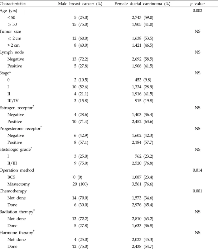
Table 2.Histopathological Characteristics and Treatment Patterns of Male Breast Cancer Compared to Female Ductal Carcinoma
Characteristics Male breast cancer (%) Female ductal carcinoma (%) p value
Age (yrs) 0.002
< 50 5 (25.0) 2,743 (59.0)
50 15 (75.0) 1,905 (41.0)
Tumor size NS
2 cm 12 (60.0) 1,638 (53.5)
> 2 cm 8 (40.0) 1,421 (46.5)
Lymph node NS
Negative 13 (72.2) 2,692 (58.5)
Positive 5 (27.8) 1,908 (41.5)
Stage* NS
0 2 (10.5) 453 (9.8)
I 10 (52.6) 1,334 (28.9)
II 4 (21.1) 1,916 (41.5)
III/IV 3 (15.8) 915 (19.8)
Estrogen receptor NS
Negative 4 (28.6) 1,403 (36.4)
Positive 10 (71.4) 2,452 (63.6)
Progesterone receptor NS
Negative 6 (42.9) 1,602 (42.3)
Positive 8 (57.1) 2,184 (57.7)
Histologic grade NS
I 3 (25.0) 762 (23.2)
II/III 9 (75.0) 2,520 (76.8)
Operation method 0.014
BCS 0 (0) 1,087 (23.4)
Mastectomy 20 (100) 3,561 (76.6)
Chemotherapy 0.001
Not done 14 (70.0) 1,573 (34.6)
Done 6 (30.0) 2,976 (65.4)
Radiation therapy NS
Not done 13 (72.2) 2,810 (63.2)
Done 5 (27.8) 1,633 (36.8)
Hormone therapy NS
Not done 4 (25.0) 2,023 (45.3)
Done 12 (75.0) 2,438 (54.7)
NS, not significant; BCS, breast-conserving surgery.
*One male with invasive cancer did not receive axillary node dissection and stage was not exactly evaluated.
Histopathologic reports of estrogen receptor/progesterone receptor and grade were not documented in 6 and 8 male patients, respectively.
We are not able to verify whether 2 and 4 male patients received radiotherapy and hormone therapy, respectively, or not.
ductal cell in origin. The most frequent histologic type was infiltrating ductal carcinoma (n = 14).
Four patients (20%) had special type ductal carcinoma; 2 had tubular carcinoma, 1 had mucinous carcinoma, and 1 had papillary carcinoma. Two patients (10%) had non-invasive carcinoma. The median size of the tumor was 1.7 cm (range, 0.5- 4 cm) and 12 cases (60%) were categorized as T1 lesions. Five of the 18 patients who underwent axillary node dissection (27.8%) showed lymph node metastasis in the axillary area. The median number of axillary lymph nodes removed was 19.5 (range, 8 - 34). The median number of axillary lymph nodes involved was 4 (range, 1 - 16) among patients with axillary lymph node metastasis. One patient had 16 axillary lymph nodes involved.
According to the TNM staging system, there were 2, 10, 4, and 3 patients in stages 0, I, II, and III, respectively. One with invasive cancer did not receive axillary node dissection and stage was not exactly evaluated. In terms of histologic grade, 3 patients had a well-differentiated tumor and 9 had a moderately to poorly differentiated tumor.
Among 14 patients available for hormone receptors assays, the estrogen receptor was positive in 10 patients (71.4%) and the progesterone receptor was positive in 8 (57.1%). The status of c-erbB-2 was available in 12 patients, 3 (25%) of whom were positive.
Treatment patterns
Treatment patterns are summarized in Table 2.
Of the 20 patients, 18 underwent modified radical mastectomy, 1 simple mastectomy, and 1 subcu- taneous mastectomy. No patients received neoad- juvant chemotherapy. After completion of surgery, adjuvant therapies were administered: 6 received chemotherapy, 4 CMF (cyclophosphamide, metho- trexate, fluorouracil) regimen, and 2 anthracycline based chemotherapy. Radiation therapy was done in 5 patients with a mean dose of 50 grays (Gy).
Twelve patients daily received hormone therapy with 20 mg of tamoxifen.
Survival
The median period of follow-up was 53 months (range, 6 - 209 months). There were no local or
regional recurrences, however, there were 2 systemic metastasis and 4 deaths during follow-up.
The 5- and 10-year disease-free survival rate was 91.7% and 82.5%, respectively (Fig. 1), whereas the 5- and 10-year overall survival rate was 85.6% and 76.0%, respectively (Fig. 2). Although survival was not significantly different, older age, larger size, axillary lymph node metastasis, advanced tumor stage, hormone receptor negativity, and higher grade showed a trend for poor overall survival in
Fig. 1. Disease-free survival curve. The dotted line represents male breast cancer and the bold line represents female ductal carcinoma.
Fig. 2.Overall survival curve. The dotted line represents male breast cancer and the bold line represents female ductal carcinoma.
Table 3.10-year Disease-free Survival and 10-year Overall Survival According to Clinicopathological Characteristics in Male Breast Cancer
Characteristics 10-yr disease-free survival 10-yr overall survival p value
Age (yrs) NS
< 50 (n = 5) 80.0 80.0
50 (n = 15) 83.3 72.0
Tumor size NS
2 cm (n = 12) 80.0 88.9
> 2 cm (n = 8) 83.3 62.5
Lymph node NS
Negative (n = 13) 83.3 90.0
Positive (n = 5) 67.7 66.7
Stage* NS
0/I (n = 12) 83.3 90.0
II/III (n = 7) 75.0 75.0
Estrogen receptor NS
Negative (n = 4) 75.0 75.0
Positive (n = 10) 66.7 100
Progesterone receptor NS
Negative (n = 6) 75.0 75.0
Positive (n = 8) 66.7 100
Histologic grade NS
I (n = 3) 100 100
II/III (n = 9) 66.7 66.7
Chemotherapy NS
Not done (n = 14) 87.5 79.5
Done (n = 6) 66.7 66.7
Radiation therapy§ NS
Not done (n = 13) 83.3 90.0
Done (n = 5) 66.7 33.3
Hormone therapy§ NS
Not done (n = 4) 100 75.0
Done (n = 12) 75.0 75.0
NS, not significant.
*One male with invasive cancer did not receive axillary node dissection and stage was not exactly evaluated.
Histopathologic reports of estrogen receptor/progesterone receptor and grade were not documented in 6 and 8 male patients, respectively.
5-yr disease-free survival rate was compared.
§We are not able to verify whether 2 and 4 male patients received radiotherapy and hormone therapy, respectively, or not.
our study (Table 3).
As mentioned above, there were 4 deaths during follow-up. The first patient was 22 years old at diagnosis. He had a history of familial BC and had a stage IIa tumor. He received adjuvant chemo- therapy with CMF regimen and radiotherapy.
However, he developed systemic metastasis and died of BC after a follow-up period of 38 months.
The second patient was 51 years old at diagnosis.
He had stage I BC and also stage IV tonsilar cancer.
He received radiation therapy to the head and neck area. After 20 months, he died probably of recurrent tonsilar cancer. The third patient was 53 years old at diagnosis and had stage I tumor. He received adjuvant hormonal therapy. However, systemic metastases in the bones and lung were found at 61 and 71 months after surgery, respec- tively. He finally died of BC at 138 months after surgery. The last patient was 63 years old at diagnosis. He did not receive axillary lymph node dissection. The tumor size was 2.5 cm. He received radiation and hormonal therapy. He died at 78 months after surgery without evidence of recur- rence.
Comparison with female ductal carcinoma To evaluate outcomes of MBC, we compared MBC to female ductal carcinoma, which is the most frequent type of female breast cancer. We retrospectively analyzed 4,648 female ductal carcinoma patients whose last follow-up data were available during the same period. The results are summarized in Table 2.
MBC patients were older at diagnosis than female ductal carcinoma patients. The median age of female ductal carcinoma patients was 47 years.
No breast-conserving surgery was performed in males. Other factors such as tumor size, lymph node, stage, steroid receptors, histologic grade, radiotherapy, and hormone therapy were not different between the 2 groups. The median tumor size of female ductal carcinoma was 2 cm and was not significantly different from that of MBC. Fewer male patients had axillary lymph node metastasis and more had estrogen receptor positivity. Chemo- therapy was more frequently employed in female ductal carcinoma, and hormone therapy was more frequently applied in MBC. The incidence rate of
stage 0 in-situ carcinoma of males was comparable to that of females.
The 5- and 10-year disease-free survival rate of female ductal carcinoma was 81.7% and 75.8%, respectively (Fig. 1), whereas the 5- and 10-year overall survival rate of female ductal carcinoma was 86.3% and 76.2%, respectively (Fig. 2). Sur- vival rates were not statistically different between MBC and female ductal carcinoma. There was no different pattern in subgroup analysis dividing female ductal carcinoma into premenopausal and postmenopausal when compared with MBC. The 10-year disease-free and overall survival rates of premenopausal ductal carcinoma patients were 74.3% and 77.1%, respectively, while the 10-year disease-free and overall survival rates of postme- nopausal ductal carcinoma patients were 78.3%
and 74.7%, respectively.
DISCUSSION
The incidence of MBC varies widely between countries. MBC accounts for 1.2% of newly diag- nosed BC in the United States and approximately 1% of all BC in Europe.9-11 However, the incidence is much higher in sub-Saharan Africa with 15% of all BC, and the relatively high incidence is attributed to possible hyperestrogenism as a result of liver damage from endemic infectious disease.10 In our study, the incidence of MBC was 0.38%, and it has been reported as 0.4 - 0.6% of all BC in Korea.3-5 It is quite low and comparable to that in Western countries despite the high prevalence of viral hepatitis B infection in men (5.1%), according to the 1998 National Health and Nutrition Survey in Korea.12 MBC accounts for 0.49% of all cases during 1975 - 1997 in Osaka, Japan.13The incidence of MBC may be lower in Asia than in the West due to biological and/or environmental factors, since the incidence of female breast cancer is much higher in Western countries.14
MBC in Western countries was presented mostly in their 60s (range, 63 - 68 years), which is 10 years later than in females.9,13,15 However, as in female breast cancer, the onset of MBC in Korea is 10 years earlier than in Western countries and appears to peak in their 50s.3,4,9 The median duration of symptoms before diagnosis was 10
months, which was a little longer than that in Western countries,16but it was significantly shorter than what was reported 18 years ago in Korea.17 The shortened symptom duration may be asso- ciated with general improvements in quality of life and awareness, which can affect the outcome of disease.
Ductal carcinoma in situ accounts for 5 - 10% of all breast tumors in men, and the most common growth pattern is papillary subtype.2,15,18 Infil- trating ductal carcinoma is the most frequent invasive carcinoma in men, accounting for 70 - 95%
of MBC, and lobular carcinoma is rare (around 1%
of all cases) due to lack of terminal lobules in the male breast.1,2,15,18,19 Our study showed that 2 out of 20 patients (10%) had non-invasive cancers and the others (90%) were invasive cancers of ductal cell origin including special type histology, but there was no case of lobular type. These results are compatible with previous studies.1,2,15,18,19
The expressions of estrogen and progesterone receptor in MBC are higher than in female breast cancer, and they have been reported to be 81 - 90.6% and 74 - 81.2%, respectively.16,20In our study, the expressions of estrogen and progesterone receptor in MBC were little higher than in female breast cancer, but it was 10 - 20% lower than that in Western populations.
The etiology of MBC is unclear, however, many risk factors, including hormonal imbalance, and genetic, environmental and epidemiologic factors, have been associated with MBC. Abnormalities in estrogen and androgen balance may play an important role in the development of disease.1,18,19 Although there is high prevalence of liver injury from viral hepatitis B infection in Korea, the inci- dence of MBC was low. Increasing age, Ashkenazi Jewish ancestry, African-American ethnicity, pre- vious chest wall irradiations, alcohol drinking, and benign breast conditions such as nipple discharge, breast cysts, and breast trauma have been sug- gested as risk factors.21-25 It is unclear whether gynecomastia is a risk factor for MBC.18,19,26
The most common surgical treatment for MBC is modified radical mastectomy, which has displaced radical mastectomy with no significant difference in survival.15,16,19 Unlike in female breast cancer, however, lumpectomy probably does not play an important role in the treatment of MBC,
because of small volume of male breast tissue.1,16 For invasive breast cancer, axillary lymph node assessment is usually performed either via sentinel node biopsy or axillary sampling/clearance.16,19As the validation, sentinel lymph node biopsy is established as an accurate and low morbid pro- cedure in female breast cancer, and sentinel node biopsy plays an important role in MBC and is being currently advocated as the standard surgical procedure.8 Recently, one patient underwent sentinel node biopsy. In our study, 2 patients did not receive axillary node dissection: One patient had intraductal papillary carcinoma, and is still alive and disease-free at more than 108 months after surgery. The other had invasive papillary carcinoma of 2.5 cm in diameter and received radiation and hormone therapy, however, he died of unknown causes at 78 months after surgery.
Although adjuvant chemotherapy and hormonal therapy have proven benefits for a subgroup of female breast cancer patients, the role of adjuvant chemotherapy in MBC is less clear.1 Adjuvant chemotherapy in MBC can usually be decided by assessing the risks and benefits in the same manner as in female breast cancer.1,15,16,27 Because of high expression rates of hormone receptor positivity in MBC, adjuvant hormone therapy with tamoxifen is theoretically the rational therapeutic strategy and should be considered in men with BC.1,15,16,27 In several retrospective studies, tamo- xifen increased disease-free survival and overall survival in MBC patients.28,29 Although aromatase inhibitors recently showed improved survival for postmenopausal women with BC,30 aromatase inhibitors alone may be problematic because 20%
of circulating estrogens are produced by testes in men independent of aromatase.15,16
In general, men with BC had a longer duration of symptoms than women, and subsequent delay in diagnosis contributes to advanced stage.1,27 Tumor size and axillary lymph node involvement are the most important prognostic factors for male and female breast cancer.20,31 When matched for age and stage, men and women have similar pro- gnoses.2,20 In Korea, it has been reported that the 5- and 10-year observed overall survival rate of female patients with breast cancer was 84.1 - 89.4%
and 70.1 - 82.9%, respectively.4,32,33 In our study, although the sample size was too small to have
an exact comparison, there was no difference in disease-free survival and overall survival between MBC and female ductal carcinoma. Although it was not statistically significant, older age, larger size, axillary lymph mode metastasis, advanced stage, higher grade, and hormone receptor negati- vity were related to poor overall survival in MBC.
It is known that men with BC have poorer overall survival rates than women, but this is probably due to older age at diagnosis, more advanced stage at presentation, and higher rates of death from comorbid disease, but not due to the biology of the disease itself.1,2,18,20
The 5- and 10-year survival rates of MBC have been reported at 57% and 28%, respectively, in 1990,17 however, those in this study were improved at 85.6% and 76.0%, respectively. The main reason for such an improvement in 20 years could be early detection and adequate treatment, resulting in shortened symptom duration, smaller tumor size, much lower incidence of axillary node involve- ment, and increased use of tamoxifen. These results suggest that early detection, adequate treat- ment, and close follow-up would be the mainstay for improving survival of MBC.
In conclusion, the tumor biology of MBC is not significantly different from that of females, however, limited public awareness and absence of adequate screening for MBC result in delayed diagnosis and poor outcomes. Therefore, educa- tion, an appropriate system for early detection, and adequate treatment are prerequisite for improving outcomes, and men presenting any breast symp- toms should be examined in the same manner and degree of urgency as in women to detect cancer at an early stage and better treatment outcomes.
REFERENCES
1. Giordano SH, Buzdar AU, Hortobagyi GN. Breast cancer in men. Ann Intern Med 2002;137:678-87.
2. Giordano SH, Cohen DS, Buzdar AU, Perkins G, Hortobagyi GN. Breast carcinoma in men: a popula- tion-based study. Cancer 2004;101:51-7.
3. The Korean Breast Cancer Society. Nationwide Korean breast cancer data of 2004 using breast cancer registra- tion program [In Korean]. J Breast Cancer 2006;9:151- 61.
4. Son BH, Kwak BS, Kim JK, Kim HJ, Hong SJ, Lee JS,
et al. Changing patterns in the clinical characteristics of Korean patients with breast cancer during the last 15 years. Arch Surg 2006;141:155-60.
5. Cho J, Han W, Ko E, Lee JW, Jung SY, Kim EK, et al.
The clinical and histopathological characteristics of male breast cancer patients [In Korean]. J Breast Cancer 2007;10:211-6.
6. Malani AK. Male breast cancer: a different disease than female breast cancer? South Med J 2007;100:197.
7. Jemal A, Siegel R, Ward E, Murray T, Xu J, Smigal C, et al. Cancer statistics, 2006. CA Cancer J Clin 2006;56:
106-30.
8. Contractor KB, Kaur K, Rodrigues GS, Kulkarni DM, Singhal H. Male breast cancer: is the scenario changing.
World J Surg Oncol 2008;6:58.
9. Sasco AJ, Lowenfels AB, Pasker-de Jong P. Review article: epidemiology of male breast cancer. A meta-analysis of published case-control studies and discussion of selected aetiological factors. Int J Cancer 1993;53:538-49.
10. Carlsson G, Hafström L, Jönsson PE. Male breast cancer. Clin Oncol 1981;7:149-55.
11. Hill TD, Khamis HJ, Tyczynski JE, Berkel HJ. Compari- son of male and female breast cancer incidence trends, tumor characteristics, and survival. Ann Epidemiol 2005;15:773-80.
12. Lee DH, Kim JH, Nam JJ, Kim HR, Shin HR. Epidemio- logical findings of hepatitis B infection based on 1998 National Health and Nutrition Survey in Korea. J Korean Med Sci 2002;17:457-62.
13. Ioka A, Tsukuma H, Ajiki W, Oshima A. Survival of male breast cancer patients: a population-based study in Osaka, Japan. Jpn J Clin Oncol 2006;36:699-703.
14. Matsuno RK, Anderson WF, Yamamoto S, Tsukuma H, Pfeiffer RM, Kobayashi K, et al. Early- and late-onset breast cancer types among women in the United States and Japan. Cancer Epidemiol Biomarkers Prev 2007;16:
1437-42.
15. Nahleh Z, Girnius S. Male breast cancer: a gender issue. Nat Clin Pract Oncol 2006;3:428-37.
16. Agrawal A, Ayantunde AA, Rampaul R, Robertson JF.
Male breast cancer: a review of clinical management.
Breast Cancer Res Treat 2007;103:11-21.
17. Chung HC, Koh EH, Roh JK, Min JS, Lee KS, Suh CO, et al. Male breast cancer-a 20-year review of 16 cases at Yonsei University. Yonsei Med J 1990;31:242-50.
18. Giordano SH. A review of the diagnosis and manage- ment of male breast cancer. Oncologist 2005;10:471-9.
19. Meguerditchian AN, Falardeau M, Martin G. Male breast carcinoma. Can J Surg 2002;45:296-302.
20. Donegan WL, Redlich PN, Lang PJ, Gall MT. Car- cinoma of the breast in males: a multiinstitutional survey. Cancer 1998;83:498-509.
21. Ko KH, Kim EK, Park BW. Invasive papillary carci- noma of the breast presenting as post-traumatic recurrent hemorrhagic cysts. Yonsei Med J 2006;47:
575-7.
22. Thomas DB, Jimenez LM, McTiernan A, Rosenblatt K, Stalsberg H, Stemhagen A, et al. Breast cancer in men:
risk factors with hormonal implications. Am J Epidemiol 1992;135:734-48.
23. Volpe CM, Raffetto JD, Collure DW, Hoover EL, Doerr RJ. Unilateral male breast masses: cancer risk and their evaluation and management. Am Surg 1999;65:250-3.
24. Crew KD, Neugut AI, Wang X, Jacobson JS, Grann VR, Raptis G, et al. Racial disparities in treatment and survival of male breast cancer. J Clin Oncol 2007;25:
1089-98.
25. Guénel P, Cyr D, Sabroe S, Lynge E, Merletti F, Ahrens W, et al. Alcohol drinking may increase risk of breast cancer in men: a European population-based case- control study. Cancer Causes Control 2004;15:571-80.
26. Weiss JR, Moysich KB, Swede H. Epidemiology of male breast cancer. Cancer Epidemiol Biomarkers Prev 2005;
14:20-6.
27. Fentiman IS, Fourquet A, Hortobagyi GN. Male breast cancer. Lancet 2006;367:595-604.
28. Ribeiro GG, Swindell R, Harris M, Banerjee SS, Cramer A. A review of the management of the male breast carcinoma based on an analysis of 420 treated cases.
Breast 1996;5:141-6.
29. Giordano SH, Perkins GH, Broglio K, Garcia SG, Middleton LP, Buzdar AU, et al. Adjuvant systemic therapy for male breast carcinoma. Cancer 2005;104:
2359-64.
30. Howell A, Cuzick J, Baum M, Buzdar A, Dowsett M, Forbes JF, et al. Results of the ATAC (Arimidex, Tamoxifen, Alone or in Combination) trial after com- pletion of 5 year' adjuvant treatment for breast cancer.
Lancet 2005;365:60-2.
31. Willsher PC, Leach IH, Ellis IO, Bourke JB, Blamey RW, Robertson JF. A comparison outcome of male breast cancer with female breast cancer. Am J Surg 1997;173:
185-8.
32. The Korean Breast Cancer Society. Survival analysis of Korean breast cancer patients diagnosed between 1993 and 2002 in Korea-a nationwide study of the cancer registry [In Korean]. J Breast Cancer 2006;9:214-29.
33. Lee WS, Lee JE, Kim JH, Nam SJ, Yang JH. Analysis of prognostic factors and treatment modality changes in breast cancer: a single institution study in Korea.
Yonsei Med J 2007;48:465-73.