1
Coronary Restenosis after Drug-Eluting Stent Implantation in Diabetic Patients
Do Sun Lim, MD
Division of Cardiology, Cardiovascular Center, Korea University Medical Center, Seoul, Korea ABSTRACT
In the era of drug-eluting stents (DESs), the angiographic rates of restenosis at later months have been drama- tically reduced, but these rates have been less prominently reduced in diabetic patients. The rate of coronary restenosis is still higher in diabetic patients, when compared with non-diabetic patients, and even after DES implantation. Diabetes remains a significant predictor of coronary restenosis even in the era of DES, and espe- cially in cases having a small baseline vessel size, a small post-PCI vessel size and a longer stent length. The use of the sirolimus-eluting stent in diabetic patients has been associated with a decreased rate of restenosis, and this suggests a reduced risk of target lesion revascularization. Diabetes still remains a major risk factor for coronary restenosis after DES implantation, and so aggressive risk factor management with the concomitant pharmaco- therapy should be done to reduce the risk of coronary restenosis. (Korean Circulation J 2006;36:1-7)
KEY WORDS:Diabetes;Coronary restenosis.
Introduction
People with diabetes mellitus are more prone to coronary heart disease, stroke and peripheral vascular disease than people without diabetes,1) and diabetes mellitus is regarded as an independent risk factor for the progression of coronary artery disease.2)3) Several studies have reported that diabetes increased the risk of cardiovascular mortality in both men and women.4)5) Moreover, diabetes has been considered to be a predic- tor of a poor prognosis after the performance of co- ronary artery bypass surgery6) and percutaneous trans- luminal coronary angioplasty(PTCA).7-9) The long-term clinical and angiographic outcomes after percutaneous coronary intervention(PCI) with BMSs have been de- monstrated to be worse in diabetic patients as com- pared with that of nondiabetic patients.10-12) Restenosis is a main clinical and angiographic concern after BMS implantation, and this is especially true in diabetic pa- tients. Diabetes is known to be a major risk factor for in-stent restenosis after bare-metal stenting.13)
As noted by several studies, with the introduction of drug-eluting stents(DESs), the angiographically-deter-
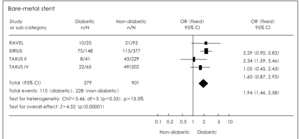
mined rates of restenosis at the later months after im- plantation have been dramatically reduced.14-16) The RAVEL study group demonstrated a significant reduc- tion in restenosis with using sirolimus-eluting stents (SESs), as compared with BMSs, among 238 patients suffering with coronary artery disease.17) The result of a subgroup analysis of the diabetic patients in the RA- VEL trial is even more surprising: among the 19 pa- tients who received SESs, the coronary restenosis rate was 0% compared with 42% for the 25 patients who received BMSs. Since the RAVEL trial included diabe- tic patients with relatively simple coronary lesions, the following SIRIUS trial enrolled patients who had more complex lesions compared with the RAVEL trial.18) Among the diabetic patients in the SIRIUS trial, the coronary restenosis rates were 18% for 131 patients with SESs and 51% for 148 patients with BMSs. In the TAXUS-IV trial, 1314 patients were prospectively ran- domized either to the slow rate-of-release paclitaxel- eluting stent(PES) group or to the BMSs group, and 318 diabetic patients were included in the study. When the TAXUS-IV investigators analyzed the 318 diabetic patients, the PESs were also highly effective in reducing clinical and angiographic restenosis in the patients with diabetes.19) The PES substantially reduced the late lumen loss, the late loss index and the coronary res- tenosis compared with the control BMS.19) The PES implantation demonstrated significantly lower rates of
Correspondence:Do Sun Lim, MD.Division of Cardiology, Cardiovascular Center, Korea University Medical Center, Anamdong 5-ga, Seongbuk- gu, Seoul 136-705, Korea
Tel: 82-2-920-5445, Fax: 82-2-927-1478 E-mail: [email protected]
target lesion revascularization(TLR) and target vessel revascularization(TVR) in diabetics, and so it reduced the need for repeat percutaneous coronary intervention and bypass surgery. The meta-analysis of four major trials that compared BMSs with DESs, 2 trials with SESs(RAVEL, SIRIUS) and 2 trials with PESs(TAX- US II, TAXUS IV), revealed that the diabetic patients, as compared with the nondiabetic patients, had a 94%
higher risk of coronary restenosis in the group that received BMSs(Fig. 1).20) The diabetic patients had a 124% higher risk of coronary restenosis compared with the nondiabetic patients in the group that received DESs(Fig. 2).20) Even with DESs, the diabetic patients showed increased rates of restenosis and a higher late loss index compared with the nondiabetic patients included in this meta-analysis. One may presume that the angiographic rates of restenosis after DES implan- tation have dramatically decreased compared with BMS implantation, but this is less prominent in diabetic patients.
Pathophysiologic Mechanisms of Coronary Restenosis
In-stent restenosis in diabetic patients is associated with complex pathophysiological processes that are not yet completely understood. Hyperplasia of smooth muscle cells in the intimal layer of the vessel wall, the so called neointimal hyperplasia, plays a major role in the process of in-stent restenosis.21) The process of coronary restenosis starts with the initial insult to coronary vessels; this is caused by both intracoronary balloon inflation and stent implantation, and the res- ponse to this initial insult is more prominent in dia- betic patients. Several studies have suggested that the pathophysiological mechanisms of coronary restenosis in diabetic patients are related to a greater degree of underlying vascular inflammation and endothelial dys- function, the elevated fractions of activated platelets with thrombus formation, the dysregulation of the growth factor expression and the elevated levels of advanced glycosylation end products.22)23) Impaired insulin sensi-
Fig. 1. Meta-analysis of the 4 trials comparing the effects of bare-metal stents on restenosis in diabetic and non-diabetic patients.
Bare-metal stent Study
or sub-category
Diabetic n/N
OR (fixed) 95% CI Non-diabetic
n/N
OR (fixed) 95% CI
RAVEL SIRIUS TAXUS II TAXUS IV
10/25 75/148
8/41 22/65 00279
21/93 115/377
43/229 49/202 000901
2.29 (0.90, 5.83) 2.34 (1.59, 3.46) 1.05 (0.45, 2.43) 1.60 (0.87, 2.93) 1.94 (1.46, 2.58) Total (95% CI)
Total events: 115 (diabetic), 228 (non-diabetic) Test for heterogeneity: Chl2=3.46, df=3 (p=0.33), p=13.3%
Test for overall effect: Z=4,52 (p<0.00001)
0.1 0.2 0.5 1 2 5 10 Non-diabetic Diabetic
Fig. 2. Meta-analysis of the 4 trials that compared the effects of drug eluting stents on restenosis in diabetic and non-diabetic patients.
0.1 0.2 0.5 1 2 5 10 Non-diabetic Diebetic Drug-eluting stent
Study or sub-category
Diabetic n/N
OR (fixed) 95% CI Non-diabetic
n/N
OR (fixed) 95% CI
RAVEL SIRIUS TAXUS II TAXUS IV
Not estimable 3.25 (1.82, 06.18) 3.28 (0.78, 13.73) 0.80 (0.29, 02.23) 2.24 (1.39, 03.61) Total (95% CI)
Total events: 31 (diabetic), 49 (non-diabetic)
Test for heterogeneity: Chl2=5.82, df=2 (p=0.05), p=85.7%
Test for overall effect: Z=3,33 (p=0.0009) 0/19 23/131
3/37 5/71 00258
0/101 24/402 6/229 19/220 000952
tivity and endothelial dysfunction also play a major role in the development of coronary restenosis.24) The coro- nary restenosis in diabetic patients results from neoin- timal hyperplasia, and this causes late luminal loss.
Macrophages and polymorphonuclear neutrophils play a major role in the process of coronary restenosis. Ma- crophages and neutrophils release chemokines, and these chemokines serve to increase the amount of matrix metalloproteinase, which leads to remodeling of the extracellular matrix and also smooth muscle cell mi- gration.25-29) In addition, smooth muscle cells are stim- ulated to increase the expression of genes that are in- volved in cell division.30) Several studies have suggested genetic factors in the pathogenesis of coronary res- tenosis, i.e., polymorphism of the gene encoding for the angiotensin-converting enzyme correlated with dif- fuse coronary restenosis.31) Diabetes are prone to coro- nary restenosis since neointimal hyperplasia is increas- ed in the diabetic coronary arteries after coronary in- tervention, which results in greater late in-stent lumen loss.32) It has been assumed that the hyperglycemic state may induce, modify or accentuate the biologic mech- anisms that contribute to the greater tissue proliferation and more diffuse restenotic pattern seen in diabetics.19) Specifically, the vascular thickening may be related to glycosylation of vascular collagen and elastin.19) Insulin could stimulate the mitogen-activated protein kinase (MAPK) signaling pathway, thereby activating the ER- K1 and ERK2 isoforms that are able to stimulate vascular smooth muscle cells, growth factors and cell migration.
In the physiological state of insulin resistance, insulin signaling through the PI3K pathway is decreased, whe- reas the MAPK pathway remains intact.33) Takagi et al.34) have shown that the neointimal index that was measured six months after coronary stenting correlated with the fasting and post-glucose load insulin levels.
Babalik et al.35) have demonstrated that hyperinsuline- mia during the OGTT was a strong predictor of res- tenosis at the six month follow-up. Insulin resistance in diabetics aggravates coronary restenosis through a direct growth factor-like effect of insulin on the vas- cular smooth muscle and neointimal cells.36)
Moreover, increased leptin levels(leptin is a hormone related to both fat metabolism and insulin resistance), have been considered as an independent predictor of restenosis. Chronic hyperleptinemia could decrease the production of nitric oxide by increasing the oxidative stress in endothelial cells.24) The role of hyperleptinemia in the process of coronary restenosis relates to its abi- lity to induce proliferation, differentiation and functional activation of vascular smooth muscle cells, endothelial cells and peripheral blood cells; all of this contributes to increased neointima formation.24) Leptin activates various signal transduction pathways in human mono- cytes and vascular cells such as the PI3K, protein kinase
A and MAPK pathways.37)
Restenosis in the Era of DES
Paclitaxel vs. sirolimusPaclitaxel and sirolimus coated stents are the currently available DESs on the market. Paclitaxel is a derivative of taxol, a compound with antimitotic properties. It inhibits microtubule depolymerization, arrests the cell cycle and stops proliferation. Since paclitaxel modulates cell mitogenesis downstream from Ras/Raf/MAP kinase and it does so independently from the PI3 kinase/
PKb/mTOR signal-transduction pathway, it may be effective for treating the diabetic patients with insulin- resistance as it inhibits both the insulin-dependent and insulin-independent pathways that mediate neointi- mal hyperplasia.38) Microtubules, in addition to their central role for cell division, they control cell signaling, cell activation, cell secretory processes and the cell mi- gration that’s involved in coronary restenosis.39)40)
Sirolimus(rapamycin) is a macrolide antibiotic that was discovered in a microorganism on Easter Island.
Sirolimus exerts antiproliferative effects on smooth muscle cells by inhibiting growth-factor induced and cytokine-induced cell division.41) The mTOR(mam- malian target of rapamycin) is an enzyme located in the nonparticulate region of the cell cytoplasm; this enzyme plays a major role in conducting extracellular signals to the intracellular pathways that adjust cell di- vision and proliferation.42) For activated smooth muscle cells to progress from the G1 phase into the S phase of the cell cycle, the mTOR dependent functions are in- terrupted by their interaction with the FKBP12: siro- limus complex. Sirolimus enters the cell and forms a complex with FKBP12. The FKBP12: sirolimus com- plex prevents the down-regulation of p27Kip1 and the activation of p70s6k, which ultimately results in arrest of the cell cycle at the G1 phase.42) Sirolimus has been coated onto the metal of a stent with an outer layer of biocompatible polymer that enable the drug to elute over a 30-day period.43)
In a recently published ISAR-DIABETES study, the use of the sirolimus-eluting stent was associated with a decrease in the late luminal loss and clinical restenosis, as compared with the use of the paclitaxel-eluting stent, in diabetic patients with coronary artery disease.44) The use of the sirolimus-eluting stent in diabetic patients was associated with a decreased rate of restenosis, and this suggests a reduced risk of target lesion revascula- rization.
Restenosis pattern
Although the rate of coronary restenosis after DES implantation is less frequent when compared with that of BMSs, the prevalence of coronary restenosis is in-
creasing due to the worldwide use of DESs in “real world” interventional cardiology. The patterns of co- ronary restenosis have been described as either diffuse (lesion>10 mm in length) or focal(lesion≤10 mm in length).45) The SIRIUS trial that dealt with relatively longer lesion lengths than those of the previous RAV- EL trial demonstrated a 9.2% rate of coronary restenosis.
The coronary restenosis noted during the SIRIUS trial occurred at the stent margins or at the site of a gap in 64.5% of the cases, and 87% of the coronary restenosis was focal.46) The observations regarding the angiogra- phic pattern of coronary restenosis after implanting SESs by Colombo A et al.47) revealed that all the res- tenotic lesions were focal. This observation was con- cordant with the findings in the SIRIUS trial. All the restenotic lesions, except for 1 multifocal restenosis that involved the distal margin of the stent, were reported to be located in the body of the stent. This finding was different from the SIRIUS and TAXUS II trials where most of the restenotic lesions were located near the stent margins or at the site of a gap between 2 stents (64.5% in the SIRIUS trial and 83.3% in TAXUS II trial).46) Moreover, 4 of 6 multifocal coronary restenoses occurred in diabetic patients.46) For the diabetic patients with coronary restenosis in the TAXUS IV trial, the diffuse type of coronary restenosis was reduced by more than 90%; therefore, most of coronary restenosis were the focal type when angiographic restenosis did occur.19) The proportion of patients with the diffuse type of co- ronary restenosis, if it occurs at all, has clearly been reduced in the era of DESs.
Predictors of Restenosis after DES Implantation in Diabetes
Several studies have demonstrated the clinical and angiographic factors of coronary restenosis after BMS implantation,48-51) and the various factors associated with coronary restenosis after BMS implantation have been reported for the diabetic populations.52)53) Several clinical and angiographic predictive factors of coronary restenosis after BMS implantation were reported by Mercado et al.,48) and diabetes was a major indepen- dent predictive factor of restenosis. Moreover, a large preprocedural reference diameter(RD) and a minimal postprocedural lumen diameter(MLD) were favorable predictive factors after BMS implantation. The stent length was found to be a significant predictor of co- ronary restenosis in BMSs, and for each millimeter of increased stent length, the risk of developing coronary restenosis increased by 4%.48) The risk factors of coro- nary restenosis after BMS implantation include the length of the stented segment and the size of the vessel, with smaller vessels having a higher rate of coronary restenosis.54) Since diabetes is known to be a major pre-
dictor of coronary restenosis after BMS implantation, several studies reported on the predictors of coronary restenosis in diabetic patients after BMS implantation.
The study published by Mazeika et al.53) has reported that poor glycemic control and the vessel size were independent predictors of coronary restenosis in diabetic patients. Another study published by West et al.52) has reported that the vessel caliber, the stented length of the vessel and a lower body mass index were predictors of restenosis in diabetic patients. One of the major risk factors for coronary restenosis is diabetes, which dou- bles the its incidence when compared to non-diabetic patients.55) Most of the studies concerning coronary restenosis after BMS implantation have emphasized the reference vessel diameter, the stent length and diabetes as major predictive factors for restenosis. However, the clinical and angiographic parameters of coronary res- tenosis in diabetic patients after DES implantation have not yet been reported on.
In our preliminary study that included stented pa- tients(n=840) with DESs from the Multicenter PCI Database Registry, we sought to identify the factors that had an influence on the likelihood of restenosis after DES implantation in diabetic patients. From this database, out of 840 patients with a minimum of six months angiographic follow-up, 211(25.1%) had dia- betes. The predictive factors of coronary restenosis were identified by performing multivariate logistic regres- sion analysis. Restenosis occurred in 92 of 629(14.6%) nondiabetic patients and in 44(20.9%) of 211 dia- betic patients(p<0.001). The factors found on multi- variate analysis for predicting restenosis in the diabetic group were current smoking, and current smoking in- creased the risk of coronary restenosis by 92%; a higher C-reactive protein(CRP) level increased the risk of coronary restenosis by 31%, the use of the paclitaxel- eluting stent(PES) increased the risk of coronary res- tenosis by 164%, a longer stent length increased the risk of coronary restenosis by 65%, a smaller reference diameter before DES implantation increased the risk of coronary restenosis by 50%, a smaller reference dia- meter increased the risk of coronary restenosis by 46%, and a minimum lumen diameter at baseline increased the risk of coronary restenosis by 45% after DES im- plantation. We could well observe that diabetes remains a significant predictor of coronary restenosis especially in the cases with a small baseline vessel size, a small post- PCI vessel size, a longer stent length, the use of the PES, a current smoker and a high level of CRP even in this era of DES.
Adjunctive Therapy
Adjunctive therapy with glycoprotein IIb/IIIa inhi- bitor, maintenance of aspirin therapy and administe-
ring a thienopyridine for longer than 1 year should be considered for those diabetic patients who have multiple predictive factor for restenosis.56) Angiotensin-convert- ing enzyme inhibitor, angiotensin receptor blocker and HMG(3-hydroxy-3-methylglutaryl)-CoA reductase in- hibitor should administered when clinically indicated.
Moreover, the thiazolidinediones may further reduce coronary restenosis in diabetic patients by activating the nuclear transcription factor peroxisome proliferator- activated receptor(PPAR)-γ.57) In addition to the op- timal adjunctive pharmacotherapy to prevent coronary restenosis in diabetic patients, instituting strict glycemic control may decrease the restenosis rate in diabetic patients.53)58) These data suggest that strict control of diabetes to achieve hemoglobin A1C levels ≤7.0% may reduce the restenosis rate and so it may improve the clinical outcomes after PCI.
Conclusions
The small reference vessel diameter with a longer lesion length in diabetic patients was noted to increase the rates of coronary restenosis after DES implanta- tion.18)19) The immediate gain in blood flow was often less optimal in diabetic patients, and this was probably due to the large plaque burden; in addition, the neoin- timal hyperplasia increased in diabetic patients after coronary intervention, resulting in greater late lumen loss.59) Using new devices such as DESs reduces the restenosis rate compared with BMSs in diabetic patients by decreasing the late luminal loss. By reducing the coronary restenosis, DESs significantly improved the major limitation of BMS implantation in diabetic pa- tients. Many studies have shown that a blockade of smooth muscle cell proliferation with DESs results in the preservation of the normal vessel phenotype and function, thereby decreasing the rate of both neointi- mal hyperplasia and in-stent restenosis.60-64) Since the introduction of paclitaxel and sirolimus-eluting stents, these devices are regarded by many physicians and scientists to be the standard of treatment for diabetic patients undergoing stent implantation. Even though DESs could lower the restenosis rates by preventing smooth-muscle cell proliferation at the stented site, atherosclerosis could progress at other coronary sites.
Therefore, combined approaches that use systemic the- rapies are required to prevent neointimal proliferation and to prevent atherosclerosis progression at the other coronary sites in diabetic patients.
BMS implantation significantly reduces the coronary restenosis rates and the target lesion revascularization (TLR) rate in the diabetic patient when compared to balloon angioplasty alone, and DESs further reduce the coronary restenosis rates and the TLR rate in the diabetic patient as compared to the BMSs. However,
the rate of coronary restenosis is still higher in diabetic patients, when compared with non-diabetic patients, and even after DES implantation. Diabetes still remains a major risk factor for coronary restenosis after DES implantation, and so aggressive risk factor management with the concomitant pharmacotherapy should be done to reduce the risk of coronary restenosis, and especially in diabetic patients who have undergone DES implan- tation.
REFERENCES
1) Laakso M. Hyperglycemia and cardiovascular disease in type 2 diabetes. Diabetes 1999;48:937-42.
2) Waller BF, Palumbo PJ, Lie JT, Roberts WC. Status of the co- ronary arteries at necropsy in diabetes mellitus with onset after age 30 years: analysis of 229 diabetic patients with and without clinical evidence of coronary heart disease and comparison to 183 control subjects. Am J Med 1980;69:498-506.
3) Kip KE, Faxon DP, Detre KM, Yeh W, Kelsey SF, Currier JW.
Coronary angioplasty in diabetic patients: The National Heart, Lung, and Blood Institute Percutaneous Transluminal Coronary Angioplasty Registry. Circulation 1996;94:1818-25.
4) Heart Outcomes Prevention Evaluation Study Investigators. Ef- fects of ramipril on cardiovascular and microvascular outcomes in people with diabetes mellitus: results of the HOPE study and MICRO-HOPE substudy. Lancet 2000;355:253-9.
5) Stamler J, Vaccaro O, Neaton JD, Wentworth D. Diabetes, other risk factors, and 12-yr cardiovascular mortality for men scre- ened in the Multiple Risk Factor Intervention Trial. Diabetes Care 1993;16:434-44.
6) Alderman EL, Corley SD, Fisher LD, et al. Five-year angiogra- phic follow-up of factors associated with progression of coronary artery disease in the Coronary Artery Surgery Study (CASS). J Am Coll Cardiol 1993;22:1141-54.
7) Jeon HS, Chae JK, Moon SK, Kim WH, Ko JK. Early and mid- term results of coronary stenting in the diabetic patient. Korean Circ J 1999;29:292-7.
8) Mick MJ, Piedmonte MR, Arnold AM, Simpfendorfer C. Risk stratification for long-term outcome after elective coronary an- gioplasty: a multivariate analysis of 5,000 patients. J Am Coll Cardiol 1994;24:74-80.
9) Stein B, Weintraub WS, Gebhart SP, et al. Influence of diabetes mellitus on early and late outcome after percutaneous translumi- nal coronary angioplasty. Circulation 1995;91:979-89.
10) Mathew V, Gersh BJ, Williams BA, et al. Outcomes in patients with diabetes mellitus undergoing percutaneous coronary inter- vention in the current era: a report from the Prevention of RES- tenosis with Tranilast and its Outcomes (PRESTO) trial. Circula- tion 2004;109:476-80.
11) Elezi S, Kastrati A, Pache J, et al. Diabetes mellitus and the clinical and angiographic outcome after coronary stent placement.
J Am Coll Cardiol 1998;32:1866-73.
12) Kim W, Jeong MH, Kim KH, et al. Long-term clinical and angiographic results of coronary stenting in diabetic patients.
Korean Circ J 2001;31:24-30.
13) Schofer J, Schluter M, Rau T, Hammer F, Haag N, Mathey DG.
Influence of treatment modality on angiographic outcome after coronary stenting in diabetic patients: a controlled study. J Am Coll Cardiol 2000;35:1554-9.
14) Stone GW, Ellis SG, Cox DA, et al. A polymer-based, paclitaxel- eluting stent in patients with coronary artery disease. N Engl J Med 2004;350:221-31.
15) Seung KB. Drug eluting stent and percutaneous coronary inter- vention. Korean Circ J 2003;33:857-60.
16) Kim KH, Jeong MH, Hong SN, et al. The clinical effects of drug- eluting stents for the treatment of coronary in-stent restenosis.
Korean Circ J 2005;35:443-7.
17) Morice MC, Serruys PW, Sousa JE, et al. A randomized com- parison of a sirolimus-eluting stent with a standard stent for coronary revascularization. N Engl J Med 2002;346:1773-80.
18) Moses JW, Leon MB, Popma JJ, et al. Sirolimus-eluting stents versus standard stents in patients with stenosis in a native co- ronary artery. N Engl J Med 2003;349:1315-23.
19) Hermiller JB, Raizner A, Cannon L, et al. Outcomes with the polymer-based paclitaxel-eluting TAXUS stent in patients with diabetes mellitus: the TAXUS-IV trial. J Am Coll Cardiol 2005;
45:1172-9.
20) Scheen AJ, Warzee F. Diabetes is still a risk factor for restenosis after drug-eluting stent in coronary arteries. Diabetes Care 2004;
27:1840-1.
21) Farb A, Weber DK, Kolodgie FD, Burke AP, Virmani R. Mor- phological predictors of restenosis after coronary stenting in humans. Circulation 2002;105:2974-80.
22) Aronson D, Bloomgarden Z, Rayfield EJ. Potential mechanisms promoting restenosis in diabetic patients. J Am Coll Cardiol 1996;27:528-35.
23) Kereiakes DJ, Young JJ. Percutaneous coronary revasculariza- tion of diabetic patients in the era of drug-eluting stents. Rev Cardiovasc Med 2005;6(Suppl 1):S48-58.
24) Piatti P, Monti LD. Insulin resistance, hyperleptinemia and en- dothelial dysfunction in coronary restenosis. Curr Opin Pharma- col 2005;5:160-4.
25) Kornowski R, Hong MK, Tio FO, Bramwell O, Wu H, Leon MB.
In-stent restenosis: contributions of inflammatory responses and arterial injury to neointimal hyperplasia. J Am Coll Cardiol 1998;31:224-30.
26) Komatsu R, Ueda M, Naruko T, Kojima A, Becker AE. Neointi- mal tissue response at sites of coronary stenting in humans:
macroscopic, histological, and immunohistochemical analyses.
Circulation 1998;98:224-33.
27) Ott I, Neumann FJ, Kenngott S, Gawaz M, Schomig A. Procoa- gulant inflammatory responses of monocytes after direct balloon angioplasty in acute myocardial infarction. Am J Cardiol 1998;
82:938-42.
28) Southgate KM, Fisher M, Banning AP, et al. Upregulation of basement membrane-degrading metalloproteinase secretion after balloon injury of pig carotid arteries. Circ Res 1996;79:1177-87.
29) Dollery CM, Humphries SE, McClelland A, Latchman DS, Mc- Ewan JR. Expression of tissue inhibitor of matrix metallopro- teinases 1 by use of an adenoviral vector inhibits smooth muscle cell migration and reduces neointimal hyperplasia in the rat model of vascular balloon injury. Circulation 1999;99:3199-205.
30) Bauters C, de Groote P, Adamantidis M, et al. Proto-oncogene expression in rabbit aorta after wall injury: first marker of the cellular process leading to restenosis after angioplasty? Eur Heart J 1992;13:556-9.
31) Amant C, Bauters C, Bodart JC, et al. D allele of the angiotensin I-converting enzyme is a major risk factor for restenosis after coronary stenting. Circulation 1997;96:56-60.
32) Kornowski R, Mintz GS, Kent KM, et al. Increased restenosis in diabetes mellitus after coronary interventions is due to exagge- rated intimal hyperplasia: a serial intravascular ultrasound study.
Circulation 1997;95:1366-9.
33) Cusi K, Maezono K, Osman A, et al. Insulin resistance differen- tially affects the PI 3-kinase- and MAP kinase-mediated signal- ing in human muscle. J Clin Invest 2000;105:311-20.
34) Takagi T, Yoshida K, Akasaka T, et al. Hyperinsulinemia during oral glucose tolerance test is associated with increased neoin- timal tissue proliferation after coronary stent implantation in nondiabetic patients: a serial intravascular ultrasound study. J Am Coll Cardiol 2000;36:731-8.
35) Babalik E, Gurmen T, Orhan L, et al. Increased secretion of in- sulin during oral glucose tolerance test can be a predictor of stent restenosis in nondiabetic patients. Catheter Cardiovasc Interv 2003;58:306-12.
36) Beckman JA, Creager MA, Libby P. Diabetes and atheroscle- rosis: epidemiology, pathophysiology, and management. JAMA 2002;287:2570-81.
37) Parhami F, Tintut Y, Ballard A, Fogelman AM, Demer LL.
Leptin enhances the calcification of vascular cells: artery wall as a target of leptin. Circ Res 2001;88:954-60.
38) le Roith D, Zick Y. Recent advances in our understanding of insulin action and insulin resistance. Diabetes Care 2001;24:
588-97.
39) Belotti D, Vergani V, Drudis T, et al. The microtubule-affecting drug paclitaxel has antiangiogenic activity. Clin Cancer Res 1996;2:1843-9.
40) Sollott SJ, Cheng L, Pauly RR, et al. Taxol inhibits neointimal smooth muscle cell accumulation after angioplasty in the rat. J Clin Invest 1995;95:1869-76.
41) Marx SO, Jayaraman T, Go LO, Marks AR. Rapamycin-FKBP inhibits cell cycle regulators of proliferation in vascular smooth muscle cells. Circ Res 1995;76:412-7.
42) Carter AJ. TOR of the cell cycle: are there important implica- tions for diabetics in the era of the drug-eluting stent? Catheter Cardiovasc Interv 2004;61:233-6.
43) Curfman GD. Sirolimus-eluting coronary stents. N Engl J Med 2002;346:1770-1.
44) Dibra A, Kastrati A, Mehilli J, et al. Paclitaxel-eluting or siro- limus-eluting stents to prevent restenosis in diabetic patients. N Engl J Med 2005;353:663-70.
45) Mehran R, Dangas G, Abizaid AS, et al. Angiographic patterns of in-stent restenosis: classification and implications for long- term outcome. Circulation 1999;100:1872-8.
46) Moses JW, Leon MB, Popma JJ, et al. Sirolimus-eluting stents versus standard stents in patients with stenosis in a native coro- nary artery. N Engl J Med 2003;349:1315-23.
47) Colombo A, Orlic D, Stankovic G, et al. Preliminary observa- tions regarding angiographic pattern of restenosis after rapamy- cin-eluting stent implantation. Circulation 2003;107:2178-80.
48) Mercado N, Boersma E, Wijns W, et al. Clinical and quantitative coronary angiographic predictors of coronary restenosis: a com- parative analysis from the balloon-to-stent era. J Am Coll Car- diol 2001;38:645-52.
49) Elezi S, Kastrati A, Neumann FJ, Hadamitzky M, Dirschinger J, Schomig A. Vessel size and long-term outcome after coronary stent placement. Circulation 1998;98:1875-80.
50) Kastrati A, Elezi S, Dirschinger J, Hadamitzky M, Neumann FJ, Schomig A. Influence of lesion length on restenosis after coro- nary stent placement. Am J Cardiol 1999;83:1617-22.
51) Bauters C, Hubert E, Prat A, et al. Predictors of restenosis after coronary stent implantation. J Am Coll Cardiol 1998;31:1291-8.
52) West NE, Ruygrok PN, Disco CM, et al. Clinical and angiogra- phic predictors of restenosis after stent deployment in diabetic patients. Circulation 2004;109:867-73.
53) Mazeika P, Prasad N, Bui S, Seidelin PH. Predictors of angio- graphic restenosis after coronary intervention in patients with diabetes mellitus. Am Heart J 2003;145:1013-21.
54) Kobayashi Y, de Gregorio J, Kobayashi N, et al. Stented segment length as an independent predictor of restenosis. J Am Coll
Cardiol 1999;34:651-9.
55) Lau KW, Ding ZP, Johan A, Lim YL. Midterm angiographic outcome of single-vessel intracoronary stent placement in dia- betic versus nondiabetic patients: a matched comparative study.
Am Heart J 1998;136:150-5.
56) Yusuf S, Zhao F, Mehta SR, Chrolavicius S, Tognoni G, Fox KK.
Effects of clopidogrel in addition to aspirin in patients with acute coronary syndromes without ST-segment elevation. N Engl J Med 2001;345:494-502.
57) Choi D, Kim SK, Choi SH, et al. Preventative effects of rosigli- tazone on restenosis after coronary stent implantation in patients with type 2 diabetes. Diabetes Care 2004;27:2654-60.
58) Corpus RA, George PB, House JA, et al. Optimal glycemic control is associated with a lower rate of target vessel revascularization in treated type II diabetic patients undergoing elective percu- taneous coronary intervention. J Am Coll Cardiol 2004;43:8-14.
59) Kornowski R, Mintz GS, Kent KM, et al. Increased restenosis in diabetes mellitus after coronary interventions is due to exagge-
rated intimal hyperplasia: a serial intravascular ultrasound study.
Circulation 1997;95:1366-9.
60) Moon KW, Lee JM, Chang K, et al. Oral everolimus reduces adventitial cell activation and neointima formation in balloon- injured rat carotid artery. Korean Circ J 2004;34:983-91.
61) Cha KS, Jeong MH, Lee SU, et al. The effects of local delivery of paclitaxel nanoparticle on porcine coronary stent restenosis.
Korean Circ J 2000;30:208-20.
62) Cho MC, Kwak NJ, Piao H, et al. Effect of paclitaxel local delivery on neointimal formation after endothelial denudation of the rat carotid artery. Korean Circ J 2000;30:198-207.
63) Chang KY, Seung KB, Kang DH, et al. The inhibition of neointi- mal hyperplasia by combination of external radiation and pa- clitaxel in a rat carotid injury model. Korean Circ J 2000;30:
758-66.
64) Fattori R, Piva T. Drug-eluting stents in vascular intervention.
Lancet 2003;361:247-9.