Received:July 6, 2016, Revised:July 28, 2016, Accepted:August 8, 2016
Corresponding to:Bin Yoo, Department of Internal Medicine, Asan Medical Center, University of Ulsan College of Medicine, 88 Olympic-ro 43-gil, Songpa-gu, Seoul 05505, Korea. E-mail:[email protected]
pISSN: 2093-940X, eISSN: 2233-4718
Copyright ⓒ 2017 by The Korean College of Rheumatology. All rights reserved.
This is a Open Access article, which permits unrestricted non-commerical use, distribution, and reproduction in any medium, provided the original work is properly cited.
Refractory Pleural Effusion in Systemic Lupus Erythematosus Treated by Pleurectomy
Sichan Kim1, Han-bit Park1, Yun Kyung Cho1, Sangyoung Yi1, Kyunghwan Oh1, Dong Kwan Kim2, Bin Yoo1
Departments of 1Internal Medicine, 2Thoracic and Cardiovascular Surgery, Asan Medical Center, University of Ulsan College of Medicine, Seoul, Korea
Pleural effusion is a common pulmonary manifestation of systemic lupus erythematosus (SLE) and often occurs as bilateral exu- dative pleural effusion. The condition usually responds quickly to corticosteroid therapy. However, massive pleural effusion refractory to immunosuppressive drugs has rarely been reported; thus, the proper therapeutic modality is largely decided on a case-by-case basis. In this case, we describe successful treatment with surgical pleurectomy for massive refractory pleural effu- sion in a patient with SLE. (J Rheum Dis 2017;24:43-47)
Key Words. Systemic lupus erythematosus, Pleural effusion, Fibrothorax, Pleurectomy
INTRODUCTION
Systemic lupus erythematosus (SLE) is an autoimmune disorder that primarily affects women and can involve vir- tually any organ, including joints, kidneys, and lungs.
Pleural effusion is the most common manifestation of pulmonary involvement in SLE, and it reportedly occurs in 45% to 60% SLE patients [1]. The pleural fluid is exu- dative in most cases and often bilateral. It is usually small or moderate (<1,000 mL) and may be asymptomatic but can also present with respiratory symptoms, such as dyspnea and chest pain [2]. Sometimes, however, mas- sive pleural effusion also has been reported [3,4].
Treatment of pleural effusion in SLE must be in- dividualized in each case because small asymptomatic ef- fusion does not require treatment [1]. Symptomatic pleu- ral effusion is usually well controlled with moderate doses of corticosteroids, which shows that most patients have responded within days to corticosteroids [3]. When the fluid volume is large and the patient is symptomatic, aspiration of pleural fluid may be required for sympto- matic control. Rarely, however, the pleural effusion in
SLE maybe refractory to treatments, including cortico- steroids and immunosuppressive drugs, thus requiring repeated therapeutic thoracentesis [2,5].
Herein, we describe a case of refractory massive pleural effusion in SLE that was successfully controlled by pleurectomy.
CASE REPORT
A 27-year-old woman with SLE presented with symp- toms of dyspnea. She was diagnosed with SLE 1 year earlier. At the time of diagnosis, she had polyarthritis, photosensitivity, and pleural effusion. Anti-nuclear anti- body titers were at 1:320, but anti-dsDNA titers were negative. Anti-Ro (SSA) and anti-La (SSB) were positive.
She had low C3 level (35.5 mg/dL; reference range, 90∼
180 mg/dL), and normal C4 level. So her SLE disease ac- tivity index-2K (SLEDAI-2K) score was 8. She was treat- ed with a low dose of corticosteroid (prednisolone, 7.5 mg/day) and hydroxychloroquine, which resulted in im- provement of joint symptoms. However, a large amount of bilateral pleural effusion was noted. Subsequently, pre-
Figure 1. Chest X-ray showing bilateral pleural effusion.
Figure 2. Chest X-ray showing persistent bilateral pleural effu- sion after pleurodesis of right pleura and rituximab admini- stration.
dnisone dosage was increased to 30 mg/day to control the pleural effusion. Four weeks later, the patient had persis- tent shortness of breath and massive pleural effusion (Figure 1). She was admitted and thoracentesis of left pleural effusion was performed, and 1,000 mL of fluid was drained. Pleural fluid analysis showed a white blood cell count of 320/mm3, with 68% lymphocytes, lactate de- hydrogenase (LDH) of 81 IU/L (serum LDH, 172 IU/L), and protein of 5.0 g/dL (serum protein, 6.7 g/dL), glucose of 109 mg/dL, adenosine deaminase (ADA) of 17.2 U/L.
Anti-nuclear antibody titers of pleural effusion were 1/320. Malignant cells were not observed on cytological examination of the pleural fluids, and Gram staining and culture results were negative. Tuberculosis (TB) culture and TB-polymerase chain reaction (PCR) were repeatedly negative. So pleural effusion was aseptic, non-malignant and had exudative features, we assumed pleural effusion was SLE-associated pleurisy. At that time, SLEDAI-2K score was 6 due to arthritis and pleurisy. Azathioprine 100 mg/day was added and the patient was followed up in the outpatient clinic. After 4 months, the patient did not show improvement in her dyspnea with pleural effusion, and she required repeated thoracentesis for symptomatic relief. Thus, she was readmitted and treated with ritux- imab 1 g at 2-week intervals. Azathioprine was ceased.
Nevertheless, pleural effusion remained uncontrolled, and mycophenolate mofetil (MMF) (1,500 mg/day) was administered in the outpatient clinic. Over the next 7 months, however, other SLE manifestations were absent but massive bilateral pleural effusion persisted, so
SLEDAI-2K score was 2 and pleural effusion still required repeated thoracentesis about monthly. Every performed thoracentesis, fluid was drained about 1.5∼2 L and pleu- ral effusion was exudate, its ADA level was below 40 U/L, malignant cells were not observed on cytology. To control the refractory pleural effusion, pleurodesis for right pleu- ral effusion was performed with minocycline as a scleros- ing agent and we discontinued MMF. Four months later from pleurodesis, rituximab 1 g was administrated again at 2-weeks interval (Figure 2).
Although the right pleural effusion was controlled, the left pleural effusion remained persistent. Therefore, she was readmitted to hospital for further investigation and treatment. On admission, her blood pressure was 132/95 mmHg, pulse rate was 107 beats per minute, respiration rate was 18 times per minute, and body temperature was 36.8oC. In the initial physical examination, auscultation revealed decreased breath sounds in the bilateral lung field but no audible crackles. Laboratory tests showed a white blood cell count of 2,700/mm3, a hemoglobin level of 12.5 g/dL, a platelet count of 295,000/mm3, a C-re- active protein level of 0.20 mg/dL, and erythrocyte sed- imentation rate of 28 mm/h. Complement components were within normal ranges (C3, 100.0 mg/dL; C4, 26.6 mg/dL), and anti-dsDNA antibody was also normal (3.8 IU/mL, 0.7). Left pleural effusion fluid analysis showed a white blood cell count of 800/mm3, with 78% lympho- cytes, LDH of 249 IU/L (serum LDH, 152 IU/L), and pro- tein of 4.3 g/dL (serum protein, 5.9 g/dL), glucose of 88
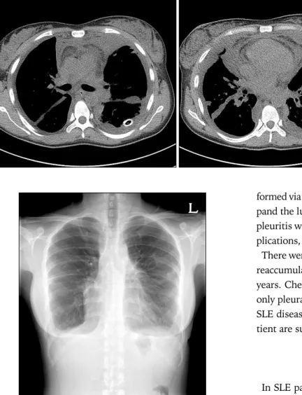
Figure 3. Chest computed to- mography showing bilateral pleural effusion with multilo- culated effusion in both hemi- thoraces.
Figure 4. Chest X-ray showing remaining bilateral cardio- phrenic angle blunting but a decreased amount of both pleural effusions after pleurectomy.
mg/dL. Culture and TB-PCR results of pleural effusion were negative. Chest X-ray showed pleural thickening with sequelae after pleurodesis in the right hemithorax and persistent massive pleural effusion on the left side.
Until then SLEDAI-2K score was continuously 2, Other SLE manifestations except pleurisy were absent.
A chest tube was inserted into the left pleural effusion and drained over 6 days. Chest computed tomography showed massive bilateral pleural effusion with multi- loculated effusion in both hemithoraces (Figure 3). After consultation with a thoracic surgeon, we decided to per- form the pleurodesis by using video-assisted thoraco- scopic surgery. Because of severe adhesions and pleural thickening in the thoracic cavity, lung injury occurred during the pleurodesis. Finally, a pleurectomy was per-
formed via an open thoracotomy for fibrosed pleura to ex- pand the lung. Decorticated left pleura showed choronic pleuritis with fibrosis. There were no postoperative com- plications, and the patient was discharged.
There were no further episodes of shortness of breath or reaccumulation of pleural effusion over a period of 2 years. Chest X-ray at 2 years after pleurectomy showed only pleural thickening of both lungs (Figure 4). Overall SLE disease activity index score and drug history of pa- tient are summarized into Figure 5.
DISCUSSION
In SLE patients, dyspnea caused by pleural effusion is uncommon and should be managed urgently. We experi- enced the case of a SLE patient with pleural effusion re- fractory to medical treatments, and who was managed with repetitive thoracentesis. Here, we report that pleu- rodesis or pleurectomy could be one of treatment option for refractory pleural effusion in patients with SLE.
Treatment of pleural effusion in SLE depends on the se- verity of symptoms. Small asymptomatic effusions may not require treatment. Nonsteroidal anti-inflammatory drugs could be useful for control of mild pleurisy, whereas corticosteroid therapy is indicated for more severe cases [1]. Hydroxychloroquine and immunosuppressive agents, such as azathioprine may be added in selected cases [2].
However, most patients with SLE-associated pleurisy re- spond within days to moderate doses of corticosteroids [3]. When the fluid volume is large, aspiration of pleural fluid may be required for the control of respiratory symp- toms [2,5]. Rarely, the effusion is refractory to therapy, but it is not clear which treatment approach is preferred in such refractory cases [3]. Immunosuppressive drugs,
Figure 5. Drug history and systemic lupus erythematosus (SLE) disease activity index-2K score. HCQ: hydroxychloroquine, MMF:
mycophenolate mofetil, SLEDAI-2K: SLE disease activity index-2K.
including azathioprine, cyclophosphamide, and metho- trexate can be considered when pleural effusion is re- fractory to corticosteroids [2]. Moreover, MMF and ritux- imab also can be considered as a viable therapeutic option in SLE treatment. MMF is an immunosuppressive drug widely used in solid organ transplantation, and it may play a role in SLE. Karim et al. [6] reported effectiveness of MMF to extra-reanl and renal manifestations of SLE by showing decrease of SLEDAI score and proteinuria. And it appears to be a safe and effective alternative im- munosuppressant for systemic manifestations of SLE not responding to conventional immunosuppressive treat- ment. And one randomized controlled study showed that MMF was more effective than intravenous cyclo- phosphamide in inducing remission of lupus nephritis and had a more favorable safety profile [7]. Another study summarized about rituximab treated SLE cases referred that 2 of 7 patients had lupus pleuritis and their clinical course showed improvement after B cell depletion [8].
For this reason, we administrated MMF and rituximab for control of SLE-associated pleurisy, but effect of these agents was limited.
So in cases refractory to systemic therapy like our pa- tient, local intervention, including pleurodesis and pleur- ectomy, may be required [5]. Some previous case reports on local therapy for pleural effusion have shown that pleurodesis with talc was effective to control the pleural effusion [9-12]. In addition, to date, there have been sev- eral reported cases of SLE patients treated with pleur- ectomy for refractory pleural effusion [5,13,14]. One of these cases reported a woman with dyspnea due to re- fractory pleural effusion of SLE, and she was treated by pleurectomy. After operation, her dyspnea, forced vital
capacity and total lung capacity were improved [5]. Other reports have shown that thoracoscopic partial pleur- ectomy combined with talc pleurodesis achieved good re- sults for control of refractory pleural effusion [13].
However, there is a report of one patient in whom pleural effusion could not be controlled by pleurodesis and pleur- ectomy [14]. In some cases, chest tube drainage has failed to expand the lung because of visceral pleural thickening;
therefore, open surgical decortication was required to ex- pand the collapsed lung and resolution of pneumothorax [15]. In our patient, pleural effusion refractory to im- munosuppressant agents including rituximab and MMF was controlled by performing open surgical pleurectomy to expand the lung because of fibrosis and adhesion.
SUMMARY
In summary, refractory pleural effusion is uncommon in SLE patients. Pleurectomy might be considered as a local treatment method for refractory pleural effusion, partic- ularly in cases where systemic disease activity is well controlled.
CONFLICT OF INTEREST
No potential conflict of interest relevant to this article was reported.
REFERENCES
1. Keane MP, Lynch JP 3rd. Pleuropulmonary manifestations of systemic lupus erythematosus. Thorax 2000;55:159-66.
2. Breuer GS, Deeb M, Fisher D, Nesher G. Therapeutic op- tions for refractory massive pleural effusion in systemic lu-
pus erythematosus: a case study and review of the literature.
Semin Arthritis Rheum 2005;34:744-9.
3. D’Cruz D, Khamashta MA, Hughes G. Pulmonary manifes- tations of systemic lupus erythematosus. 6th ed.
Philadelphia, Lippincott: Williams & Wilkins, 2002, p.
663-84.
4. Murin S, Wiedemann HP, Matthay RA. Pulmonary manifes- tations of systemic lupus erythematosus. Clin Chest Med 1998;19:641-65.
5. Sharma S, Smith R, Al-Hameed F. Fibrothorax and severe lung restriction secondary to lupus pleuritis and its success- ful treatment by pleurectomy. Can Respir J 2002;9:335-7.
6. Karim MY, Alba P, Cuadrado MJ, Abbs IC, D'Cruz DP, Khamashta MA, et al. Mycophenolate mofetil for systemic lupus erythematosus refractory to other immunosuppressive agents. Rheumatology (Oxford) 2002;41:876-82.
7. Ginzler EM, Dooley MA, Aranow C, Kim MY, Buyon J, Merrill JT, et al. Mycophenolate mofetil or intravenous cy- clophosphamide for lupus nephritis. N Engl J Med 2005;
353:2219-28.
8. Ng KP, Leandro MJ, Edwards JC, Ehrenstein MR, Cambridge G, Isenberg DA. Repeated B cell depletion in treatment of
refractory systemic lupus erythematosus. Ann Rheum Dis 2006;65:942-5.
9. Glazer M, Berkman N, Lafair JS, Kramer MR. Successful talc slurry pleurodesis in patients with nonmalignant pleural effusion. Chest 2000;117:1404-9.
10. Rodriguez-Panadero F, Antony VB. Pleurodesis: state of the art. Eur Respir J 1997;10:1648-54.
11. Ukale V, Agrenius V, Hillerdal G, Mohlkert D, Widström O.
Pleurodesis in recurrent pleural effusions: a randomized comparison of a classical and a currently popular drug. Lung Cancer 2004;43:323-8.
12. Weissberg D, Ben-Zeev I. Talc pleurodesis. Experience with 360 patients. J Thorac Cardiovasc Surg 1993;106:689-95.
13. Elborn JS, Conn P, Roberts SD. Refractory massive pleural effusion in systemic lupus erythematosus treated by pleu- rectomy. Ann Rheum Dis 1987;46:77-80.
14. Sherer Y, Langevitz P, Levy Y, Fabrizzi F, Shoenfeld Y.
Treatment of chronic bilateral pleural effusions with intra- venous immunoglobulin and cyclosporin. Lupus 1999;8:
324-7.
15. Passero FC, Myers AR. Hemopneumothorax in systemic lu- pus erythematosus. J Rheumatol 1980;7:183-6.