http://www.ecevr.org/ 35
CLINICAL
EXPERIMENTAL VACCINE
RESEARCH
Introduction
Bordetella pertussis is the main pathogen causing whooping cough (pertussis) that oc- curs after transmission of the bacteria in the form of airborne droplets between indi- viduals. The incidence of this infection is highest in children under 5 years old, and the disease causes severe illness and death among neonates and infants. Pertussis vaccines
© Korean Vaccine Society.
This is an Open Access article distributed under the terms of the Creative Commons Attribution Non-Com- mercial License (http://creativecommons.org/licenses/
by-nc/4.0) which permits unrestricted non-commercial use, distribution, and reproduction in any medium, pro- vided the original work is properly cited.
K O R E A N V A C C I N E S O C I E T Y
K O R E A N K O R E A N A C C I N E O C I E T Y V
S
Clin Exp Vaccine Res 2019;8:35-42 https://doi.org/10.7774/cevr.2019.8.1.35 pISSN 2287-3651 • eISSN 2287-366X
Chulmin Park1, Dong Ho Huh1, Seung Beom Han1,2, Gi Sub Choi3, Kyu Ri Kang1, Ji Ahn Kim1, Jin Han Kang1,2
1Vaccine Bio Research Institute, 2Department of Pediatrics, College of Medicine, The Catholic University of Korea, Seoul; 3Research Center, GC Pharma, Yongin, Korea
Received: December 27, 2018 Revised: January 14, 2019 Accepted: January 18, 2019
Corresponding author: Jin Han Kang, MD, PhD Department of Pediatrics, Seoul St. Mary’s Hospi- tal, College of Medicine, The Catholic University of Korea, 222 Banpo-daero, Seocho-gu, Seoul 06591, Korea
Tel: +82-2-2258-6183, Fax: +82-2-537-4544 E-mail: [email protected]
G. S. Choi is an employee of the Green Cross Corporation, but he was not involved in data acquisition and analysis. All other authors declare no conflict of interest. This work was supported by GC Pharma (Yongin, Republic of Korea, grant No. 5-2017-D0083-0002).
This work was supported by Korea Ministry of Food and Drug Safety (MFDS) (grant No.
16172MFDS269).
Purpose: There is no standard method for confirming the immunogenicity of acellular pertus- sis vaccines. We tried to develop a local standard method for evaluating the immunogenicity of the three-component of acellular pertussis vaccines which was developed by a Korean lo- cal company.
Materials and Methods: The developed pertussis antigens (pertussis toxin, filamentous hemagglutinin, pertactin) were evaluated by in-house enzyme-linked immunosorbent assay (ELISA) using 189 negative sera, 25 positive sera, and 73 paired sera (pre- and post-Tdap [teta- nus, diphtheria, and acellular pertussis] vaccinated sera). ELISA units were calculated by the reference line method, compared with World Health Organization reference sera, and the cut- off value was calculated using negative sera.
Results: When compared to National Institute for Biological Standards and Control control antigen (NIBSC) control antigens, the developed pertussis toxin (PT) and filamentous haemag- glutinin (FHA) antigens were 203.48 and 61.60 IU/μg, respectively. Each in-house ELISA was established by validating the coefficients of variation % (PT, 11.53%; FHA, 8.60%; pertactin [PRN], 9.86%) obtained from the results of inter- and intra-assay variation. Also, the cut-off values of PT, FHA, and PRN were 11.65, 38.95, and 5.66 EU/mL, respectively. The distributions of antibody levels in paired showed that 93.15% (68/73) in anti-PT IgG, 97.26% (72/73) in anti-FHA IgG, and 100% in anti-PRN IgG were higher than a 100% increase after vaccination. Addition- ally, the values of 89.04% (65/73) in anti-PT IgG, 97.26% (72/73) in anti-FHA IgG, and 100% in anti-PRN IgG were below each cut-off point.
Conclusion: We established an in-house ELISA method using self-developed antigens, and these immunoassays have provided a way to standardize measuring the immunogenicity of newly developed vaccines, through single- and dual-serology.
Keywords: Pertussis vaccine, Pertussis toxin, FHA protein, Pertactin, Enzyme-linked immuno- sorbent assay, Serology
Development and implementation of standardized method for
detecting immunogenicity of acellular pertussis vaccines in Korea
1 / 1 CROSSMARK_logo_3_Test
2017-03-16 https://crossmark-cdn.crossref.org/widget/v2.0/logos/CROSSMARK_Color_square.svg
have been very effective for over 40 years. However, re-emer- gence of pertussis was recently reported even in countries with high vaccination coverage [1-3]. This increased inci- dence of adolescents and adults and familial transmission have been reported in many countries [4,5]. Recently several risk causal factors have been suggested to contribute to the re- emergence of pertussis. First, protection after natural pertus- sis infection exposure is not life-long (typically 7 years) of the decreased immunity of pertussis vaccines may explain the shift in the incidence peak from school-age to adolescents/
adults [2,6]. Additionally, the genetic changes in circulating strains of B. pertussis may have caused the re-emergence of pertussis [2,7,8]. Thus, active immunization using pertussis vaccines is strongly recommended and a novel pertussis vac- cine for overcoming these problems should be developed.
To prevent pertussis, diphtheria, and tetanus, diphtheria tox- oid and acellular pertussis (DTaP) vaccines are administered to children under 7 years of age. DTaP vaccines consist of two, three, or five components of pertussis antigens (pertussis tox- in, filamentous hemagglutinin, agglutinogens 1, 2) to reduce adverse events. Additionally, tetanus, diphtheria, and acellu- lar pertussis (Tdap) vaccines which contain reduced doses of diphtheria and pertussis have been used as booster vaccines in adolescents and adults. In Korea, these acellular pertussis (aP) vaccines have been used since the 1990s, and all DTaP and Tdap vaccines are imported from foreign countries. How- ever, pertussis vaccines are costly and the supply is unstable.
Three-component acellular vaccines (DTaP and Tdap) have been developed by a domestic company to solve these prob- lems. Phase I and IIa clinical studies using the newly devel- oped Tdap vaccine were conducted in 2017.
Currently, there is no standard method for confirming the immunogenicity of aP vaccines. Thus, a standard method should be developed by each manufacturer or country while developing novel aP vaccines. Here, we developed a local standard method for evaluating the immunogenicity of the three-component (pertussis toxin, filamentous hemaggluti- nin, and pertactin [PRN]) in Tdap vaccine (GC Tdap) which was developed by GC Pharma (Yongin, Korea).
Materials and Methods
Antigens and sera
Reference serum (World Health Organization [WHO] Inter- national Standard Pertussis Antiserum, National Institute for Biological Standards and Control control antigen [NIBSC]
06/140), reference pertussis toxin (PT; NIBSC JNIH-5) [9], and filamentous haemagglutinin (FHA; NIBSC JNIH-4) anti- gens were purchased from the NIBSC (Hertfordshire, UK).
PRN antigen was supplied by GC Pharma. The PT, FHA, and PRN antigens of the GC Tdap vaccine were compared to ref- erence antigens by immunological analysis with reference serum. For immunological evaluation of these antigens, 189 negative sera obtained from healthy volunteers, 25 sera from patients with pertussis (confirmed by culture and polymerase chain reaction), and 73 paired sera from pre- and post-Tdap vaccinated sera (46 paired sera obtained from GC Tdap-vac- cinated cases, group A; 27 paired sera obtained from GSK Tdap-vaccinated cases, group B) were used.
Immunological analysis
To evaluate and compare the pertussis antigens of the GC Tdap vaccine with the reference serum and antigens, an in- house sample diluted by 7-fold was examined by enzyme linked immunosorbent assay (ELISA) using a modified proto- col [10] as followings. Micro-titer plates (Nunc immuno plate, Thermo Fisher Scientific, Waltham, MA, USA) were coated with 100 μL PT (0.1 μg/mL), FHA (1.0 μg/mL), or PRN (1.0 μg/mL) in carbonate buffer (0.05 M Na2HCO3, pH 9.6). The plates were sealed and incubated at ±28°C overnight (16-24 hours). All sera were diluted in incubation buffer (phos phate buffered saline pH 7.2 with 0.5% Tween 20 and 0.5% bovine serum albumin). Reference sera and control sera were diluted by two-fold (PT, l/240-1/30,720; FHA, 1/60-1/7,680; PRN, 1/120-15,360). After coating, the plates washed 3 times with wash buffer (0.145 M NaCl, 0.5% Tween 20), and then incu- bated with incubation buffer for 1 hour. Reference, control, and sample sera (50 μL) were incubated for 2 hours at ±28°C.
Goat serum anti-human IgG conjugated to alkaline phospha- tase (AP; Kirkegaard and Perry, Gaithersburg, MD, USA) di- luted in incubation buffer was added and incubated at 28°C overnight. After washing, the AP substrate PNPP (Sigma-Al- drich, St. Louis, MO, USA) in 1 M Tris-HCl pH 9.8 with 0.3 mM MgCl2 was added, after which the reaction was stopped by adding 5 N NaOH. Absorbance was measured at 405 nm with a SpectraMax 190 (Molecular Devices, Sunnyvale, CA, USA).
All results were validated according to the following criteria:
reagent control (blank) must be <0.200, antibody control must be <0.150, and standard curve must have a fit of r2≥0.98.
The variance in the ELISA results was used to calculate the various coefficients of variation (CV), defined as the respec- tive standard deviation (SD) divided by the overall mean, and
then the CV was used to determine the reproducibility of ELISA. When the inter-assay CV against each pertussis anti- gen was below 15%, the ELISA was accepted as an evaluation method.
Calculation of ELISA unit and determination of cut-off value A dose-response curve was calculated from the natural loga- rithm of the absorbance value (A405) against the log2 serum dilution. Additionally, ELISA units were calculated using the reference line method as previously described [11]. The lower limit of detection was defined as the lowest amount of anti- body detected and was determined to be 1 ELISA unit (EU).
To calculate the cut-off value of each antigen, sera from nor- mal volunteers (n=189) were used. The cut-off value was cal- culated as the mean±2SD as previously described [12]. By comparison with control antigens (NIBSC PT and FHA anti- gen), antigens in the GC Tdap vaccine were assessed using each calculated cut-off value.
Comparison by single-sample serology and dual-sample serology Clinical sera (n=25) were tested by in-house ELISA and with a Bordetella pertussis IgG ELISA kit (IBL International GmbH, Hamburg, Germany). Based on the results (positive, equivo- cal, and negative) using the cut-off values, in-house ELISA re- sults were compared to those of the commercial kit. Based on a ≥100% increase in antibody concentration, dual sample se- rology was tested to determine the sensitivity and specificity of antigens [13,14]. In each in-house ELISA, 73 paired sera from pre- and post-vaccinated sera were used, and then each result was compared to those of NIBSC antigens.
Statistical analysis
Data were compared by the unpaired t test. All hypotheses were two-tailed and considered significant at the p<0.01 level.
Ethics statement
The study protocol was approved by the Institutional Review Board of the Catholic Medical Center (IRB No. KC12TNS10283 and XC09TIMI008K). Informed consent was confirmed by the IRB.
Results
Comparing pertussis antigens by reference serum, establish- ing in-house ELISA, and determining cut-off values
The concentration of antigens in the GC Tdap was deter-
mined from a standard curve using reference international standard pertussis antiserum (06/140). The final concentra- tions were 0.1 μg/mL of PT, 1 μg/mL of FHA, and 0.5 μg/mL of PRN.
Using the reference line method with WHO international standard pertussis antiserum (06/140) and NIBSC control antigens, the PT and FHA antigens of the GC Tdap vaccine were 203.48 and 61.60 IU/μg, respectively, compared to those of NIBSC antigens. However, PRN antigens could not be compared because of no controls were available. Each in- house ELISA was established by validating the CV% (PT CV%, 11.53%; FHA, 8.60%; PRN, 9.86%) obtained from the results of 15 consecutive tests including analysis of inter- and intra-as- say variation.
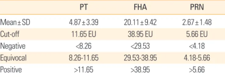
Each cut-off value was determined from the results of in- house ELISA for the sera of the negative groups (n=189). The cut-off values of PT, FHA, and PRN were 11.65, 38.95, and 5.66 EU/mL, respectively (Table 1). In this population, the mean concentration of anti-FHA IgG in healthy volunteers was higher than in those of anti-PT and PRN IgG. The interpreta- tion criteria (negative, equivocal, and positive) were deter- mined from the cut-off value and SD (Table 1). There was no significant difference in results compared to the in-house ELI- SA using NIBSC antigens (only PT and FHA).
Verification of established in-house ELISA
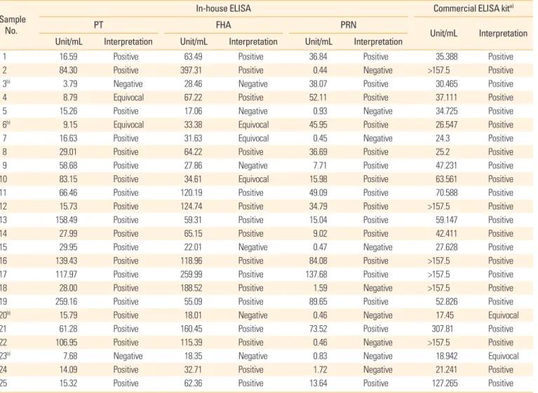
The in-house ELISA established in this study was verified by comparison of the results obtained using commercial ELISA kit for 25 clinical sera obtained from patients with pertussis to detect IgG against B. pertussis PT, FHA, and lipopolysaccha- ride (LPS). Among the samples (n=23) showing positive re- sults for the commercial kit, 21 samples showed anti-PT or/
and anti-FHA positivity in the in-house ELISA results (Table 2). However, the results of equivocal samples (n=2) did not coincide (Table 2); this may be because of the activity of other
Table 1. Cut-off values and interpretation of each in-house ELISA results obtained from the sera of volunteer groups (n=189)
PT FHA PRN
Mean±SD 4.87±3.39 20.11±9.42 2.67±1.48
Cut-off 11.65 EU 38.95 EU 5.66 EU
Negative <8.26 <29.53 <4.18
Equivocal 8.26-11.65 29.53-38.95 4.18-5.66
Positive >11.65 >38.95 >5.66
ELISA, enzyme-linked immunosorbent assay; PT, pertussis toxin; FHA, filamentous haemagglutinin; PRN, pertactin; SD, standard deviation; EU, ELISA unit.
antigens (anti-LPS). Anti-PRN IgG detection by the in-house ELISA showed a positivity of 68.0% (17/25) in patients with pertussis, although comparison with the commercial kit or a control antigen was not possible. These results suggest that the cut-off values determined in this study can be used as cri- teria for determining antibody production at the single serol- ogy level in vaccine studies.
Evaluation of in-house ELISA in vaccine study
Each in-house ELISA was validated using vaccinated sera and then compared according to the type of antigen (PT &
FHA from NIBSC & GC Tdap vaccine). In anti-PT IgG analy- sis, there were no significant differences between the results for the type of antigens (NIBSC & GC Tdap vaccine) (Table 3).
However, the anti-FHA IgG results showed significant differ-
ences in the pre-vaccinated sera (p<0.001) and an increased ratio (p=0.001) after vaccination, but not in the post-vacci- nated sera (p=0.470), between each method (Table 3).
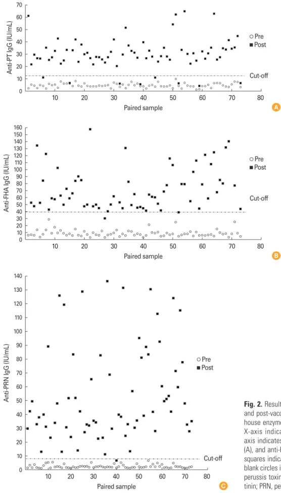
In a dual-serology study (pre- and post-vaccinated sera by Tdap of A & B company), there were a significant increase (p<0.001) in individual immunogenicity after vaccination (Fig. 1). The distributions of antibody levels in paired sera showed that 6.85% (5/73) in anti-PT IgG, 1.37% (1/73) in anti- FHA IgG, and 0% in anti-PRN IgG were lower than a 100% in- crease after vaccination. Additionally, the values of 10.96%
(8/73) in anti-PT IgG, 1.37% (1/73) in anti-FHA IgG, and 0%
in anti-PRN IgG were below each cut-off point (Fig. 2). These results suggest that the in-house method is valuable for vac- cine studies using clinical specimens with dual-serology or single-serology.
Table 2. Comparison of in-house ELISA and commercial ELISA kit using 25 clinical sera obtained from patients with pertussis
Sample No.
In-house ELISA Commercial ELISA kita)
PT FHA PRN
Unit/mL Interpretation Unit/mL Interpretation Unit/mL Interpretation Unit/mL Interpretation
1 16.59 Positive 63.49 Positive 36.84 Positive 35.388 Positive
2 84.30 Positive 397.31 Positive 0.44 Negative >157.5 Positive
3b) 3.79 Negative 28.46 Negative 38.07 Positive 30.465 Positive
4 8.79 Equivocal 67.22 Positive 52.11 Positive 37.111 Positive
5 15.26 Positive 17.06 Negative 0.93 Negative 34.725 Positive
6b) 9.15 Equivocal 33.38 Equivocal 45.95 Positive 26.547 Positive
7 16.63 Positive 31.63 Equivocal 0.45 Negative 24.3 Positive
8 29.01 Positive 64.22 Positive 36.69 Positive 25.2 Positive
9 58.68 Positive 27.86 Negative 7.71 Positive 47.231 Positive
10 83.15 Positive 34.61 Equivocal 15.98 Positive 63.561 Positive
11 66.46 Positive 120.19 Positive 49.09 Positive 70.588 Positive
12 15.73 Positive 124.74 Positive 34.79 Positive >157.5 Positive
13 158.49 Positive 59.31 Positive 15.04 Positive 59.147 Positive
14 27.99 Positive 65.15 Positive 9.02 Positive 42.411 Positive
15 29.95 Positive 22.01 Negative 0.47 Negative 27.628 Positive
16 139.43 Positive 118.96 Positive 84.08 Positive >157.5 Positive
17 117.97 Positive 259.99 Positive 137.68 Positive >157.5 Positive
18 28.00 Positive 188.52 Positive 1.59 Negative >157.5 Positive
19 259.16 Positive 55.09 Positive 89.65 Positive 52.826 Positive
20b) 15.79 Positive 18.01 Negative 0.46 Negative 17.45 Equivocal
21 61.28 Positive 160.45 Positive 73.52 Positive 307.81 Positive
22 106.95 Positive 115.39 Positive 0.46 Negative >157.5 Positive
23b) 7.68 Negative 18.35 Negative 0.83 Negative 18.942 Equivocal
24 14.09 Positive 32.71 Positive 1.72 Negative 21.241 Positive
25 15.32 Positive 62.36 Positive 13.64 Positive 127.265 Positive
ELISA, enzyme-linked immunosorbent assay; PT, pertussis toxin; FHA, filamentous haemagglutinin; PRN, pertactin.
a)Bordetella pertussis IgG ELISA kit (IBL International GmbH, Hamburg, Germany).
b)Different data between the interpretation of in-house ELISA (anti-PT and anti-FHA IgG) and commercial kit.
Discussion
aP vaccines have been used since the late 1990s worldwide because they are comparatively safer than whole cell pertus- sis vaccines. The three components in aP vaccines are an at- tenuated toxin component such as PT, a protein associated with bacterial adhesion such as FHA or PRN. PT is a secretory toxin that can be divided into five subunits, largely the A and B parts [15,16]. Part B attaches to the host cell and serves to transport part A into the host cell. Part A can destroy the function of the host cell through its ADP-ribosylating activity [17,18]. This toxin is not secreted by B. parapertussis strains.
FHA, a major adhesin molecule, contributes to pathogenicity
by attaching to sugar components, heparin, integrin CR3 site, macrophages, and epithelial cells [19-21], and PRN plays an important role in macrophage adhesion [22,23]. These fac- tors are involved in host antibody formation for protection.
Therefore, a standardized ELISA assay method is critical for vaccine evaluation, as it can measure the effect of a vaccine by analyzing the antibodies of PT, FHA, and PRN.
No control antigen or standard antibody is available in South Korea. Additionally, a standard PRN antigen has not established worldwide. Thus, we developed an in-house ELI- SA method using NIBSC standard control antigens (NIBSC PT & FHA), NIBSC standard control serum, and the PRN an- tigen of a domestic developed acellular vaccine in this study.
Table 3. Comparison of results of each in-house ELISA in paired sera (pre- and post-vaccinated sera) using NIBSC and GC Tdap vaccine anti- gens
Antigen GC Tdapa)
No. Preb) Postc) Ratiod)
Anti-PT (IU/mL)
NIBSC 31 4.761±3.202 29.498±9.965 8.652±7.180
GC Pharma 46 4.715±1.549 28.630±10.641 7.103±4.960
p-value 0.9343 0.720 0.266
Anti-FHA (IU/mL)
NIBSC 31 20.990±6.252 74.070±31.191 3.716±1.691
GC Pharma 46 9.314±4.901 86.203±89.363 10.900±11.278
p-value <0.001 0.470 0.001
Values are presented as mean±SD.
ELISA, enzyme-linked immunosorbent assay; NIBSC, National Institute for Biological Standards and Control; PT, perussis toxin; FHA, filamentous haemagglutinin; SD, standard deviation.
a)These sera were obtained from volunteers vaccinated with GC Tdap.
b)Pre-vaccinated sera.
c)Post-vaccinated sera.
d)Increased ratio after vaccination.
Fig. 1. Analysis of anti-pertussis antigens (A, PT; B, FHA; C, PRN), IgG in the pre- and post-vaccinated sera group, and increased ratio between A and B Tdap vaccination group. PT, perussis toxin; FHA, filamentous haemagglutinin; PRN, pertactin; Tdap, tetanus, diphtheria, and acellular pertussis.
35 30 25 20 15 10 5
0 Pre Post Ratio
Anti-PT (IU/mL)
A p<0.001
A Tdap B Tdap
90 80 70 60 50 40 30 20 10
0 Pre Post Ratio
Anti-PRN (IU/mL)
C p<0.001
A Tdap B Tdap 160
140 120 100 80 60 40 20
0 Pre Post Ratio
Anti-FHA (IU/mL)
B p<0.001
A Tdap B Tdap
Fig. 2. Results of dual-serology (paired pre- and post-vaccinated sera) by established in- house enzyme-linked immunosorbent assay.
X-axis indicates paired sample, while y- axis indicates anti-PT IgG (A), anti-FHA IgG (A), and anti-PRN IgG (C), respectively. Black squares indicate post-vaccinated sera, while blank circles indicate pre-vaccinated sera. PT, perussis toxin; FHA, filamentous haemagglu- tinin; PRN, pertactin
70 60 50 40 30 20 10 0
160 150 140 130 120 100110 90 80 70 60 50 40 3020 10 0
140 130 120 110 100 90 80 70 60 50 40 30 20 10 0
10 20 30 40 50 60 70 80
10 20 30 40 50 60 70 80
10 20 30 40 50 60 70 80 Paired sample
Paired sample
Paired sample
Anti-PT IgG (IU/mL)Anti-FHA IgG (IU/mL)Anti-PRN IgG (IU/mL)
A
B
C
Pre Post
Pre Post
Pre Post
Cut-off
Cut-off
Cut-off
In these assays, the CVs of PT, FHA, and PRN were 11.53%, 8.60%, and 9.86%, respectively. Because all measured CVs were below 15%, this method is reproducible. This estab- lished method was used to determine the negative and posi- tive criteria of each pertussis antigen by measuring the cut- off level using normal human samples. These cut-off levels against each antigen were applied to the sera of 25 patients with pertussis. Although conventional commercial kits are limited to differentiate the positivity of each test pertussis an- tigen, the results were significantly matched except for in 4 cases when these results were compared by using conven- tional pertussis diagnostic test kit approaches (Table 2). The cut-off point is a reference criterion for the immunological diagnosis of pertussis in countries with a high vaccination coverage [14], but it is also an important factor in determin- ing the effect of the Tdap vaccine in adults. While there were no significant differences in anti-PT IgG analysis, the anti- FHA IgG results between the type of antigens (NIBSC & GC Tdap vaccine) showed significant differences in the pre-vac- cinated sera and an increased ratio (Table 3). This may be be- cause of differences in the detection ability at low concentra- tions of anti-FHA IgG. However, because there was no signifi- cant difference in the post-vaccinated group (anti-FHA IgG, p=0.470) (Table 3), it is possible to verify the vaccine effect Table 4. Comparison of results of in paired sera (pre- and post-vacci- nated sera) from groups A and B
GC Tdapa)
(n=46) GSK Tdapb)
(n=27) p-value
PT
Prec) 4.715±1.549 4.595±2.030 0.776
Postd) 28.630±10.641 33.244±15.350 0.135
Ratioe) 7.103±4.960 8.261±5.223 0.349
FHA
Prec) 9.314±4.901 9.049±5.309 0.829
Postd) 86.203±89.364 145.733±148.966 0.036 Ratioe) 10.900±11.278 17.944±16.998 0.037 PRN
Prec) 2.626±1.259 2.683±1.353 0.856
Postd) 80.428±131.954 65.577±31.087 0.568 Ratioe) 26.289± 29.519 27.279±12.821 0.869 Values are presented as mean±SD.
PT, pertussis toxin; FHA, filamentous haemagglutinin; PRN, pertactin; SD, standard deviation.
a)These sera were obtained from volunteers vaccinated with GC Tdap.
b)These sera were obtained from volunteers vaccinated with GSK Tdap.
c)Pre-vaccinated sera.
d)Post-vaccinated sera.
e)Increased ratio after vaccination.
using the same approach as used in single serology.
In a dual-serology study (pre- and post-vaccinated sera by Tdap of A & B company), the antibody titers of pertussis anti- gens before vaccination were all below the cut-off value, and there were a significant increase (p<0.001) in individual im- munogenicity after vaccination (Figs. 1, 2). There was no dif- ference in the anti-PT and anti-PRN IgG formation in the Tdap vaccine group of the two companies (p=0.135 and p=0.568, respectively), and there was a difference in anti- FHA IgG immunogenicity, but the difference was not signifi- cant (p>0.01) (Table 4). In the distribution of anti-pertussis antigen IgG in pre- and post-vaccinated sera, the cut-off point can be a useful tool for evaluating immunogenicity after vac- cination with a single serology test. These results suggest that the established in-house ELISA method and cut-off points in this study are valuable for confirming individual immunoge- nicity of each pertussis antigen after vaccination.
In conclusion, we established an in-house ELISA method using self-developed vaccine raw materials by comparing immunogenicity test methods with the WHO standard anti- body and control antigen. The developed test and cut-off point were verified by using pertussis-positive sera and vac- cine sera. There are some limitations to using the standard antigen and standard serum. Particularly, because a standard antigen for PRN is not available, studies are needed to estab- lish the standard antigens and standard antibodies in South Korea. Verification of pertussis antigens of B. pertussis strains with polymorphisms and undergoing epidemiological chang- es should be conducted after some time. When the immuno- dynamic changes in pertussis are large, a new standard anti- body and control antigen should be developed to re-establish the reference unit, cut-off limit, and so on. Our method can be used to establish a domestic standard pertussis immuno- assay using the sera of patients with pertussis and vaccine se- ra and suggests the necessity of establishing domestic stan- dard sera and an antigen.
ORCID
Chulmin Park https://orcid.org/0000-0001-9147-0478 Dong Ho Huh https://orcid.org/0000-0001-5395-7197 Seung Beom Han https://orcid.org/0000-0002-1299-2137 Gi Sub Choi https://orcid.org/0000-0002-5733-9799 Kyu Ri Kang https://orcid.org/0000-0001-9135-1890 Ji Ahn Kim https://orcid.org/0000-0002-1200-5330 Jin Han Kang https://orcid.org/0000-0003-1610-6742
References
1. Celentano LP, Massari M, Paramatti D, Salmaso S, Tozzi AE; EUVAC-NET Group. Resurgence of pertussis in Eu- rope. Pediatr Infect Dis J 2005;24:761-5.
2. Cherry JD. Epidemic pertussis in 2012: the resurgence of a vaccine-preventable disease. N Engl J Med 2012;367:785- 7.
3. Chiappini E, Stival A, Galli L, de Martino M. Pertussis re- emergence in the post-vaccination era. BMC Infect Dis 2013;13:151.
4. Bechini A, Tiscione E, Boccalini S, Levi M, Bonanni P.
Acellular pertussis vaccine use in risk groups (adoles- cents, pregnant women, newborns and health care work- ers): a review of evidences and recommendations. Vac- cine 2012;30:5179-90.
5. Zepp F, Heininger U, Mertsola J, et al. Rationale for pertus- sis booster vaccination throughout life in Europe. Lancet Infect Dis 2011;11:557-70.
6. Kmietowicz Z. Pertussis cases rise 10-fold among older children and adults in England and Wales. BMJ 2012;345:
e5008.
7. Cherry JD. Why do pertussis vaccines fail? Pediatrics 2012;
129:968-70.
8. Healy CM, Rench MA, Castagnini LA, Baker CJ. Pertussis immunization in a high-risk postpartum population. Vac- cine 2009;27:5599-602.
9. Xing D, Wirsing von Konig CH, Newland P, et al. Charac- terization of reference materials for human antiserum to pertussis antigens by an international collaborative study.
Clin Vaccine Immunol 2009;16:303-11.
10. European Centre for Disease Prevention and Control.
Guidance and protocol for the serological diagnosis of hu- man infection with Bordetella pertussis. Stockholm: Euro- pean Centre for Disease Prevention and Control; 2012.
11. Reizenstein E, Hallander HO, Blackwelder WC, Kuhn I, Ljungman M, Mollby R. Comparison of five calculation modes for antibody ELISA procedures using pertussis se- rology as a model. J Immunol Methods 1995;183:279-90.
12. Baughman AL, Bisgard KM, Edwards KM, et al. Establish- ment of diagnostic cutoff points for levels of serum anti-
bodies to pertussis toxin, filamentous hemagglutinin, and fimbriae in adolescents and adults in the United States.
Clin Diagn Lab Immunol 2004;11:1045-53.
13. Andre P, Caro V, Njamkepo E, Wendelboe AM, Van Rie A, Guiso N. Comparison of serological and real-time PCR assays to diagnose Bordetella pertussis infection in 2007. J Clin Microbiol 2008;46:1672-7.
14. Guiso N, Berbers G, Fry NK, et al. What to do and what not to do in serological diagnosis of pertussis: recommen- dations from EU reference laboratories. Eur J Clin Micro- biol Infect Dis 2011;30:307-12.
15. Tamura M, Nogimori K, Murai S, et al. Subunit structure of islet-activating protein, pertussis toxin, in conformity with the A-B model. Biochemistry 1982;21:5516-22.
16. Stein PE, Boodhoo A, Armstrong GD, Cockle SA, Klein MH, Read RJ. The crystal structure of pertussis toxin.
Structure 1994;2:45-57.
17. Katada T, Ui M. ADP ribosylation of the specific mem- brane protein of C6 cells by islet-activating protein associ- ated with modification of adenylate cyclase activity. J Biol Chem 1982;257:7210-6.
18. Locht C, Coutte L, Mielcarek N. The ins and outs of per- tussis toxin. FEBS J 2011;278:4668-82.
19. Funnell SG, Robinson A. A novel adherence assay for Bor- detella pertussis using tracheal organ cultures. FEMS Mi- crobiol Lett 1993;110:197-203.
20. Ishibashi Y, Nishikawa A. Bordetella pertussis infection of human respiratory epithelial cells up-regulates intercellu- lar adhesion molecule-1 expression: role of filamentous hemagglutinin and pertussis toxin. Microb Pathog 2002;33:
115-25.
21. Melvin JA, Scheller EV, Miller JF, Cotter PA. Bordetella per- tussis pathogenesis: current and future challenges. Nat Rev Microbiol 2014;12:274-88.
22. Carbonetti NH. Bordetella pertussis: new concepts in pathogenesis and treatment. Curr Opin Infect Dis 2016;29:
287-94.
23. Hovingh ES, Mariman R, Solans L, et al. Bordetella pertus- sis pertactin knock-out strains reveal immunomodulatory properties of this virulence factor. Emerg Microbes Infect 2018;7:39.