Antioxidant and Antidiabetic Activities of Aralia elata Seeds
Weicheng Hu, Mee Jung Jung, Seong-Il Heo, and Myeong-Hyeon Wang*
School of Biotechnology, Kangwon National University, Chuncheon 200-701, Republic of Korea Received February 28, 2008; Accepted June 19, 2008
Aralia elata seeds were successively extracted with water, methanol, ethanol, acetone and chloroform. The crude extracts were investigated for antioxidant and antidiabetic activities. The antioxidant properties of various extracts were evaluated by antioxidant tests, such as DPPH free radical-scavenging activity, hydroxyl radical-scavenging assay, metal-chelating activity, lipid peroxidation inhibition activity and reducing power assay. The 70% methanol extract exhibited the highest activity in the in vitro models of DPPH free radical-scavenging activity, metal-chelating activity, and reducing power assay. Acetone extract showed good effects on lipid peroxidation inhibition and hydroxyl radical-scavenging assay at a low concentration. In addition, the α- glucosidase inhibition assay showed that 70% methanol extract had the highest activity. These results indicate the high possibility of using A. elata seeds for medical application due to their efficient antioxidant properties.
Key words: Aralia elata seeds, antioxidant, metal chelating, α-glucosidase
Free radicals are known as the major cause of oxidative damage of biological molecules in the human body, including coronary heart disease, aging, cancer and dementia [Cheng et al., 2003]. Therefore, antioxidants play an important part in the inhibition of cellular damage by the free radicals [Brash and Havre, 2002]. Antioxidants in biological systems have diverse functions, including defending against oxidative damage and participating in the major signalling pathways of cells [Wojtaszek, 1997].
One major action of antioxidants in cells is to prevent damage caused by the action of the ROS [Schinella et al., 2002]. Several synthetic antioxidants such as BHA, BHT, and TBHQ are universally being used. However, their use has been questioned because of their toxicity. Some toxicological studies have implicated the use of these synthetic antioxidants in promoting the development of cancerous cells in rats [Huang and Wang, 2004]. Plants (fruits, vegetables, medicinal herbs) contain a wide variety
of free radical-scavenging molecules, such as phenolic compounds, flavonoid compounds, vitamins, terpenoids, and some other endogenous metabolites, which are rich in antioxidant activity [Pietta, 2000]. Flavonoid compounds may reduce the risk of cancer, cardiovascular disease and many other diseases with high content of antioxidative phytochemicals [McCarty, 1999].
Diabetes mellitus is a major chronic disease caused by an improper balance of glucose homeostasis and has a significant impact on the health, quality of life, as well as the health care system [Hanngle, 1990]. One of the therapeutic ways is to retard the absorption of glucose by the inhibition of carbohydrate-hydrolysing enzymes, for example α-amylase and α-glucosidase in the digestive organs [Tiwari and Rao, 2002]. Diabetes can increases the production of ROS by glucose autoxidation. Thus, many natural resources have been investigated with respect to the suppression of glucose production [Ye et al., 2002].
Aralia elata is one of the most popular edible vegetable in Korea. The bark and roots of A. elata have been used in treating cancer, cough ulcer, diabetes, cataractogenesis and schizophrenia [Chung and Jung, 2003]. However up to date, only few studies have investigated the bioactivities of A. elata seeds. The purpose of the present study was to evalute the antioxidant and antidiabetic bioactivities of A.
elata seeds, such as free radical-scavenging activity, hydroxyl radical-scavenging assay, metal-chelating activity, lipid peroxidation inhibition, and α-glucosidase inhibition assay.
*Corresponding author
Phone: 82-33-250-6486; Fax: 82-33-241-6480 E--mail: [email protected]
Abbreviations: BHA, butylated hydroxyanisole; BHT, butylated hydroxytoluene; DPPH, 1,1-diphenyl-2-picrylhydrazyl radical;
MDA, malondialdehyde; pNPG, 4-nitrophenyl glucopyranoside;
ROS, reactive oxygen species; TBA, thiobarbituic acid; TBHQ,
tert-buthylhydroquinone; TCA, thrichloroacetic acid doi:10.3839/jabc.2008.020
Material and Methods
Preparation of the extracts. A. elata seeds were collected from Chuncheon, Korea, dried in a shade at room temperature, and powdered. Fifty grams of the seed powder was extracted separately with water, absolute MeOH, 70% MeOH, absolute EtOH, 70% EtOH, absolute acetone and chloroform at 70oC for 3 h. The extracts were filtered through filter paper (Whatmen 70 mm) and evaporated using a vacuum rotary evaporator (EYELA, CCA-1110). Finally, the samples were dried by freezing in a high vacuum (EYELA, FD-5N) for 2 days to obtain the crude extracts. Dried samples were weighed and kept at 4oC for further analysis.
Chemicals. L-Ascorbic acid, DPPH ·, 2-deoxy-D- ribose, ferrous chloride, 2N folin-ciocalteu’s phenol reagent, iron (II) sulfate heptahydrate, tannic acid, α- tocopherol, TCA, BHT, and EDTA disodium dehydrate were purchased from Sigma (Sternheim, Germany). TBA was purchased from Alfa Aesar (A Johnson Matthey Company, Karlsruhe, Germany). Hydrogen peroxide, gallic acid, and sodium carbonate were purchased from Junsei (Junsei Chemical Co., Ltd., Tokyo, Japan) Iron (III) chloride hexahydrate was purchased from Cica- reagent (Kanto Chemical Co., Ltd., Tokyo, Japan). All other unlabelled chemicals and reagents were purchased from Sigma Chemical Co. (St. Louis, MO).
DPPH radical-scavenging activity. The free radical- scavenging activity was determined by DPPH test according to the method of Kilani et al. [2005]. Briefly, 0.5 mL of 0.25 mM DPPH solution (in MeOH) was added to the 1.5 mL test tubes containing 0.5 mL of different concentration (0.01-0.4 mg/mL) of the extract using L-ascorbic acid as the positive control. The mixture was shaken vigorously for 1 min and kept at room temperature for 30 min in the dark. The absorbances of all sample solutions were measured at 517 nm. Each measurement was carried out in triplicate. The capability to scavenge the DPPH radical was calculated using the following equation:
I (%)=[1−(Ai−Aj )/Ac]×100%
where Ac is the absorbance of DPPH solution without the sample (0.5 mL DPPH solution+0.5 mL of methanol), Ai is the absorbance of the test sample mixed with DPPH solution (0.5 mL sample+0.5 mL DPPH solution), and Aj isthe absorbance of the sample without DPPH solution (0.5 mL sample+0.5 mL methanol).
Scavenging ability on hydroxyl radical. The scavenging abilities of the test extracts on ·OH were determined using the deoxyribose assay [Hou et al., 2003]. The reaction mixture containing FeSO4 (10 mM, 0.2 mL), EDTA (10
mM, 0.2 mL), H2O2 (10 mM, 0.2 mL), 2-deoxy-D-ribose (10 mM, 0.2 mL) was mixed with or without other extracts into 1 mL of final reaction volume to make a phosphate buffer (0.1 M NaH2PO4-Na2HPO4, pH 7.4) solution. The mixture was incubated in the boiling water for 10 min, followed by the addition of 1 mL each of 2.8% TCA and 1% TBA solution. Finally, the reaction mixture was cooled and centrifuged at 800×g for 10 min.
The absorbance of the supernatant was measured at 532 nm.
Scavenging percentage (%)=[1−(As−Ac)/Ab]×100%
whereAs, denotes in the presence of deoxyribose and sample; Ab, in the presence of deoxyribose but without test compounds; and Ac, in the presence of test compounds but without deoxyribose.
Metal-chelating activity. The metal-chelating activities of different solvent extracts were estimated by the method of Gulcin et al. [2004]. In brief, 1 mL each of A. elata extract at different concentrations was mixed with 3.7 mL of absolute MeOH and 0.1 mL of 1 mM FeCl2. The reaction was initiated by the addition of 0.2 mL of 5 mM ferrozine, followed by vigorous shaking and the mixture was left to react at room temperature for 10 min. These extracts are comparable to that of EDTA and α-tocophenol, which were used as the positive control. Each test was replicated three times. The absorbance was measured at 562 nm.
Lipid peroxidation inhibition activity. The inhibition of lipid peroxidation activity was assayed by the method of Veigas et al. [2007] with some modifications. Two grams of the pig liver were homogenized in 10 mL of 200 mM Tris-HCl buffer (pH 7.2). The liver homogenate (0.2 mL) was incubated with the sample (0.05 mL), 4 mM FeCl2 (0.05 mL) and 0.1 mM ascorbic acid (0.05 mL) at 37oC for 1 h, followed by the addition of the TBA reagent (2 mL of 0.6% TBA). The final solution was heated at 100oC in the boiling water for 10 min, and 5 mL of n- butyl alcohol (n-BuOH) was added to the solution. The mixture was then shaken vigorously, and the n-BuOH layer was separated by centrifugation at 3,000×g for 10 min. Absorbance of the supernatant was measured at 532 nm.
Reducing power assay. The reducing power was determined according to the method of Nandita and Rajini [2004]. Various concentrations of the extracts (1 mL) were mixed with 2.5 mL sodium phosphate buffer (NaH2PO4-Na2HPO4, 0.2 M, pH 6.6) and 2.5 mL of 0.1%
potassium ferricyanide. The mixture was incubated at 50 for 20 min. After adding 2.5 mL of 10% TCA, the mixture was centrifuged at 3000×g for 10 min. The upper layer (2.5 mL) was mixed with 2.5 mL of distilled water
and 0.5 mL of 0.1% of ferric chloride, and the absorbance was measured at 700 nm. α-Tocopherol was used as a positive control.
Rat intestinal α-glucosidase inhibitory activity. The inhibitory activity A. elata seed extracts against the rat intestinal α-glucosidase was determined by measuring the formation of 4-nitrophenol by α-glucosidase after the reaction with pNPG as described by Kim et al. [2004].
The inhibitory activities of various concentrations (100, 500 and 1000µg/mL) were measured in the 96-well plates. Fifty microliters of α-Glucosidase (0.0075 unit) was mixed with 50µL extract in phosphate buffer (0.2 M KH2PO4-K2HPO4, pH 6.8). After pre-incubation at 37oC for 15 min, 50µL pNPG (3 mM) was added to the mixture as a substrate and incubated at 37oC for 10 min;
acarbose was used as a positive control. Fifty microliters of the sodium carbonate (0.1 M) was added to stop the reaction. Absorbances of the reactants were measured at 405 nm.
Determination of total phenolic and flavonoid contents. Total phenolic content was determined using the Folin-Denis reagent [Yin and Wang, 2007]. The results were expressed as tannic acid equivalents. One milliliter sample was mixed with 2 mL Folin-Denis reagent and 2 mL 35% Na2CO3. The mixture volume was made up to 10 mL and shaken vigorously. After incubation at room temperature for 30 min, the absorbance was measured at 765 nm. The results were expressed as mg of tannic acid per gram of the extract.
Total flavonoid content was estimated according to the method of Ordonez et al. [2006]. To 0.5 mL of each sample, 0.5 mL of 2% AlCl3 ethanol solution was added.
After standing for 1 h at room temperature, the absorbance was measured at 420 nm. Total flavonoid content was calculated as quercetin from a standard curve.
Statistical analysis. All experimental data were expressed as mean±standard derivation. Data analyses were performed using the SPSS 7.5 (Window Version 7.5 Software Inc., New York, NY) and p<0.05 was considered as significant.
Result and Discussion
Effect of different solvent types on yields of A. elata seeds. Yields of A. elata seeds obtained from various solvents were as follows: MeOH gave the highest yield (13.43%), followed by water (13.09%), 70% MeOH (11.08%), 70% EtOH (9.54%), EtOH (8.77%), acetone (4.31%) and chloroform (3.75%).
DPPH radical-scavenging activity. DPPH is a stable free radical compound, which has been widely used as a substrate to evaluate the antioxidative action of the
antioxidants [Molyneux, 2004]. The DPPH radical- scavenging activity of the extract has been attributed to the ability of these extracts in pairing with the odd electron of the DPPH radical [Jorge et al., 2007]. The radical-scavenging activities of A. elata seeds are shown in Table 1 expressed in 50% inhibition concentration (IC50). The 70% MeOH extract exhibited the highest radical-scavenging activity (IC50 49.8µg/mL), followed by 70% EtOH (IC50 57.7µg/mL) and water (IC50 95.3µg/
mL) when compared with a positive control, L-ascorbic acid (IC50 3.7µg/mL). In comparison, other extracts showed weak free radical-scavenging activities. It has been found that compounds such as cystine, glutathione, ascorbic acid, tocopherol, flavonoids, and tannins can scavenge DPPH by their hydrogen-donating ability [Jung
et al., 2008]. Although the DPPH radical-scavenging activities of different extracts of the A. elata seeds were significantly lower than that of L-ascorbic acid, it was evident that the 70% MeOH extract have hydrogen- donating ability and could serve as free radical inhibitors or scavengers, possibly acting as the primary antioxidants.
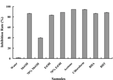
Scavenging ability of hydroxyl radicals. The hydroxyl radicals-scavenging abilities of the extracts are shown in Fig. 1. Except those from water and 70% MeOH, all extracts exhibited inhibitory effects higher than 80% at 1 mg/mL. The positive control, BHA and BHT eliminated 86.59% and 88.41% of the hydroxyl radicals at 500µg/
mL, respectively.
Hydroxyl radical is the most reactive free radical and can be formed from superoxide anion and hydrogen peroxidate, in the presence of metal ions, such as iron.
Hydroxyl radicals react with lipid, polypeptides, proteins, and DNA [Girrotti, 1998]. The hydroxyl radicals- scavenging ability may be related with the inhibition of Table 1. Effect of different solvent types on yield and DPPH free radical- scavenging activity of extracts obtained from different solvents of A. elata seeds
Samples Yield (%) DPPH radical activity(IC50: µg/mL)
70% MeOH 11.08 049.8
70% EtOH 09.54 057.7
Water 13.09 095.3
Acetone 04.31 239.1
MeOH 13.43 249.2
EtOH 08.77 348.6
Chloroform 03.75 516.2
L-Ascorbic acid 003.7
IC50: The effective concentration at which DPPH radicals were scavenged by 50%. L-Ascorbic acid was used as a positive control.
lipid peroxidation observed in the present study.
Metal-chelating activity. The metal-chelating activity is based on the chelating of Fe2+ ions by the reagent ferrozine, which is a quantitative formation of a complex with Fe2+ ions [Lindley, 1998]. Table 2 shows the results of metal-chelating activity of A. elata seeds. Here, 70%
MeOH extract (IC50 128.2µg/mL), water extract (IC50 172.7µg/mL), 70% EtOH extract (IC50 247.4µg/mL), and MeOH extract (IC50 398.5µg/mL) showed high chelating activities as demonstrated by their efficacity in inhibiting the formation of ferrous and ferrozine complexes. These activities are comparable to those of EDTA (IC50 39.9µg/mL) and α-tocopherol (IC50 458.6
µg/mL), which were used as a positive controls.
Lipid peroxidant assay. In present study, the inhibition of lipid peroxidation in pig liver homogenate, which was induced by FeCl2-ascorbic system, was studied. Lipid peroxidation is an oxidation of the unsaturated fatty acid
in the cell membranes that produce numerous degradation products [Spiteller, 1996]. MDA has been studied widely as an index of lipid peroxidation and a marker of oxidative stress. The lipid peroxidation inhibition activity of the extract was measured and compared with that of α- tocopherol (Fig. 2). The data indicated that acetone extract (70.85%) was the most effective against liver lipid peroxidation at 500µg/mL. The control substance, α- tocopherol showed 77.06% inhibition at the same concentration. It has been reported that the lipid peroxidation inhibition could be caused by the scavenging of the hydroxyl radicals [Spiteller, 1996].
Reducing power assay. The reducing powers of different extracts of A. elata increased with increasing Fig. 1. Hydroxyl radical-scavenging activity of A. elata
seeds according to the types of extracting solvent.
Concentration of the extracts was 1,000µg/mL. BHA (500µg/mL) and BHT (500µg/mL) were used as the positive controls. Each value represents the mean±SD (n=3).
Table 2. Metal chelation effect of extracts obtained from A. elata seeds in different solvents
Extracts Metal chelating (IC50: µg/mL)
70% MeOH 0128.2
Water 0172.7
70% EtOH 0247.4
MeOH 0398.5
EtOH 1369.2 Acetone 1375.1 Chloroform 2009.5
EDTA 0039.9
α-Tocopherol 0458.6
EDTA and α-Tocopherol were used as positive controls.
Fig. 2. Effect of A. elata seed extracts and α-tocopherol on Fe2+/L-ascorbic acid- induced lipid peroxidation in pig liver homogenate. Concentration of the extracts was 500µg/mL. α-Tocopherol (500µg/mL) was used as the positive control. Each value represents the mean±SD (n=3).
Fig. 3. Reducing power of the A. elata seeds extracts at different concentrations. Concentrations of the extracts were 100 (□), 500µg/mL (▧), and 1,000µg/mL (▥), with α-tocopherol used as a positive control. Values are means of three determinations±standard deviation (n=3).
concentration (Fig. 3). The reducing powers of the different extracts and standard compounds followed the order of α-tocopherol (0.233-0.887)>70% MeOH extract (0.123-0.606)>70% EtOH extract (0.098-0.499)>MeOH extract (0.052-0.395)>water extract (0.079-0.379)> EtOH extract (0.074-0.323)>acetone extract (0.028-0.169)>
chloroform extract (0.022-0.142). The reducing powers of various extracts arise from the hydrogen-donating ability, as described by Lee et al. [2007].
Rat intestinal α-glucosidase inhibitory activity. In the intestinal α-glucosidase inhibitory activity assay of the rat, 70% MeOH extract at 1 mg/mL revealed high inhibiton on α-glucosidase by 73.86% (Table 3). Therat intestinal α-glucosidase inhibitory activity order was as follows: 70% MeOH extract>70% EtOH extract>MeOH extract>EtOH extract>acetone extract>chloroform extract
>water extract. Acarbose, an anti-diabetic drug used to treat type 2 diabetes mellitus and, in some countries, prediabetes, was used as a positive control. It is sold in Europe under the brand name Glucobay® (Bayer AG), in
North America as Precose® (Bayer Pharmaceuticals), and in Canada as Prandase® (Bayer AG). It is an inhibitor of
α-glucosidase, an enteric enzyme that releases glucose from carbohydrates.
α-Glucosidase, located in the bush-border surface membrane of the intestinal cells, is the key enzyme stimulating the final step in the digestive process of carbohydrates. Hence, α-glucosidase inhibitors can retard the liberation of D-glucose from the complex dietary carbohydrates and delay glucose absorption [Schmidt et
al., 1977].
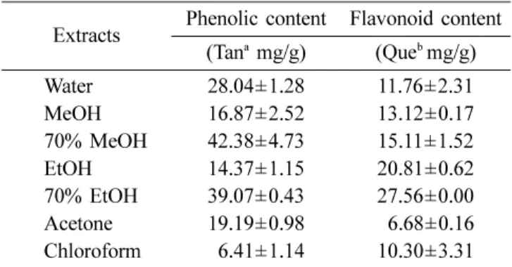
Determination of total phenolic and flavonoid contents. Table 4 shows the total phenolic and flavonoid contents of the A. elata seeds. The total phenolic contents of water, MeOH, 70% MeOH, EtOH, 70% EtOH, acetone and chloroform extracts were 28.04, 16.87, 42.38, 14.37, 39.07, 19.19 and 6.41 mg/g tannic acid equivalent, respectively. The contents of total flavonoids were 70% EtOH extract (27.56 mg/g quercetin equivalent)
>EtOH extract (20.81 mg/g quercetin equivalent)>70%
MeOH extract (15.11 mg/g quercetin equivalent)>MeOH extract (13.12 mg/g quercetin equivalent)>water extract (11.76 mg/g quercetin equivalent)>chloroform extract (10.30 mg/g quercetin equivalent)>acetone extract (6.68 mg/g quercetin equivalent).
In conclusion, A. elata seeds can be utilized as a source of natural antioxidant and antidiabetic agent. In the present study, 70% MeOH had good effect on DPPH free radical-scavenging, metal-chelating, and reducing power activities. The use of natural components in the seeds with antioxidant and antidiabetic activities to present various radical injuries in pathological condition in vivo is underway.
Table 3. Effect of A. elata seed extracts on rat intestinal
α-glucosidase inhibition assay
Extracts Concentration (µg/mL) Inhibition (%)
Water 100 -
500 08.80±1.44
10000 15.56±2.48
MeOH 100 11.95±2.66
500 38.96±2.97
10000 52.09±4.77
70% MeOH 100 18.92±0.49
500 61.24±1.37
10000 73.86±1.76
EtOH 100 10.88±2.79
500 29.70±1.01
10000 48.58±5.19
70% EtOH 100 13.94±1.80
500 61.24±2.78
10000 72.02±1.57
Acetone 100 09.31±4.30
500 15.36±2.57
10000 34.44±4.54
Chloroform 100 -
500 -
10000 16.73±1.67
Acarbose 0.01 58.85±2.14
0.1 89.88±0.32
Acarbose at 0.01 and 0.1µg/mL was used as the control.
Values represent the means±SD (n=3).
Table 4. Total phenolic and flavonoid contents of A.
elata seeds extracts obtained from different solvents Extracts Phenolic content Flavonoid content
(Tana mg/g) (Queb mg/g)
Water 28.04±1.28 11.76±2.31
MeOH 16.87±2.52 13.12±0.17
70% MeOH 42.38±4.73 15.11±1.52
EtOH 14.37±1.15 20.81±0.62
70% EtOH 39.07±0.43 27.56±0.00
Acetone 19.19±0.98 06.68±0.16
Chloroform 06.41±1.14 10.30±3.31
aTannic acid (Tan) was used as a standard for measuring the total phenolic content. bQuercetin (Que) was used as a standard for measuring the total flavonoid content. Values are means of three determinations±standard deviation (n=3).
Acknowledgments. This research was supported by the Institute of Bioscience & Biotechnology, in Kangwon National University.
References
Brash DE and Havre PA (2002) New careers for antioxi- dants. Proc Natl Acad Sci USA 99, 13969-13971.
Cheng HY, Lin TC, Yu KH, Yang CM and Lin CC (2003) Antioxidant and free radical scavenging activities of Ter- minalia chebula. Biol Pharm Bull 26, 1331-1335.
Chung CK and Jung ME (2003) Ethanol fraction of Aralia elata Seemann enhances antioxidant activity and lowers serum lipids in rats when administered with benzo(α)pyrene. Biol Pharm Bull 26, 1502-1504.
Girrotti AW (1998) Lipid hydroperoxide generation, turn- over, and effector action in biological systems. J Lipid Res39, 1529-1542.
Gulcin I, Sat IG, Beydemir S and Elmastas M (2004) Com- parison of antioxidant activity of clove (Eugenia caryo- phylata Thund) buds and lavender (Lavandula stoechas L.). Food Chem 87, 393-400.
Hanngle YJ (1992) Glucose toxicity. The Endocrine Society
13, 415-431.
Hou WC, Lin RD, ChengKT, HungYT and ChoCH (2003) Free radical-scavenging activity of Taiwanese native plants. Phytomedicine 10, 170-175.
Huang HL and Wang BG (2004) Antioxidant capacity and lipophilic content of seaweeds collected from the Qingdao coastline. J Agric Food Chem 52, 4993-4997.
Jorge R, Claudio OA, Cristina C, Ester N, Tomas DC, Jorge SD and Ramiro AM (2007) Antioxidant properties and free radical-scavenging reactivity of a family of hydrox- ynaphthalenones and dihydroxyanthracenones. Bioorg Med Chem 15, 7058-7065.
Jung MJ, Heo SI and Wang MH (2008) Free radical scav- enging and total phenolic contents from methanolic extract of Ulmus davidiana. Food Chem108, 482-487.
Kilani S, Ammar RB, Bouhlel I, Abdelwahed A, Hayder N, Mahmoud A, Ghedira K and Chekir-Ghedira L (2005) Investigation of extracts from (Tunisian) Cyperus rotun- dus as antimutagens and radical scavengers. Environ Toxicol Pharmacol 20, 478-484.
Kim YM, Wang MH and Rhee HI (2004) A novel α-glu- cosidase inhibitor from from pine bark. Carbohydr Res
339, 715-717.
Lee YL, Huang GW, Liang ZC and Mau JL (2007) Antioxi- dant properties of three extracts from Pleurotus citrino- pileatus. Swiss Soc of Food Sci Technol 40, 823-833.
Lindley MG (1998) The impact of food processing on anti- oxidants in vegetable oils, fruits and vegetables. Trends Food Sci Technol9, 336-340.
McCarty MF (1999) Vegan proteins may reduce risk of can- cer, obesity, and cardiovascular disease by promoting increased glucagon activity. Med Hypotheses 53, 459- Molyneux P (2004) The use of the stable free radical diphe-485.
nyl-picrylhydrazyl (DPPH) for estimating antioxidant activity. Songklanakarin J Sci Technol 26, 211-219.
Nandita S and Rajini PS (2004) Free radical scavenging activity of an aqueous extract of potato peel. Food Chem
85, 611-616.
Ordonez AAL, Gomez JD, Vattuone MA and Isla MI (2006) Antioxidant activities of Sechium edule (Jacq.) swartz extracts. Food Chem97, 452-458.
Pietta PG (2000) Flavonoids as antioxidants. J Nat Prod 63, 1035-1042.
Schinella GR, Tournier HA and Prieto JM (2002) Antioxi- dant activity of anti-inflammatory plant extracts. Life Sci
70, 1023-1033.
Schmidt DD (1977) α-Glucosidase inhibitors. Naturwissen- schaften 64, 535.
Spiteller G (1996) Enzymic lipid peroxidantion-A conse- quence of cell injury? Free Radic Biol Med 21, 1003- 1009.
Tiwari AK and Rao JM (2002) Diabetes mellitus and multi- ple therapeutic approaches of phytochemicals: Present status and future prospects. Curr Sci83, 30-38.
Veigas JM, Narayan MS, Laxman PM and Neelwarne B (2007) Chemical nature, stability and bioefficacies of anthocyanins from fruit peel of Syzygium cumini Skeels.
Food Chem 105, 619-627.
Vessal M, Hemmati M and Vasei M (2003) Antidiabetic effects of quercetin in streptozotocin-induced diabetic rats. Comp Biochem Physiol C Pharmacol Toxicol Endo- crinol 135, 357-364.
Wojtaszek P (1997) Oxidative burst: An early plant response to pathogen infection. Biochem J 322, 681-692.
Ye F, Shen Z and Xie M (2002) α-Glucosidase inhibition from a Chinese medical herb (Ramulus mori) in normal and diabetic rats and mice. Phytomedicine 9, 161-166.
Yen GC, Duh PD and Tsai CL (1993) Relationship between antioxidant activity and maturity of peanut hulls. J Agric Food Chem 41, 67-70.
Yin Y and Wang MH (2007) Antioxidant and anticancer activity of fractions from Picrasma qassioides (D. Don) Benn methanolic extract. Kor J Med Crop Sci 5, 329- 334.