• 검색 결과가 없습니다.

Buddleja officinalis prevents the normal cells from oxidative damage via antioxidant activity

N/A
N/A
Protected

Academic year: 2021

Share "Buddleja officinalis prevents the normal cells from oxidative damage via antioxidant activity"

Copied!
8
0
0

로드 중.... (전체 텍스트 보기)

전체 글

(1)

Introduction

Oxidative DNA damage is mediated by reactive oxygen species (ROS) and is though to play an important role in mutagenesis, aging and carcinogenesis (Barja, 2004). Among DNA damage causing cancer development, approximately 80% of the damage is caused by the reactive oxygen species (ROS) such as hydrogen peroxide (H

2

O

2

), singlet oxygen (

1

O

2

), and hydroxyl radical (

.

OH) (Ghosal et al., 2005).

Cancer pathogenesis is a multi-step process involving mutations in critical genes required for maintaining cellular homeostasis and the clonal expansion of these mutated cells (Armitage and Doll, 1954). The foremost is the ability to induce DNA damage that can cause to mutation if replication proceeds without proper repair. Oxidative DNA damage can lead to mutations and be suspected to be a major cause of cancer (Schwarz et al., 1984). Furthermore, persistent oxidative DNA damage can alter signaling cascades, gene expression, induce or arrest transcription, and increase replication errors and genomic instability, all of which have

been described in the progression of cancer development (Powell et al., 2005). Of the ROS, hydroxyl radical is the most reactive oxygen radical formed via Fenton reaction in living systems. In general, this radical is considered to be a harmful byproduct of oxidative metabolism, which can cause molecular damage in living system, and also, play a critical role in initiating and catalyzing a variety of radical reactions in the presence of oxygen (Livingstone, 2001).

The flowers of Buddleja officinalis are used to treat sore and damaged eyes, a condition which is similar to skin wounds (Houghton, 1984). However, whether it has any protective effect on oxidative DNA damage and cell death induced by hydroxyl radical remains unclear. In the present study, protective effect of water of Buddleja officinalis on oxidative DNA damage and apoptosis induced by hydroxyl radical was investigated in non-cellular system and cellular system.

Materials and Methods

Sample preparation

One kilogram of Buddleja officinalis was ground and then

Buddleja officinalis prevents the normal cells from oxidative damage via antioxidant activity

Se Chul Hong, Jin Boo Jeong and Hyung Jin Jeong*

School of Bioresources, Andong National University, Andong 760-749, Korea

Abstract - The flowers of Buddleja officinalis are used to treat sore and damaged eyes, a condition which is similar to skin wounds. However, whether it has any protective effect on oxidative DNA damage and cell death induced by hydroxyl radical remains unclear. In this study, we evaluated the protective effects of the extracts against oxidative DNA and cell damage caused by hydroxyl radical. DPPH radical, hydroxyl radical, hydrogen peroxide and intracellular ROS scavenging assay, and Fe

2+

chelating assay were used to evaluate the antioxidant properties. phi X 174 RF I plasmid DNA and intracellular DNA migration assay were used to evaluate the protective effect against oxidative DNA damage. Lastly, MTT assay and lipid peroxidation assay were used to evaluate the protective effect against oxidative cell damage. It was found to prevent intracellular DNA and the normal cells from oxidative damage caused by hydroxyl radical via antioxidant activities.

These results suggest that Buddleja officinalis may exert the inhibitory effect on ROS-induced carcinogenesis by blocking oxidative DNA damage and cell death.

Key words - Reactive Oxygen Species (ROS), Oxidative DNA damage, Oxidative Cell Death, Carcinogenesis, Lipid peroxidation

* Corresponding author. E-mail : [email protected]

(2)

extracted with 3 L of 80% methanol with shaking for 24 hours. After 24 hours, the methanol-soluble fraction was filtered, concentrated by a vacuum evaporator, and fractioned in a separating funnel with the ethyl acetate. The ethyl acetate fraction was separated, evaporated by a vacuum evaporator, prepared aseptically, and kept in refrigerated for the further assay.

DPPH radical scavenging activity

The antioxidant activity of the extracts was evaluated first by monitoring its ability in quenching the stable free radical DPPH (Hus et al., 2006). Reaction mixture containing 40 l μ of test samples (4 mg/ml dissolved in DMSO) and 760 l of μ 300 M DPPH ethanol solution in micro tube were incubated μ at 37 ℃ for 30 min and absorbance was measured at 515 nm according to the increasing concentrations of the extracts.

The DPPH quenching ability was calculated from the log-dose inhibition curve. All determination was carried out in triplicate. Ascorbic acid was used as a positive control.

Hydrogen peroxide scavenging assay

One hundred micro liter of 0.1 M phosphate buffer (pH 5.0), 40 l of test samples and 60 l of 1 mM hydrogen μ μ peroxide were mixed, and then incubated for 5 min at 37 . ℃ After 5 min, 400 l of 1.25 mM ABTS and 400 l peroxidase μ μ (1unit/ml) were added to the mixtures, and then incubated for 10 min at 37 . After 10 min, the absorbances were read at ℃ 405 nm. Hydrogen peroxide scavenger ability was calculated from the log-dose inhibition curve. All determination was carried out in triplicate.

Hydroxyl radical scavenging assay

Hydroxyl radical scavenger ability was measured according to a literature procedure (Smirnoff and Cumbes, 1989) with a few modifications. Hydroxyl radical was generated from Fenton reaction between 1.5 mM FeSO

4

and 6 mM H

2

O

2

(1.4:1, v/v) at 37 ℃ for 30 min before the assay and detected by their ability to hydroxylate salicylate. The reaction mixture (1 ml) contained 760 µl of hydroxyl radical, 40 µl of varying concentrations of the extracts and 300 µl of sodium salicylate (20 mM). After a reaction for 1 hour at 37 , the ℃ absorbance of the hydroxylated salicylate complex was measured at 562 nm. Hydroxyl radical scavenger ability was

calculated from the log-dose inhibition curve. All determi- nation was carried out in triplicate.

Fe

2+

-chelating activity assay

This assay was measured according to a literature procedure (Hus et al., 2006) with a few modifications. The reaction mixture (800 µl) contained 120 µl of 2 mM FeCl

2

, 40 µl of varying concentrations of the extracts and 640 µl of distilled water. The mixture was shaken vigorously and left at room temperature for 5 min. After 5 min, 200 µl of 5 mM ferrozine was added and mixed. The absorbance of the Fe

2+

-ferrozine complex was measured at 562 nm. Fe

2+

- chelating activity assay was calculated from the log-dose inhibition curve. All determination was carried out in triplicate. Deferoxamine was used as a positive control.

Intracellular ROS scavenging activity

Intracellular ROS scavenging activity was carried by DCF-DA used to detect the levels of intracellular ROS (Rosenkranz et al., 1992). NIH 3T3 cells were seeded on six-well plate containing a coverslip at 5×10

4

cells/well.

Sixteen hours after plating, the cells were treated with varying concentrations of the extracts from Buddleja officinalis and 30 min later, 20 mM of H

2

O

2

was added to the plate for 1 hour. After 1 hour, the media was changed, 300 µM of DCF-DA was added to each well and then the plate was incubated for an additional 30 min at 37 . Next, after ℃ washing with PBS, the stained cells were mounted onto microscope slide in mounting medium. The images were collected using a confocal microscope.

Phi X-174RF I plasmid DNA cleavage assay

Conversion of the supercoiled form of plasmid DNA to the

open-circular and further linear forms has been used as an

index of DNA damage (Jung and Surh, 2001). For DNA

cleavage assay by hydroxyl radical and ferrous iron, reaction

mixtures (90 µl) contained 10 µl of phi X-174 RF I plasmid

DNA, 4 l of varying concentrations of the extracts, 76 µl of μ

hydroxyl radical generated from Fenton reaction between

250 µl of 1.5 mM FeSO

4

and 175 µl of 6 mM H

2

O

2

. The

mixtures were incubated at 37 ℃ for 30 min. After 30 min, 10

µl of a solution containing 50% glycerol (v/v), 40 mM EDTA

and 0.05% bromophenol blue was added to stop the reaction

(3)

and the reaction mixtures was electrophoresed on 1% agarose gel. The DNA in the gel was visualized and photographed under ultraviolet light after ethidium bromide staining.

Intracellular DNA damage assay

NIH 3T3 cells (2 × 10

6

) were cultured in 6-well plates for 24 hours at 37 ℃ in an incubator with a humidified atmosphere of 5 % CO

2

. After 24 hours, the cells were treated with the varying concentration of the extracts from Buddleja officinalis for 30 min and then added with 5 mM FeCl

2

and 5 mM H

2

O

2

for 1 hour. After 1 hour, each cell was harvested and then the supernatant was discarded. Each cell was resuspended with 20 l of lysis buffer (50 mM Tris-HCl, pH μ 8.0, 10 mM EDTA, 0.5% SDS and 0.5 mg/ml proteinase K) by pipetting cells to ensure complete lysis and then incubated at 55 ℃ for 60 min. After 60 min, each cell was centrifuged, 5 l of RNase A was added to the supernatant, and each cell μ

was incubated at 55 ℃ for another 60 min. After 60 min, each cell was spun briefly to remove any further cell debris and collect the supernatant. Each lysate was heated at 70 ℃ for a few minutes and mixed with 10 l of loading buffer (50% μ glycerol (v/v), 40 mM EDTA and 0.05% bromophenol blue).

the reaction mixtures was electrophoresed on 2% agarose gel, and the DNA in the gel was visualized and photographed under ultraviolet light after ethidium bromide staining.

Cell viability assay

NIH 3T3 cells (5×10

3

cells/well) were cultured in a 96-well plate at 37 ℃ for 24 hours. After 24 hours, the varying concentrations of the extracts from Buddleja officinalis were treated to each, and then incubated at 37 ℃ for 30 min. After 30 min, 10 mM H

2

O

2

was applied to each well and then incubated at 37 ℃ for 24 hours. After 24 hours, 50 l of MTT μ solution (1 mg/ml) was treated to each well for 4 hours, the supernatant was removed, and then 100 l of DMSO was μ treated to each well. The absorbances were measured with a micro plate reader at 540 nm.

Lipid peroxidation assay

This assay was carried according to literature procedure with some modifications. The NIH 3T3 cells were cultured in a 6-well plate at 2×10

6

cells/well for 16 hours. Sixteen hours after plating, the cells were treated with the varying con-

centrations of the extracts from Buddleja officinalis for 30 min. After 30 min, 1 mM H

2

O

2

and FeSO

4

were added to the plate and then the cells were incubated for 12 hours. The cells were then washed with cold phosphate-buffered saline (PBS), harvested, and homogenized in an ice-cold 1.15% KCl. One hundred micro liter of the cell lysate was mixed with 0.1 ml of 8.1% sodium dodecylsulfate, 0.75 ml of 20% acetic acid (adjusted to pH 3.5), and 0.75 ml of 0.8% thiobarbituric acid (TBA). The mixtures were made up to a final volume of 4 ml with distilled water and heated to 95 ℃ for 2 hours. After cooling to room temperature, 2.5 ml of an n-butanol/pyridine mixture (15:1, v/v) was added and the mixtures were shaken.

After centrifugation at 1000 g for 10 min, the supernatant fractions were isolated and the absorbance was measured spectrophotometrically at 532 nm.

Results

Antioxidant activities of the extracts from Buddleja officinalis

The antioxidant activities of the extracts from Buddleja officinalis were evaluated by DPPH radical, hydroxyl radical, hydrogen peroxide and intracellular ROS scavenging assay, and Fe

2+

chelating assay. The extracts removed DPPH radical by 2% at 0.32 g/ml, 10 % at 1.6 g/ml, 41% at 8 g/ml, 76% μ μ μ at 40 g/ml, 91% at 200 g/ml, respectively (Fig. 1). In hydrogen μ μ

Fig. 1. DPPH radical scavenging activity and hydrogen peroxide scavenging activity of the extracts from Buddleja officinalis. The absorbance values were converted to scavenging activity (%) and data plotted as the means of replicate scavenging activity (%) values

± 1 S.D. (n=3) against the extract concentration in mg extract per ml

reaction volume

(4)

peroxide scavenging assay (Fig. 1), the extracts removed hydrogen peroxide by 22% at 0.32 g/ml, 61% at 1.6 g/ml, μ μ 67% at 8 g/ml, 72% at 40 g/ml, 90% at 200 g/ml, μ μ μ respectively. In hydroxyl radical scavenging assay, the extracts removed hydroxyl radical by 20% at 0.32 g/ml, μ 51% at 1.6 g/ml, 56% at 8 g/ml, 74% at 40 g/ml, 84% at μ μ μ 200 g/ml, respectively μ (Fig. 2). In Fe

2+

chelating assay, the extracts chelated Fe

2+

ions by 1% at 0.32 g/ml, 10% at 1.6 g/ml μ μ , 31% at 8 g/ml, 75% at 40 g/ml, 71% at 200 g/ml, μ μ μ respectively (Fig. 2). Also, intracellular ROS scavenging activity of the extracts was done by using DCF-DA staining (Fig. 3A and 3B). The intracellular ROS scavenging activity was 23% at 8 g/ml, 35% at 40 g/ml and 59% at 200 g/ml, μ μ μ respectively (Fig. 3A). As shown in Fig. 3B, the treatment of the extracts reduced the red fluorescence upon H

2

O

2

treatment alone, which reflects a reduction of ROS generation.

Protective effect of the extracts from Buddleja officinalis on oxidative DNA damage in non-cellular and cellular system

Protective effect of the extracts from Buddleja officinalis on oxidative DNA damage was evaluated by the plasmid DNA cleavage assay using phi X-174 RF I plasmid DNA in non cellular system and intracellular DNA migration in the

cellular system. In the plasmid DNA cleavage assay using phi X-174 RF I plasmid DNA (Fig, 4A), induction of single strand breaks to supercoiled double stranded plasmid DNA leads to formation of open circular DNA, while the formation of a linear form of DNA is indicative of double strand breaks (Li et al., 1993). Fig. 4A shows gel electrophoretogram of the cleavage of the plasmid DNA by hydroxyl radical. As observed in Fig. 4A, the plasmid DNA was mainly of the

Fig. 2. Hydroxyl radical scavenging activity and Fe

2+

chelating activity of the extracts from Buddleja officinalis. The absorbance values were converted to chelating activity (%) and data plotted as the means of replicate chelating activity (%) values ± 1 S.D. (n=3) against the extract concentration in mg extract per ml reaction volume.

(A) (B)

Fig. 3. Intracellular ROS scavenging activity of the extracts fromBuddleja officinalis. (A) The intracellular ROS generated was detected by

the DCF-DA method. (B) Representative confocal images illustrate the increase in red fluorescence intensity of DCF produced by ROS in

H

2

O

2

treated cells as compared to the control and the lowered fluorescence intensity in the cells treated with the extracts in presence of H

2

O

2

.

(5)

supercoiled form in the absence of hydroxyl radical (untreated group). When in addition of hydroxyl radical without the extracts from Buddleja officinalis, the supercoiled was converted into open circular form. In presence of hydroxyl radical, however, addition of the extracts from Buddleja officinalis inhibited the conversion of the supercoiled form into the open-circular or linear form by 18% at 1.6 g/ml, 41% at μ 8 g/ml, 70% at 40 g/ml and 80% at 200 g/ml, respectively. μ μ μ DNA migration assay is a sensitive biomarker of the DNA damage. In DNA migration assay (Fig. 4B), the extracts from Buddleja officinalis inhibited DNA migration induced by hydroxyl radical in a does-dependent manner.

Protective effect of the extracts from Buddleja officinalis on oxidative cell damage

The effect of the extracts from Buddleja officinalis on the oxidative cell damage was evaluated by MTT assay and lipid peroxidation assay (Fig. 5). In MTT assay (Fig. 5A), the cells treated with hydroxyl radical alone induced the cell death by

approximately 50% compared with the untreated cells (control), while the addition of the extracts in presence of hydroxyl radical inhibited the oxidative cell death. We observed a significant dose-dependent inhibition of the cell death. Hydroxyl radical can react with a number of target molecules including proteins, membrane lipids and DNA.

Moreover, oxidation of lipids induced by the hydroxyl radical can generate products, such as malondialdehyde and unsaturated aldehydes, that can bind to DNA to generate mutagenic adducts (Chaudhary et al., 1994). In lipid peroxidation assay (Fig. 5B), the extracts inhibited the lipid peroxidation by 36% at 1.6 g/ml, 41% at 8 g/ml, 50% at 40 g/ml and μ μ μ 70% at 200 µg/ml.

Discussion

Cellular systems generate a variety of ROS such as superoxide, hydrogen peroxide and hydroxyl radical.

Hydroxyl radical is an extremely reactive species, reacting

(A) (B)

Fig. 4. Protective effect of the extracts fromBuddleja officinalis on DNA oxidation induced by hydroxyl radical using cleavage of phi X-174

RF I plasmid DNA (A) and gel electrophoretogram for DNA migration induced by hydroxyl radical (B).

(6)

with virtually all known bio-molecules at diffusion-limited rates of reactions (~10

7

-10

10

M/s). Moreover, this radical has been shown to per-oxidize lipids, oxidize protein, and promote DNA strand scission (Grisham, 1992). This DNA damage by hydroxyl radical has been shown to play a key role in the carcinogenesis (Barreto et al., 2005). In addition, a possible mechanism of oxidative DNA damage induced by metals is thought to be the formation of hydroxyl radicals through Fenton-type reduction with hydrogen peroxide, which is naturally present in cells (Moriwaki et al., 2008).

The role of an antioxidant is to remove free radicals. The mechanism for antioxidants to remove free radical involves donating hydrogen to a free radical and hence its reduction to an unreactive species through removing the odd electron feature which is responsible for radical reactivity (Wang et al., 2008). Hydroxyl radical scavenging is an important antioxidant activity because of very high reactivity of hydroxyl radical which enables it to react with a wide range of molecules found in living cells such as sugars, amino acids, lipids and nucleotides (Stohs and Bagchi, 1995). Iron and copper are essential transition metal elements in the human body required for the activity of a large range of enzymes and for some proteins involved in cellular respiration, O

2

transport and redox reactions. Unfortunately, they contain unpaired electrons that enable them to participate in one-electron transfer reactions. Hence, they are powerful

catalysts of autoxidation reactions (Lloyd et al., 1997). This study demonstrate that the extracts from Buddleja officinalis could efficiently scavenge DPPH, hydroxyl radical and hydrogen peroxide, and chelate Fe

2+

ion in non-cellular system. In addition, it could scavenge intracellular ROS in the cellular system. ROS damage can be affected by two factors:

(1) scavenging of radicals formed during reactions and (2) inhibiting the radical generation (Leonard et al., 2006). The scavenging activities of hydroxyl radical and hydrogen peroxide and the chelating activity of Fe

2+

ion by the extracts from Buddleja officinalis mean that Buddleja officinalis is valuable as the agent scavenging formed radical and inhibiting the radial generation. ROS-induced oxidative DNA damages promote the initiation phase among the processes of carcinogenesis. Therefore, the inhibition of oxidative DNA damage is important to a chemoprevention of the carcinogenesis. The present investigation also examined the ability of the extracts from Buddleja officinalis to inhibit DNA damage in phi X-174 RF I plasmid DNA cleavage and intracellular DNA migration from exposure to hydroxyl radical. The result of the present study indicates that the extracts from Buddleja officinalis can inhibit DNA damage caused by hydroxyl radical. Another aspect of ROS damage involves injury to cellular membranes. Measurement of lipid peroxidation is used as an indicator of membrane damage (Leonard et al., 2006). Lipid peroxidation can cause a

(A) (B)

Fig. 5. Inhibitory effect of the extracts from Buddleja officinalis on the oxidative cell death. (A) The viability of NIH 3T3 cells against the

treatment of hydroxyl radical was evaluated by a MTT assay. (B) The effect of the extracts from Buddleja officinalis on the inhibition of

lipid peroxidation evaluated by measuring the amount of TBARS formation.

(7)

cascade effect of lipid-derived radicals, thereby causing additional membrane damage. The products of lipid peroxidation, malondialdehyde and other groups of aldehyde products may also cause DNA damage (Vaca et al., 1988). It has also been proposed that free radicals derived from lipid peroxidation function as tumor initiators (Esterbauer, 1982). Our result showed that the extracts from Buddleja officinalis are effective in inhibiting lipid peroxidation and oxidative cell death of cellular membranes.

The results of the present investigation indicate the following: (1) the extracts from Buddleja officinalis scavenged DPPH radical, hydrogen peroxide and hydroxyl radical, and chelating Fe

2+

ion in non-cellular system. In the cellular system, the extracts from Buddleja officinalis scavenged intracellular ROS; (2) DNA damage from hydroxyl radical was inhibited by the extracts from Buddleja officinalis; (3) cell death and lipid peroxidation in cell membranes caused by hydroxyl radical were inhibited by the extracts from Buddleja officinalis;. Together, these data indicate that Buddleja officinalis possesses a spectrum of antioxidant and DNA- protective properties common to anti-cancer agents.

Acknowledgement

This work was supported by a support work for Human Resources Development and GB-Regional Innovation Research Program (2008), Korea

Literature Cited

Armitage P., Doll, R., 1954. The age distribution of cancer and a multi-stage theory of carcinogenesis. Br. J. Cancer. 8: 1-12.

Barja, G., 2004. Free radicals and aging, Trends Neurosci. 27:

595 600. –

Barreto, R., Kawakita, S., Tsuchiya, J., Minelli, E., Pavasuthipasit, K., Helmy, A., Marotta, F., 2005. Metal-induced oxidative damage in cultured hepatocytes and hepatic lysosomal fraction: beneficial effect of a curcumin/absinthium compound.

Chinese Journal of Digestive Diseases. 6: 31 36. –

Chaudhary, A.K., Nokubo, M., Reddy, G.R., Yeola, S.N., Morrow, J.D., Blair, I.A., Marnett, L.J., 1994. Detection of endogenous malondialdehyde-deoxyguanosine adducts in

human liver. Science. 265; 1580-1582.

Esterbauer, H., 1982. In: McBrien, D.C.H., Slater, T.F. (Eds), Free Radicals, Lipid Peroxidation and Cancer. Academic Press, New York, pp 101-128.

Ghosal, D., Omelchenko, M.V., Gaidamakova, E.K., Matrosova, V.Y., Vasilenko, A., Venkateswaran, A., Zhai, M., Kostandarithes, H.M., Brim, H., Makarova, K.S., Wackett, L.P., Fredrickson, J.K., Daly, M.J., 2005. How radiation kills cells: survival of Deinococcus radiodurans and Shewanella oneidensis under oxidative stress. FEMS Microbiol. Rev.

29: 361 375. –

Grisham, M.B., 1992. Reactive oxygen metabolism. In: Grisham M.B., ed. Reactive metabolites of oxygen and nitrogen in biology and medicine. Austin: RG Landers Company. pp 39.

Houghton, P.J., 1984. Ethnopharmacology of some Buddleja species, Journal of Ethnopharmacology. 11: 293 308. – Hus, B., Coupar, I.M., Ng, K., 2006. Antioxidant activity of hot

water extract from the fruit of the Doum palm. Hyphaene thebaica. Food Chem. 98: 317-328.

Jung, Y, Surh, Y, 2001. Oxidative DNA damage and cytotoxicity unduced by copper-stimulated redox cycling of salsolinol, a neurotoxic tetrahydroisoquinoline alkaloid. Free Radic. Biol.

Med. 30: 1407-1417.

Kang, K.A., Zhang, R., Piao, M.J., Ko, D.O., Wang, Z.H., Kim, B.J., Park, J.W., Kim, H.S., Kim, D.H., Hyun, J.W., 2008.

Protective effect of irisolidone, a metabolite of kakkalide, against hydrogen peroxide induced cell damage via antioxidant effect. Bioorganic & Medicinal Chemistry. 16;

1133-1141.

Leonard, S.S., Keil, D., Mehlman, T., Proper, S., Shi, X., Harris, G.K., 2006. Essiac Tea: Scavenging of reactive oxygen species and effects on DNA damage. Journal of Ethnopharmacology. 103: 288-296.

Livingstone, D.R., 2001. Contaminant-stimulated reactive oxygen species production and oxidative damage in aquatic organisms, Mar. Pollut. Bull. 42: 656 666 –

Li, Y., Trush, M.A., 1993. Oxidation of hydroquinone by copper: chemical mechanism and biological effects. Arch.

Biochem. Biophys. 300: 346-355.

Lloyd, R.V., Hanna, P.M., Mason, R.P., 1997. The origin of the hydroxyl radical oxygen in the Fenton reaction. Free Radic.

Biol. and Med. 22: 885-888

Powell, C.L., Swenberg, J.A., Rusyn, I., 2005. Expression of

base excision DNA repair genes as a biomarker of oxidative

DNA damage. Cancer Letters. 229: 1-11.

(8)

Rosenkranz, A.R., Schmaldienst, S., Stuhlmeier, K.M., Chen, W., Knapp, W., Zlabinger, G.A., 1992. Microplate assay for the detection of oxidative products using 2',7'-dichlorofluorescin- diacetate. J. Immunol. Methods. 156: 39-45.

Schwarz, S.M., Peres, G., Kunz, W., Furstenberger, G., Kittstein, W., Marks, F., 1984. On the role of superoxide anion radicals in skin tumour promotion. Carcinogenesis. 5: 1663-1670.

Smirnoff, N., Cumbes, Q.J., 1989. Hydroxyl radical scavenging activity of compatible solutes. Phytochemistry. 28: 1057-1060.

Stohs, S.J., Bagchi, D., 1995. Oxidative mechanism in the

toxicity of metal ions. Free Radic. Biol. and Med. 18: 321-336

Wang, H., Gao, X.D., Zhou, G.C., Cai, L., YaO, W.B., 2008. In

vitro and in vivo antioxidant activity of aqueous extract from

Choerospondias axillaris furit. Food Chemistry. 106: 888-895

(Received 29 October 2008 ; Accepted 27 November 2008)

수치

Fig. 1. DPPH radical scavenging activity and hydrogen peroxide scavenging activity of the extracts from Buddleja officinalis
Fig. 3. Intracellular ROS scavenging activity of the extracts fromBuddleja officinalis
Fig. 4. Protective effect of the extracts fromBuddleja officinalis on DNA oxidation induced by hydroxyl radical using cleavage of phi X-174 RF I plasmid DNA (A) and gel electrophoretogram for DNA migration induced by hydroxyl radical (B).
Fig. 5. Inhibitory effect of the extracts from Buddleja officinalis on the oxidative cell death

참조

관련 문서

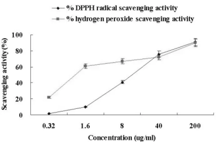
To measure antioxidant effects of the yuzu seed oil, DPPH radical scavenging and lipid peroxidation were investigated... DPPH radical scavenging

The characteristics of typhoons were examined via synoptic analysis and track examination, and the damage types and scales were analyzed to contribute to

In chlorination, the transition state resembles the reactant (small radical character), therefore the tertiary radical is slightly more stable than the primary one. In

Fresh samples were immediately tested for antioxidant activity, and antioxidant activity was compared according to gestational age and measured the difference

DPPH radical scavenging activity of Yanggaeng added with different levels of Molokhia( Corchorus olitorius L.) powder.. Bars with different letters

According to the result of comparative study on nutrient components and antioxidant activity in Abelmoschus esculentus depending on drying methods amino

Antiinflammatory and superoxide radical scavenging activities of procyanidins containing extract from the bark of Pinus pinaster Sol.. and

Effect of hydrogen peroxide on photooxidation rate of aquouse Phenol and Chlorophenols solution in continuous flow system... Total ion chromatogram by