학술강좌
약물 유발성 심혈관계 질환
안현영 삼성서울병원 약제부
초고령 사회의 진입을 눈앞에 두고 있는 만큼 심혈 관계 질환의 유병률이 증가할 것으로 예상된다. 약물 에 의한 심혈관계 이상반응이나 질환을 줄이고 기저 심혈관계 질환의 치료를 방해하지 않기 위해 약물 유 발성 심혈관계 질환의 종류, 기전, 원인, 위험인자, 예방 및 치료 등에 대하여 근거중심의 정리 및 검토 가 필요하다.
1. 약물 유발성 부정맥
심장의 전기적 자극의 전도 통로는 동방결절(Sino- Atrial node, SA node), 방실결절(Atrio-Ven- tricular node, AV node), 히스다발(his bun- dle), 방실다발갈래(bundle branches), 푸르킨 예섬유(purkinje fibers) 순으로 전도된다(Fig.
1).
1)심전도는 심장박동과 관련된 전위를 신체 표면 에서 도형으로 기록한 것으로 3차원의 구조물인 심 장의 정확한 벡터를 알기 위해 12유도 심전도(12- lead electrocardiogram, 12-lead ECG)가 사 용된다. P파는 동방결절에서 시작된 심방의 탈분극, QRS complex는 방실결절 이후의 심실의 탈분극, T파는 심실의 재분극으로 발생한다. PR 간격은 P파 시작부터 QRS complex 시작까지의 간격으로 방실 결절 전도 시간을 말하며 방실전도 지연시에 간격이 연장된다. QRS complex의 간격은 심실이 탈분극 하는 시간으로 심실 내 전도 시간을 말하며, QT 간격 은 심실 총 재분극 시간이다(Fig. 1).
1)부정맥은 심 박수에 따라 서맥성과 빈맥성, 발생위치에 따라 상심 실성과 심실성으로 분류된다.
1) 서맥성 부정맥(Bradyarrhythmias)
A. 동성서맥(Sinus bradycardia), 방실차단(AV nodal block)
서맥이란 분당 심박수 60회 미만으로 정의하며 동
방결절(SA node)에서의 전기 자극의 생성과 전도
이상, 또는 방실전도계의 이상에 의해 발생한다. 방
실차단은 심방과 심실 사이의 자극 전도가 방해되어
나타나는 결과로 그 정도에 따라 1도, 2도, 3도 즉
완전 차단으로 구분된다.
2)1도 차단은 1도 방실 지연
이 좀더 정확히 나타내는 용어일 수 있는데 일반적으
로 방실의 실제 차단은 있지 않고 전기적 자극의 지연
이 방실 결절 내에서 이루어지는 것이 보통으로, PR
간격이 0.2초 이상으로 넓어진다. 2도 방실 차단은
심방과 심실사이에서 일부 전기적 자극이 전도되지
않음으로써 나타나고 양상에 따라 Mobitz type I과
Mobits type II로 구분된다. 3도 방실 차단은 심방
에서 심실로 어떠한 전기적 자극의 전도도 없는 것으
로 완전 방실차단(complete AV block) 으로 불리
기도 한다.
3)서맥과 방실차단의 발생 메커니즘은 동
방결절의 자동능(automaticity)과 전도 억제 또는
재분극 지연, 방실결절을 통한 전도 억제로 설명하고
있다.
2)Table 1에서 동성서맥, 방실차단을 유발할
수 있는 약물을 확인할 수 있다.
4)이러한 서맥성 부정
맥을 예방하기 위해 2도 또는 3도 차단이 있으면서 심
장박동조율기(pacemaker)가 없는 환자에서는 유
발 약물의 사용을 피하고, 1도 차단 환자의 경우 동방
결절 또는 방실결절 억제제 투여가 절대 금기는 아니
므로 투여가능하나 서맥이 악화되지 않는지 보기 위 해 PR 간격 모니터링이 필요하다. 유발약물의 신,간 기능 부전에 따른 적절한 용량 조정이 이루어지도록 하고 CYP3A4 억제제 등과의 상호작용에 의해 혈중 약물 농도 상승이 예상되는 약물과의 병용을 피한다.
또한 고위험 약물을 2제 이상 병용할 경우 서맥의 빈 도나 중증도가 증가하므로 심박수의 면밀한 모니터링 이 필요하다. 서맥 또는 방실차단이 발생한 경우, 원 인 약물의 중단과 다른 약물로의 대체를 검토하고 어 쩔 수 없이 투여 유지가 필요한 경우 용량 감량을 고 려한다.
4)2도, 3도 방실차단이 있는 경우, 방실전도 를 향상시키고, 심실박동수를 증가시켜 증상을 개선 시킬 적절한 약물로 부교감신경 억제제인 atropine 0.5 mg 을 3-5분 간격으로 최대 3 mg까지 투여 한다. Atropine에 반응이 없는 경우 관상동맥 허 혈 가능성이 낮다면 isoproterenol, dopamine, dobutamine, 또는 epinephrine과 같은 베타효 능제 투여를 고려할 수 있고, 불응성인 경우 임시 경
피적 박동조율기(temporary transcutaneous pacing)를 사용할 수 있다.
2)동방결절, 방실결절 차단 약물이 과용량 투여된 경 우, 복용 시간에 따라 위세척 또는 활성탄을 사용하여 위장관 흡수 전 제거를 기대할 수 있으며, 베타차단제 (beta blocker, BB)나 칼슘통로차단제(calcium channel blocker, CCB)의 과용량으로 인한 서맥 관련 증상이 나타나거나 혈역학적으로 불안정한 서맥 이 나타날 경우, glucagon 3-10 mg을 일시주입 (bolus) 후 3-5 mg/h 투여속도로 지속정맥점적 주 입할 수 있다. 불응성 서맥에 대하여 심박수 상승과 혈역학적 안정을 위해 고용량 regular insulin(1 unit/kg 정맥 내 bolus 후 0.5 unit/kg/h)을 고 려할 수 있고 이때 저혈당 위험 문제로 dextrose와 함께 투여하면서 저칼륨혈증 등이 발생하지 않는지 잘 관찰하여야 한다.
5)2) 빈맥성 부정맥(Tachyarrhythmia)
Fig. 1 Cardiac conduction system and ECG wave
1)Table 1 Drugs that may cause/exacerbate sinus bradycardia/atrioventricular block
4)Mechanism Drug class Drug Incidence, % or
Odds Ratio
Acetylcholinesterase inhibitor
Anesthetic
Antidepressant
Antihypertensive
Inotrope Antiarrhythmic
Donepezil 0.6–48
Citalopram 0.1–2.4
Escitalopram …
Fluoxetine …
Diltiazem 4.2–16
Verapamil 0–11
Digoxin 0–7
Flecainide 2–13.2
Clonidine 5–17.5
Pyridostigmine …
Bupivacaine 2–32
Propofol 14.7
Adenosine 1–8
Amiodarone 3–20
Disopyramide 0–4
Dronedarone 0.7–2.3
Ivabradine 3.7–15.7
Propafenone 0.7–10
Sotalol 1.5–17.1
Neostigmine
β-Blockers (in- cluding eye drops)
OR 2.7 (95% CI 1.4–5.4)
0.6–25
Stimulation of activity of the parasympathetic nervous system, leading to inhibition of automaticity
of sinus node
Atrioventricular node, HPS inhibition; sinoatrial node inhibition in patients with
sinus node dysfunction Reduction in sympathetic activity
Sinoatrial/atrioventricular node inhibition
Inhibition of If channels in the sinus node
Sinoatrial/atrioventricular node inhibition
Sinoatrial/atrioventricular node inhibition
Na and Ca inhibition
Stimulation of central α2-receptors, reducing / release of norepinephrine
β-Blockers and non-DHP CCBs:
inhibition of automaticity of sinus node
Increased vagal tone Sinoatrial/atrioventricular node
inhibition
Sinoatrial/atrioventricular node, HPS inhibition
Sinoatrial/atrioventricular node inhibition
Ca indicates calcium; CCB, calcium channel blocker; CI, confidence interval; DHP, dihydropyridine; HPS, His-Purkinje system;
If, hyperpolarization-activated cyclic nucleotide-gated funny channel; Na, sodium; OR, odds ratio; and, unknown.
빈맥성 부정맥은 발생 위치에 따라 상심실성(su- praventricular)과 심실성(ventricular) 빈맥으 로 나뉘는데 상심실성 빈맥으로 발작성 상심실성 빈 맥(paroxysmal supraventricular tachycar- dia, PSVT), 심방빈맥(atrial tachycardia), 심방조동(atrial flutter), 심방세동(atrial fi- brillation) 등이 있으며 심실성 빈맥으로 심실빈 맥(ventricular tachycaridia, VT), 심실세동 (ventricular fibrillation) 등이 있다. 일반적으 로 심방성 빈맥은 심실성 빈맥보다 환자들이 잘 견 뎌내지만 심실성 빈맥은 빈맥의 발생과 동시에 혈압 이 떨어지고 좌심실기능이 급격히 악화되어 심한 혈 역학적인 이상 반응을 동반하기 때문에 응급을 요하 는 부정맥이다.
6)여기서는 약물에 의한 QTc 간격 (corrected QT interval) 연장 위주로 다뤄본다.
A. 심실빈맥(Ventricular tachycardia, VT) 심실빈맥은 분당 100회 이상의 빠르기로 심실에 서 기원하여 정상 전도로를 경유하지 않는 연속된 3회 이상의, 심실에서 유래한 박동으로 정의하며 QRS complex가 120 msec 이상으로 넓다. 지속 시간 30초 기준으로 지속성과 비지속성, QRS 형태 (morphorlogy)에 따라 단일형(monomorphic) 과 다형(polymorphic), 빈맥의 발작 당시 혈역 학적 상태에 따라 안정형과 불안정형으로 구분하는 데 증상이 경증부터 급성심장사(sudden cardiac death, SCD)까지 나타날 수 있다.
7)심실빈맥은 심 실세동으로 이행될 경우 혈역학적인 허탈(collapse) 로 진행되어 즉시 의식을 소실하고 응급심폐소생술을 시행하지 않으면 환자가 사망할 수 있는 매우 응급한 부정맥이다. 단일형 심실빈맥 유발 약물은 Table 2 와 같다.
4)약물 유발성 심실빈맥을 예방하기 위해 고 위험 약물의 사용이 적절한 환자를 잘 선택하고 용량 조절이 필수적이다. 항부정맥 약물 중 class IC 계 열, 예를 들어 flecainide, propafenon 등은 이 전에 심근경색 또는 심근병증이 있는 환자에게 사용 하지 않아야 하며 치료역이 좁은 digoxin, theo- phylline과 같은 약물은 혈중약물농도 모니터링이 필요하며 특히 신부전환자, 저마그네슘혈증 또는 저 칼륨혈증 환자에서 필수적이다. 약물 유발성 심실빈
맥 발생시에는 혈역학적 안정 여부에 따라 불안정한 경우 QRS complex의 R파에 맞추어 전기적 충격 을 가하는 synchronized cardioversion (동기화 심율동전환)을 긴급히 실시하고 혈역학적으로 안정 한 경우라면 amiodarone, lidocaine 정맥주사가 가능하다.
7)B. Torsades de Pointes (TdP)
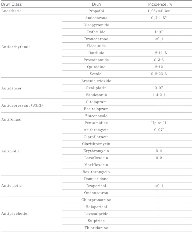
약물 유발성 부정맥으로 대표적인 것이 QT연장에 의한 TdP 발생으로, 약 200개 이상의 약물이 TdP 유발약물로 보고된다(Table 3).
8)심전도에서 QRS complex 시작과 T파 종료까지의 간격을 QT 간격 으로 정의하는데 심박수가 느려질수록 간격이 연장되 고 심박수가 빨라질수록 즉, RR 간격이 짧아질수록 QT 간격이 짧아지므로 심박수로 보정한 QT (cor- rected QT, QTc) 간격을 통해 좀더 정확한 평가가 이루어질 수 있다(Fig. 2).
9)TdP는 QTc 간격이 연 장된 상태에서 일어나는 다형(polymorphic)의 심 실빈맥으로 보통 분당 160-250회 속도로 QRS 진 폭(amplitude)이 wax and wane 양상으로 나타 나며, 일시적이고 저절로 소실될 수 있지만 심실세동 과 급성심장사까지 진행할 수 있어 생명을 위협하는 응급 부정맥이다(Fig. 3).
11)약물에 의한 QTc 간격 연장은 이렇게 생명을 위협할 수 있는 부정맥으로 연 결될 수 있기 때문에 약물 유발성 QTc 연장에 대한 이해와 예방, 조치가 필요한 이유이다. 일반인구에 서 TdP 발생률은 낮은 보고율 때문에 정확히 알기 어 려우나 1백만 인년(person-year)당 남성 2.5명, 여성 4명의 보고가 있으며, 연간 10만 명당 4명까지 높게 보고한 자료도 있다.
13),14)중환자에서 발생률은 0.07%으로 높았으며, 심정지의 6%가 TdP에 의해 발생하기도 했다.
15)약물 유발성 QTc 연장의 위험인자로 이미 QTc가
500 msec 이상으로 연장되어 있거나 약물시작 전에
비해 QTc가 60 msec 이상 연장된 경우, 여성, 65
세 이상, 급성심근경색, 저칼륨혈증, 저마그네슘혈
증, 저칼슘혈증, 서맥, 심실 비대 등 기저 구조적 심
질환이 있다.
11)약물 측면에서 위험 인자로는 QTc
연장 고위험 약물의 혈중 농도를 상승시킬 수 있는 상
황, 예를 들어 고용량 또는 고농도로 투여, CYP450
억제제의 병용 등 상호작용에 의해 QTc 연장 고위험 약물의 혈중 농도 증가, 신,간부전 환자에서 적절한 용량 조정이 이루어지지 않은 경우, 빠른 정맥내 주 입 등이 있으며, 2가지 이상의 QTc 연장 약물의 병 용하는 경우나, 전해질불균형을 유발할 수 있는 이뇨 제, 특히 K, Mg 저하와 관련이 있는 loop 이뇨제 사용시에 QTc 연장의 위험이 증가할 수 있다.
11),16)QTc 연장을 예방하기 위해 고위험 약물 시작 전의 기 저 12-유도 ECG를 확인하여 이미 QTc 간격이 연 장되어 있는 경우 위험 약물을 투여하지 않고, 어쩔 수 없이 투여가 필요한 경우 ECG를 모니터링하면서 QTc 간격 500 msec 미만으로 유지되도록 하며, 투 여 중 500 msec 이상으로 연장된 경우 약물 중단을 고려한다.
17)위험 약물 시작 후나 증량된 경우, 또는 Table 2 Drugs that may cause/exacerbate monomorphic VT
4)Mechanism
Drug class Drug Incidence, %
Anesthetic
β2-agonist
Phosphodiesterase inhibitor
Vasodilator Sympathomimetic Inotrope
Antiarrhythmic
Bupivacaine …
Ropivacaine …
Adenosine Up to 5
Amiodarone …
Flecainide 0–13
Ibutilide 0–9.8 Propafenone 0–10
Terbutaline 0–15
Theophylline …
Levosimendan …
Dobutamine 0–15.7
Milrinone 0–9.5 Phosphodiesterase inhibition
Methamphetamine … Sympathetic stimulation Digoxin Up to 7
Inhibition of sodium channel conductance
Stimulation of β2-receptors, leading to ventricular ectopic activity
Calcium sensitization, phosphodiesterase inhibition Stimulation of β2-receptors, leading
to ventricular ectopic activity Inhibition of the sodium-potassi- um-ATP pump, leading to increased intracellular calcium concentrations, resulting in afterdepolarizations and
ventricular ectopic activity
Phosphodiesterase inhibition, leading to elevated concentrations of cAMP, causing increased intracellular
calcium concentrations, resulting in afterdepolarizations and ventricular
ectopic activity Inhibition of sodium channel
conductance
cAMP, indicates cyclic adenosine monophosphate; VT, ventricular tachycardia; and …, unknown.
Table 3 QT Interval-Prolonging Drugs Known to Cause TdP
4),8)Drug Class Drug Incidence, %
Anesthetic
Anticancer
Antidepressant (SSRI)
Antifungal
Antibiotic
Antiemetic
Antipsychotic Antiarrhythmic
Propofol 1.93/million
Amiodarone 0.7–1.5*
Disopyramide …
Dofetilide 1–10
†Dronedarone <0.1
Flecainide …
Ibutilide 1.2–11.5
Procainamide 0.3–6
Quinidine 2–12
Sotalol 0.2–23.6
Arsenic trioxide …
Oxaliplatin 0.07
Vandetanib 1.4–2.1
Citalopram …
Escitalopram …
Fluconazole …
Pentamidine Up to 21
Azithromycin 0.97
‡Ciprofloxacin …
Clarithromycin …
Erythromycin 0.4
Levofloxacin 0.2
Moxifloxacin …
Roxithromycin …
Domperidone …
Droperidol <0.1
Ondansetron …
Chlorpromazine …
Haloperidol …
Levosulpride …
Sulpiride …
Thioridazine …
*0.7% to 0.8% with oral administration; 1.5% associated with intravenous administration
†Incidence is higher in patients with heart failure with reduced ejection fraction and can be as high as 10% if higher-than-recom- mended doses are administered or if dose is not appropriately adjusted in patients with kidney disease. ‡ 0.97% of patients with heart rate–corrected QT interval >450 milliseconds during azithromycin therapy …; unknown
과용량 투여된 경우 매일 12유도 ECG, 간단하게 단 일유도 ECG strip (single lead ECG strip)으 로 확인하고 QTc 간격 모니터링 기간은 QTc 연장 고위험 약물의 치료기간과 그 약물의 반감기에 따라 달라지겠다. 약물 농도의 상승에 의해 QTc 연장 위 험이 증가하므로 농도를 상승시킬 수 있는 유의한 약 물 상호작용이 있는지 검토하고 신장 및 간부전에 따 른 적절한 용량 조정이 이루어지도록 한다. 혈중 K
> 4.0 mEq/L, Mg > 2.0 mEq/L로 유지하고 무 엇보다 중요한 것은 QTc 연장 위험 약물로 알려져 있 는 약물을 2제 이상 병용하는 것을 피해야 한다. 만약 QTc 연장으로 인한 TdP가 발생할 경우, 원인 약물 을 중단하고, 저칼륨혈증, 저마그네슘혈증, 저칼슘 혈증을 교정한다. 혈역학적으로 불안정한 경우, 제
세동을 실시하는 것이 우선이며, 혈중 마그네슘 농도 와 상관없이 1-2 g의 마그네슘을 정맥주사하고 필요 시 반복함으로써 혈역학적으로 안정한 TdP를 종료 시킬 수 있다. 마그네슘 정주에 불응성인 서맥과 연 관된 TdP가 반복되는 경우 조율(pacing)이나 베타 효능제인 isoproterenol을 지속점적정맥 주입하면 심박수를 증가시키고 QT 간격을 짧게 함으로써 TdP 종결이 가능하다.
7)2. 약물 유발성 심부전
약물 유발성 심부전을 유발하거나 악화시키는 기 전은 직접적인 심독성 유발, negative inotropic effect (음성변력작용) 또는 negative chrono- Fig. 3 ECG Example of TdP. In this example, note the prolongation of the QT interval
before the arrhythmia initiation, short-long-short complexes
12)Fig. 2 The ECG waveform and segments in a normal cardiac cycle
10)tropic effect (음성변시작용), 과도한 수분과 Na 저류, 고혈압 악화, 부정맥 유발 등으로 설명되며, 단독 또는 약물상호작용에 의해 이미 치료중인 심부 전 약물의 효과를 방해하기도 한다.
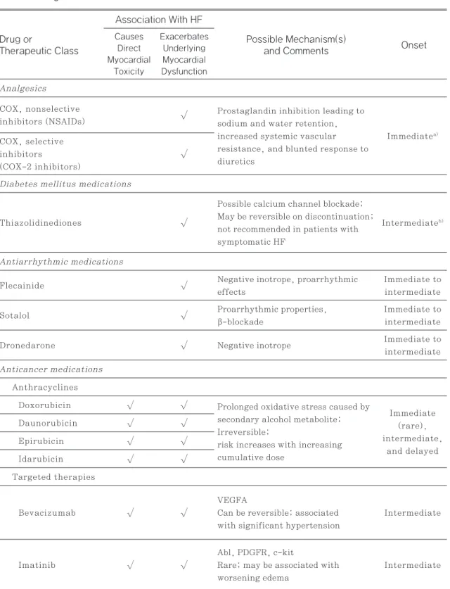
18)심부전 발현시 기를 투여 시작 후 1주 이내에 즉시 나타나는 경우, 수주에서 수개월 이내, 또는 1년 이후에 지연성으로 나타나는 것으로 발생 시기를 구분하기도 하며 직접 적인 심근 독성이 있다고 알려져 있는 약물 목록은 Table 3과 같다.
18)1) Anthracyclines
Anthracycline계 항암제는 doxorubicin, da- unorubicin, epirubicin, idarubicin이 있으며 용량 의존성 심근 손상을 유발하여 좌심실 기능 이 상과 심부전까지 유발할 수 있다. 유방암, 폐암환자 630명이 포함된 연구에서 doxorubicin 치료기간 동안 심부전 발생률은 누적용량 400 mg/m
2일 때 5%에서 700 mg/m
2일 때 48%으로 기하급수적으 로 증가하였다.
19)좌심실박출률(left ventricular ejection fraction, LVEF)의 변화를 모니터링하 는 것이 심독성 여부를 확인하는 것의 기본이며, 치 료시작 전 이미 기저 좌심실 부전이 있다면 anthra- cycline계 약물 투여로 인한 심부전 발생 위험의 강 력한 표지가 된다.
20)이러한 직접적인 심독성 예방 을 위해 anthracycline계 약물의 누적 용량을 400 mg/m
2미만으로 제한하는 것이 적절하며, dox- orubincin에 비해 epirubicin이 심부전 발생률이 낮다는 일부 근거가 있으나 동일한 용량 비교시에는 낮으나 대부분의 치료 레지멘에서 epirubicin이 보 다 고용량을 사용하므로 동일한 효능의 종양살해 용 량에서 anthracycline계 약물 간 임상적으로 중요 한 심독성 차이를 확인할 근거는 충분하지 않다.
21)Liposomal doxorubicin은 항종양 효과는 거의 비슷하면서 심부전 발생이나 좌심실 기능의 감소가 낮은 것이 확인되었다.
22)유의한 LVEF 감소가 있거 나 심부전이 발생한 경우 anthracycline계 약물 투 여를 지속할지에 대한 주의깊은 결정이 필요하고 치 료를 유지할 경우 angiotensin converting en- zyme inhibitor (ACEI)/angiotensin recep- tor blocker (ARB)와 BB를 투여하며 치료를 지속
하는 것이 가능할 수 있다.
20)LVEF가 감소한 무증상 환자에게는 ACEI/ARB 단독 또는 BB와 병용하는 가이드라인 근거의 심부전 치료를 고려해야 한다.
23)Anthracycline계 약물이 포함된 항암치료로 심독 성이 발생한 226명을 5년 추적 관찰했을 때, enal- april 또는 enalapril과 BB의 병용요법을 실시한 환자의 18%가 회복이 없었지만 11%는 기저 LVEF 까지 완전회복, 나머지는 LVEF의 부분 회복을 보였 다.
24)급성백혈병, 골수이식의 혈액암 환자 대상으 로 1차 예방으로서의 enalapril과 carvedilol 투 여군과 위약군을 비교했을 때 항암화학요법에 의한 심부전 발생이나 사망의 발생률의 유의한 차이가 관 찰되기도 하였으나(6.7 vs. 22%, p=0.036),
25)심부전 위험 요인이 없거나 심부전이 없는 상태에서 anthracyclin계 항암 치료시에 1차 예방으로서의 ACEI/ARB, BB의 선제적 투여는 대부분 소규모 연 구로 근거가 부족하므로 일반적 접근법은 아니다.
20)고위험 환자에서 심독성의 초기 표지 확인과 1차 예 방으로서 심보호 처방의 이득을 더 잘 정의하기 위해 추가적인 연구가 필요하겠다.
2) Tumor necrosis factor-alfa (TNF-α) in- hibitor (TNF-α 억제제)
TNF-α 억제제는 자가면역 질환 등의 치료약물
로 사용되고 있으며 infliximab, etanercept,
adalimumab 등이 있다. 심부전 관련 부작용은 무
작위 대조군 연구와 시판 후 조사에서 확인되었는데
ATTACH 연구에서 NYAH (New York Heart
Association) 기능등급 III 또는 IV단계이면서
LVEF < 35%인 환자 150명을 대상으로 inflix-
imab 5 mg/kg, 10 mg/kg, 위약군으로 나누어
0, 2, 6주차에 투여한 후 28주간 추적 관찰한 결과,
심부전 악화로 인한 사망이나 입원률이 저용량, 고용
량군 각각 8%, 26%였으며, 고용량 10 mg/kg 투여
군은 위약군에 비해 그 위험이 유의하게 증가하였다
(Hazard ration(HR) 2.84; 95% confidential
interval(CI), 1.01-7.97).
26)FDA에서 발표한
etanercept 또는 infliximab과 관련된 심부전 사
례 47명 중 38명이 새롭게 발생한 경우로 이 중 19
명은 관상동맥질환, 고혈압, 심근경색 병력, 당뇨와
Table 4 Drug induced Heart failure
18)Onset Drug or
Therapeutic Class
Association With HF Causes
Direct Myocardial
Toxicity
Exacerbates Underlying Myocardial Dysfunction
Possible Mechanism(s) and Comments Analgesics
Diabetes mellitus medications
Antiarrhythmic medications
Anticancer medications Anthracyclines
Targeted therapies COX, nonselective inhibitors (NSAIDs)
Thiazolidinediones
Flecainide
Doxorubicin Daunorubicin Epirubicin Idarubicin
Bevacizumab
Imatinib Sotalol
Dronedarone COX, selective inhibitors
(COX-2 inhibitors)
√
√
√
√ √
√ √
√ √
√ √
√ √
√ √
√
√
√
Prostaglandin inhibition leading to sodium and water retention, increased systemic vascular resistance, and blunted response to diuretics
Possible calcium channel blockade;
May be reversible on discontinuation;
not recommended in patients with symptomatic HF
Negative inotrope, proarrhythmic effects
Prolonged oxidative stress caused by secondary alcohol metabolite;
Irreversible;
risk increases with increasing cumulative dose
VEGFA
Can be reversible; associated with significant hypertension
Abl, PDGFR, c-kit
Rare; may be associated with worsening edema
Proarrhythmic properties, β-blockade
Negative inotrope
Immediate
a)Intermediate
b)Immediate to intermediate
Immediate (rare), intermediate,
and delayed
Intermediate
Intermediate
Immediate to
intermediate
Immediate to
intermediate
같은 위험인자가 없었고 10명은 50세 미만이었다.
27)류마티스 관절염 치료 가이드라인에 따르면 심부전 환자에서 TNF-α 억제제는 다른 치료 방법이 없을 경우에만 그것도 보상된(compensated) 심부전일 경우에만 사용하도록 권장한다.
28)Infliximab 투여 용량이 5 mg/kg을 넘지 않도록 하고 심부전 환자에 게 이 약을 투여하기로 결정하였다면 치료기간 동안
면밀히 모니터링 해야 하며 투여기간 중 심부전의 증 상이 악화되거나 새로운 심부전 증상이 나타나는 경 우 이 약의 투여를 중지하도록 권고하고 있다.
28),29)3) Dipeptidyl peptidase-4 (DPP-4) inhibi- tors (DPP-4 억제제)
Onset Drug or
Therapeutic Class
Association With HF Causes
Direct Myocardial
Toxicity
Exacerbates Underlying Myocardial Dysfunction
Possible Mechanism(s) And comments
Lapatinib
Trastuzumab
√ √
√ √
ErbB2
Can be reversible
ErbB2, antibody dependent Cytotoxicity
Can be reversible with temporary cessation of therapy or institution of HF medications
Intermediate
Intermediate
Taxanes Paclitaxel
Docetaxel
√ √
√ √
Potentiation of Anthracyclines;
Can separate administration of the anthracycline from the taxane
Intermediate
Hematologic medications
Pulmonary medications Anagrelide
Bosentan Epoprostenol Cilostazol
√
√
√
√
Possible inhibition of PD IV
Unknown
Unknown; Contraindicated in HF Inhibition of PD III resulting in Arrhythmias; Contraindicated in HF patients
Immediate to Delayed
c)Delayed Immediate
Unknown
Abl,Abelson murine leukemia viral oncogene; c-kit, tyrosine protein kinase kit; COX-2, cyclooxygenase-2;
Erb-B2, Erb-B2 receptor tyrosine kinase 2; HF, heart failure; NSAID, nonsteroidal anti-inflammatory drug;
NYHA, New York Heart Association; PD, phosphodiesterase; PDGFR, platelet-derived growth factor receptor;
VEGFA, vascular endothelial growth factor-A ligand; VEGFR, vascular endothelial growth factor receptor a) Immediate: Effect is demonstrated within 1 wk of drug administration
b) Intermediate: Effect is demonstrated within weeks to months of drug administration.
c) Delayed: Effect is demonstrated within ≥1 y of drug administration
당뇨 치료약 중 DPP-4 억제제 계열 약물과 심 부전 유발 간에서는 혼재된 결과를 보인다. SA- VOR-TIMI 53 연구에서 16,492명의 제2형 당뇨 환자를 대상으로 일반적인 당뇨치료에 각각 sax- agliptin 투여군과 위약군으로 무작위 배정한 연구 에서 중앙값 2.1년 후 심혈관사망, 심근경색, 뇌졸 중 위험은 양군 간 차이가 없었으나 saxagliptin 투여 군에서 심부전 입원률이 유의하게 높았다(HR 1.27; 95% CI, 1.07–1.51).
30)이 연구에서 BB 투여여부에 따라 차이가 나타났는데 BB가 함께 투여 된 환자에서 saxagliptin 투여로 인한 심부전 위험 은 10,162명 중 18% 발생인 반면, BB를 투여 받 지 않은 환자에서는 6,330명 중 81%에서 심부전 위 험이 증가하는 경향이 관찰되었다(HR 1.18; 95%
CI, 0.97–1.43). 이는 심부전 발생의 여러 기전 중 교감신경계 활성화로 베타 수용체 시그널링을 자극하 는 것이 관여된다는 추정을 뒷받침한다.
31)반면 si- tagliptin, linagliptin, alogliptin을 각각 위약 군과 비교한 대규모 무작위 대조군 연구에서는 심부 전으로 인한 입원 위험에 대한 통계적 유의성이 관찰
되지 않았다.
32)-34)2020년 미국 당뇨협회 가이드라 인에 따르면 심부전 환자에서 saxagliptin 사용을 피하고 이를 제외한 나머지 DPP-4 억제제는 심부 전 환자에서 사용이 고려될 수 있다.
35)2019년 국 내 당뇨병 진료지침에 따르면 심부전의 병력 또는 중 등도, 중증의 신장애 등 심부전으로 인한 입원의 위 험 요소가 있는 환자에게 saxagliptin 사용을 주의 하도록 하고, NYHA 기능등급 III, IV단계에서 임 상 경험없이 alogliptin, linagliptin 사용은 권장 하지 않고 있다.
36)3. 약물 유발성 고혈압
약물 유발성 고혈압 약물은 Table 5와 같다.
37)1) Vascular endothelial growth factor (VEGF) inhibitor (혈관내피성장인자 억제제)
Bevacizumab은 VEGF 억제제로 고혈압 발생은 VEGF 매개의 혈관 이완이 억제되며 혈관 저항의 증 Table 5 Drug induced hypertension
37)Drugs Mechanism
NSAIDs/COX-2 inhibitors
Calcineurin inhibitors (cyclosporine, tacrolimus)
Corticosteroids (esp. fludrocortisone, hydrocortisone)
Estrogen-containing oral contraceptives
Amphetamine/amphetamine derivatives, caffeine, cocaine, ergot alkaloids, phenylephrine, pseudoephedrine, triptans, venlafaxine
Erythropoiesis-stimulating agents
Prostaglandin inhibition resulting in sodium/water retention
Sympathetic simulation, vascular smooth muscle con- traction, sodium/water retention
Mineralocorticoid receptor stimulation resulting in fluid-volume expansion
Increased production of angiotensinogen stimulating the RAA system
Sympathetic nervous system stimulation
Fluid-volume expansion by increased RBC production
COX, cyclooxygenase; NSAID, nonsteroidal anti-inflammatory drug; RAA, renin-angiotensin-aldosterone; RBC, red blood cell
가를 주된 기전으로 설명하고 있다.
38)-40)Bevaci- zumab 치료를 받은 환자에서 CTCAE (National Cancer Institute’s Common Terminology Criteria for Adverse Events (CTCAE) (Na- tional Cancer Institute, 2010) 기준, 모든 등 급의 고혈압이 36%에서 관찰되었으며,
41)20개의 phase II와 phase III의 임상연구를 포함시킨 메타 분석에서 3-4단계의 고혈압 위험은 5.28배까지 증 가한 것으로 보고하였다.
42)Bevacizumab 투여 후 혈압 상승까지의 시간은 다양하게 나타나나 보통 첫 번째 주기 내에 관찰되며 저용량(<10 mg/kg) 투여 군에서 고혈압 위험이 3배 증가한 것에 비해 고용량 (≥10 mg/kg) 투여군에서는 7.5배 증가되어 용량 의존적으로 그 위험이 증가하는 결과를 보였고
41)누 적용량과도 관련이 있는 것으로 나타났다.
43)Beva- cizumab으로 인한 혈압 상승의 위험인자로 이미 고 혈압이 있는 경우 가장 강력한 인자였으며 60세 이 상 고령, BMI≥25일 경우 bevacizumab 고혈압 발생과 연관성이 있었다.
44)-46)2-3주간격으로 혈압 을 모니터링하고 고혈압으로 진행되거나 혈압의 유 의한 상승이 관찰되는 경우 항고혈압 약제를 시작하 는 것이 권장된다.
47)Sorafenib과 sunitinib 또 한 multikinase inhibitor로서 VEGF 수용체 억 제 기전이 있으며 TARGET trial에서 sorafenib 치료 관련 고혈압이 17%에서 발생하였고
48)메타분 석에서는 위약과 비교하여 고혈압 발생과 관련된 상 대위험이 증가하였다(Relative risk (RR) 2.93;
95% CI,1.52– 5.66).
49)고혈압 발생은 VEGF 신 호전달체계를 억제하는 모든 약물에서 발생하는 약물 군 효과(class effect)로 보고있다.
37),48)2) Calcineurin inhibitor (CNI)
면역억제제 중 CNI 계열 약물은 고혈압 유발 약물 로 잘 알려져 있다. 신장 이식 후 1년째에 cyclo- sporine 연관성의 고혈압 발생률은 32.7%에서 81.6%까지 연구마다 다양하게 보고된다.
50),51)골수 이식 환자에서 cyclosporine 치료환자의 57%에 서 고혈압이 발생한 반면 methorexate 치료 군에 서는 4%가 발생하여 대조를 보이고,
52)한 연구에서 심장 이식환자에서 cyclosporine 연관성의 고혈압
발생은 거의 100%에 가까웠다.
53)Cyclosporine 에 의한 고혈압 발생은 전신 및 신장의 혈관수축, 신 장에서의 Na 저류, 직접적인 신독성 등의 기전으로 설명하고 있다.
54)보통 약물을 중단할 경우 혈압은 떨어지게 되지만 완전히 이전 상태로 회복되지 않을 수 있다. CCB의 투여로 성공적으로 혈압을 낮추나 cyclosporine의 혈중농도를 상승시킬 수 있는 상 호작용이 알려져 있다.
55)또 다른 CNI 계열 약물인 tacrolimus도 고혈압 발생과 관련이 있으나 그 상 승 정도가 덜하므로 cyclosporine 유발성 고혈압 이 문제가 되는 경우 tacrolimus로 변경을 고려해 볼 수 있다.
37)3) 기타
Nonsteroidal antiinflammatory drugs (NSAIDs)에 의한 혈압 상승기전은 Na와 수분 저 류로 전신 말초 저항 증가, prostaglandin 생성 억제로 신장의 endothelin-1 합성 증가를 기전으 로 보고 있으며,
56)NSAIDs 투여시 이뇨제, ACEI, ARB, BB 등의 고혈압 치료약물의 혈압 저하 효과를 감소시키나 CCB의 효과는 방해하지 않는 것으로 알 려져 있다.
57)전신 코르티코스테로이드인 fludro- cortisone, hydrocortisone, prednisolone, methylprednisolone, dexamethasone 또한 혈압을 올릴 수 있는데 이는 고용량 투여시, 그리고 기저 심장 또는 신장질환이 있는 경우 발생하기 쉽 다.
58)기전이 명확히 밝혀져 있지 않으나 체액 과부 하가 주 기전이므로 스테로이드 투여로 인한 혈압 상 승 시에는 이뇨제 사용을 고려할 수 있다.
37)4. 심혈관 위험의 증가와 관련된 약물
1) Hormone replacement therapy (HRT, 호르 몬대체요법)
Women’s Health Initiative (WHI)에서 진
행한 연구에서 건강한 폐경 후 여성에서 에스트로겐
conjugated equine estrogen 0.625 mg, 프
로게스틴 medroxyprogesteron acetate 2.5
mg 복합정을 매일 복용한 군과 위약군으로 나누어
위험과 유익성을 비교한 연구에서 평균 추적 관찰기 간 5.2년 동안 관상심장 질환 사고의 누적위험이 호 르몬 치료군에서 29% 유의하게 높았고(HR 1.29;
nominal 95% CI (nCI), 1.02-1.63) 뇌졸중 위험 또한 호르몬 치료군에서 41% 높았으며(HR 1.41; 95% nCI, 1.07-1.85) 폐색전증의 위험 도 2배 이상 높았다(HR 2.13; 95% nCI, 1.39- 3.25).
59)에스트로겐과 프로게스틴 병합 치료시 관 상심장질환의 위험만 분석한 연구에서는 그 위험비가 위약군 대비 1.24로 높은 경향을 나타내었으며, 특 히 추적 기간을 1년 단위로 보았을 때 치료시작 후 1 년째에 위험비가 1.8배로 유의하게 가장 높고, 이후 위험비는 더 적은 상승을 보이면서 기복이 나타났으 나 5년까지는 위험비가 높은 경향을 유지하였다.
60)따라서 이러한 호르몬 대체요법은 유효한 가장 낮은 용량으로 필요한 최단기간 만큼만 쓰는 것이 권고되 고, 특히 시작 후 1년 이내에 그 위험이 가장 높으므 로 심혈관 사고 발생에 대한 세심한 모니터링과 주의 가 필요하다.
2) Erythropoiesis-stimulation agent (ESA, 적혈구생성자극제)
ESA는 내인성적혈구 생성인자와 같은 작용을 하는 당단백질로 만성신장질환에서 빈혈 교정 목적으로 투 여한다. CHOIR 연구는 만성신장질환 환자에서 빈 혈 교정을 위해 ESA 중 epoetin alfa를 헤모글로 빈 목표 13.5 g/dl와 11.3 g/dl로 치료한 군으로 나누어 사망, 심근경색, 심부전으로 인한 입원, 뇌졸 중의 심혈관 사고의 누적발생률을 카플란 마이어 추 정으로 분석한 결과, 고헤모글로빈군에서 위험이 유 의하게 높았으며(HR 1.34; 95% CI, 1.03-1.74) 특히 심부전으로 인한 사망과 입원이 고헤모글로빈 군에서 높은 경향이 나타났다(HR 1.41; 95% CI, 0.97–2.05).
61)TREAT 연구는 당뇨, 만성신장질 환, 빈혈이 동반되어 있는 환자에게 ESA 중 darb- epoetin alfa를 헤모글로빈 목표 13 g/dl로 투여 한 군과 9 g/dl를 목표로 하여 9 g/dl 미만으로 떨 어질 때만 darbepoetin alfa를 투여한 군으로 나누 어 심혈관 사건 결과를 비교하였다.
62)다른 심혈관계 종말점은 유의한 차이가 관찰되지 않았으나 고헤모
글로빈 군에서 뇌졸중이 발생하기 쉬웠고(5.0% vs.
2.6%, p<0.001) 정맥혈전색전 사고도 darbepo- etin alfa 투여군에서 유의하게 높은 발생률을 나타 내었다(2.0% vs. 1.1%, p=0.02). 이는 급성 허 혈성 뇌졸중 초기에 erythropoietin의 투여가 위약 군에 비해 더 많은 사망을 나타내어 ESA 와 stroke 사이에 부정적인 관계를 보여준 이전 연구와 유사한 결과로(16.4% vs. 9.0%),
63)TREAT 연구를 통 해 ESA 사용과 정맥혈전색전증 발생 위험 증가 관 련성을 더 공고히 할 수 있었다. KDIGO (Kidney Disease: Improving Global Outcomes) 가이 드라인에 따르면 ESA는 헤모글로빈 9 g/dl 미만으 로 떨어지지 않도록 하기 위해 9.0-10.0 g/dl일 때 투여를 시작하여, 11.5 g/dl까지만 투여하고, 특히 13.5 g/dl 이상 의도적으로 올리기 위해 사용하지 않는 것을 권고한다.
64)국내 급여 기준 또한 헤모글 로빈 10 g/dl 이하인 경우 투여를 시작하여 11 g/dl 까지 요양급여를 인정하고 있다.
65)3) NSAIDs
NSAIDs의 심근경색, 뇌졸중 증가 등 심혈관 부작
용 위험이 높은 이유는 cyclooxygenase (COX)-2
억제와 관련되어 있다. 혈관 내피세포는 COX-2를
통해 혈관이완을 유도하는 prostacyclin을 만들어
내는데 COX-2 선택적 NSAIDs는 이를 저해해 혈
관이완 효과를 낮추지만, 혈전을 유발하는 throm-
boxane A2 생성에 관여하는 COX-1은 거의 영
향이 없거나 억제되지 않아 혈소판 응집 및 혈전생
성 쪽으로 작용하게 된다.
66)그러나 일부 연구에서
는 COX-2 선택성과 심혈관 위험 증가 사이의 뚜렷
한 관계를 밝혀내지 못하고, 혈압, 신장기능, 내피세
포, nitric oxide 생성과 같은 다른 요인 또한 중요
한 것으로 나타나기도 하였다.
67)COX-2 선택적 억
제제인 rofecoxib과 valdecoxib은 골관절염과 류
마티스 관절염 치료제로 승인을 받았지만 심혈관 위
험 문제로 각각 2004년, 2005년 시장에서 철수되었
으나 최근 celecoxib은 심혈관 위험과 관련하여 비
열등성을 증명하였다. PRECISION trial은 골관절
염 또는 류마티스 관절염이 있는 24,081명의 환자
를 대상, 이중 약 75%가 기저 심혈관 위험이 없고 나
머지는 중등도 내지 중증 위험인자를 갖고 있는 환자 에게 celecoxib 100-200 mg 1일 2회, naprox- en 375-500 mg 1일 2회, 또는 ibuprofen 600- 800 mg 1일 3회 투여를 무작위 배정하여 심혈관 사 망, 심근경색, 뇌졸중의 일차 복합 종료점(primary composite outcome)의 비열등성을 평가하였다.
평균 치료기간은 20.3개월, 평균 추적 기간은 34개 월로 각 약물군에서 일차복합종료점의 유의한 차이 는 관찰되지 않았다(celecoxib vs. naproxen, HR 0.93; 95% CI, 0.76-1.13, celecoxib vs.
ibuprofen, HR 0.85; 95% CI, 0.70-1.04).
68)NSAIDs 계열 내 약물 간에도 심혈관 위험의 차이 가 있는 것으로 보이는데 여러 관찰 연구에서 di- clofenac은 다른 NSAIDs에 비해 심혈관 위험이 높은 것이 비교적 일관되게 증명되었다.
69),70)덴마크 에서 실시한 국가등록 자료를 활용한 연구에서는 이 전 심근경색이 있었던 환자에게 NSAIDs를 사용하 는 것이 사망, 심근경색 재발과 유의한 관련이 있었 으며(HR 1.45; 95% CI, 1.46-1.64) 개별 약물 분석 결과 diclofenac이 가장 높은 위험을 나타냈다 (HR 3.26; 95% CI, 2.57-3.86).
69)NSAIDs의 국내 허가 내 경고사항으로 모든 NSAIDs에 대하여 중대한 심혈관계 혈전 반응, 심근경색증 및 뇌졸중 등 심혈관계 위험이 증가하는 것을 경고하고 있고 특히 기저 심혈관계 질환 또는 심혈관계 질환의 위험 인자 가 있는 환자이거나 장기 복용, 고용량 투여한 환자 일수록 심혈관계 이상반응 발생가능성이 증가함을 경 고하고 있다. 심혈관계 이상반응에 대한 잠재적 위험 을 최소화하기 위해 최저 유효용량으로 가능한 최단 기간 동안 사용해야 한다.
4) Antipsychotics (항정신병약)
항정신병약물은 QTc 연장 고위험 약물로 잘 알 려져 있다.
4)2000년부터 2009년까지 심실부정맥 (ventricular arrhythmia, VA) 또는 급성심장 사(sudden cardiac death, SCD) 의심 진단을 받 은 17,718명의 대규모 환자를 대상으로 환자-교차 연구(case-crossover study)를 진행하였다. 항 정신병 약물의 사용은 14일 이내 VA 또는 SCD 발생 위험을 1.53배 증가시켰고(95% CI, 1.38-1.70),
1세대와 2세대 항정신병 약물로 구분했을 때, 1세 대 약물사용이 2세대 약물보다 VA, SCD 위험이 증가했다(adjusted OR[AOR]=1.66; 95% CI, 1.43-1.91 for FGAs, AOR=1.36; 95% CI, 1.20-1.54 for SGAs).
71)이는 이전 발표된 연구 들과도 맥락을 같이하는 결과들이다.
72),73)개별 약물 로 분석하면 위험 증가와 연관된 약물은 haloperi- dol (AOR=1.46; 95% CI, 1.17-1.83), olan- zapine (AOR=1.64; 95% CI, 0.98-2.72), quetiapine (AOR=1.29; 95% CI, 1.07-1.56), risperidone (AOR=1.39; 95% CI, 1.13-1.72) 등 이었다.
71)이 연구에서 ziprasidone은 위험증 가 약물로 통계적 유의성이 없었는데 대상 환자에서 ziprasidone 사용 환자가 적어 통계적 파워가 충분 하지 않았던 것으로 해석하고 있다. 막 항정신병약물 을 시작한 환자는 그 위험이 가장 높은 시기이므로 처 음 투여를 시작할 때 심전도 모니터링을 하면서 위험 과 유익성의 세심한 평가가 필요하다.
결론
약물 유발성 심혈관계 질환으로 QTc 연장에 의한
TdP 등 부정맥, 심부전, 고혈압 등이 발생가능하며
본문에서 다루지 않은 기타 부정맥, 저혈압, 심낭염
등도 가능하여 이는 심혈관계 사건의 증가로 연결된
다. 일시적, 가역적으로 발현되기도 하지만, 만성질
환으로도 진행되는 경우도 있으며, 약제로 인한 심혈
관계 부작용으로 인해 그 약물이 원래 목표로 했던 치
료를 제한하는 요소가 되거나, 동반된, 기저 심부전
이나 고혈압 등의 치료를 방해하는 원인이 되기도 한
다. 약물 유발성 심혈관계 질환의 종류, 기전, 원인,
위험인자, 예방 및 치료 등에 대하여 이해하여 약제
사용시에 관련하여 유발될 수 있는 심혈관계 질환이
나 부작용을 미리 예측하고, 고위험 약물, 고위험 환
자에서 집중적인 모니터링과 함께 선제적으로 대체
약물을 고려하거나 위험요인을 줄일 수 있는 방법을
모색해야 한다. 원인 약물 감별을 위해 환자의 기저
질환, 의심 약물을 포함한 기타 약제의 시작 시기, 투
여기간, 용량, 상호작용, 신기능, 간기능 부전 등을
면밀하게 검토하여 인과성 평가가 필요하며, 원인 약
물로 확인된 경우 약물의 중단 또는 감량, 대체약물
로의 변경, 치료제의 투여를 고려한다.
참고문헌
1) Master the ECG & Echo Today: ECG Book; Introduction to ECG Interpreta- tion; Clinical ECG Interpretaion; as- sessed January 20, 2021. Available from https://ecgwaves.com/topic/introduc- tion-electrocardiography-ecg-book/
2) Kusumoto FM, Schoenfeld MH, Barrett C et al. 2018 ACC/AHA/HRS guideline on the evaluation and management of pa- tients with bradycardia and cardiac con- duction delay: a report of the American College of Cardiology/American Heart Association Task Force on Clinical Prac- tice Guidelines and the Heart Rhythm Society. Circulation. 2019;140(8):e382–
e482.
3) Atrioventricular block type. In: Mary BC. Understanding Elctrocardiography.
8th ed. Missouri: Mosby. 2003:216-30.
4) James ET, Mina KC, Kristen BC et al.
Drug-Induced Arrhythmias: A Scientific Statement From the American Heart Association. Circulation. 2020;142(15):e 214–e233.
5) Engebretsen KM, Kaczmarek KM, Mor- gan J et al. High-dose insulin thera- py in beta-blocker and calcium chan- nel-blocker poisoning. Clin Toxicol (Phila). 2011;49(4):277–83.
6) Olshansky B, Chung M, Pogwizd S. Arr- hythmia Essentials. Sudbury: MA Jones and Bartlett learning. 2012:186-213.
7) Al-Khatib SM, Stevenson WG, Ack- erman MJ et al. 2017 AHA/ACC/HRS guideline for management of patients with ventricular arrhythmias and the prevention of sudden cardiac death: a
report of the American College of Cardi- ology/American Heart Association Task Force on Clinical Practice Guidelines and the Heart Rhythm Society. Circulation.
2018;138(13):e272-391.
8) Woosley RL, Heise CW, Gallo T, Tate J, Woosley D, Romero KA. QT drugs List, AZCERT, Inc, 1822 Innovation Park Dr., Oro Valley, AZ 85755. Accessed June 10, 2020. Available from https://www.
CredibleMeds.org
9) Funck-Brentano C, Jaillon P. Rate-cor- rected QT interval: techniques and limi- tations. Am J Cardiol. 1993;72(6):17-22.
10) Zheng J, Zhang J, Danioko S et al. A 12-lead electrocardiogram database for arrhythmia research covering more than 10,000 patients. Scientific Data.
2020;7(1):48.
11) Drew BJ, Ackerman MJ, Funk M et al.
on behalf of the American Heart Asso- ciation Acute Cardiac Care Committee of the Council on Clinical Cardiology, the Council on Cardiovascular Nursing, and the American College of Cardiology Foundation. Prevention of torsade de pointes in hospital settings: a scientif- ic statement from the American Heart Association and the American College of Cardiology Foundation. Circulation.
2010;121(8):1047–60.
12) Aimee L, David P. Basic cardiac eletro- physiology and common drug induced arrhythmia. Crit Care Nurs Clin N Am.
2016;28(3):357-71.
13) Sarganas G, Garbe E, Klimpel A et al.
Epidemiology of symptomatic drug-in- duced long QT syndrome and torsade de pointes in Germany. Europace. 2014;
16(1):101-8.
14) Darpö B. Spectrum of drugs prolonging
QT interval and the incidence of tor- sades de pointes. Eur Heart J Suppl.
2001;3:70-80.
15) Pickham D, Helfenbein E, Shinn JA et al. High prevalence of corrected QT interval prolongation in acutely ill pa- tients is associated with mortality: re- sults of the QT in Practice (QTIP) Study.
Crit Care Med. 2012;40(2):394–9.
16) Zeltser D, Justo D, Halkin A et al.
Torsade de pointes due to noncardiac drugs: most patients have easily iden- tifiable risk factors. Medicine (Balti- more). 2003;82(4):282–90.
17) Sandau KE, Funk M, Auerbach A et al.; on behalf of the American Heart Association Council on Cardiovascular and Stroke Nursing; Council on Clini- cal Cardiology; and Council on Cardio- vascular Disease in the Young. Update to practice standards for electrocar- diographic monitoring in hospital set- tings: a scientific statement from the American Heart Association. Circula- tion. 2017;136(19):e273–e344.
18) Page RL, O'Bryant CL, Cheng D et al.
Drugs that may cause or exacerbate heart failure: a scientific statement from the American Heart Association.
Circulation. 2016;134(6):e32-e69.
19) Swain SM, Whaley FS, Ewer MS.
Congestive heart failure in patients treated with doxorubicin: a retrospec- tive analysis of three trials. Cancer.
2003;97(11):2869–79.
20) Henriksen PA. Anthracycline cardi- otoxicity: an update on mechanism, monitoring and prevention. Heart.
2018;104(12):971-7.
21) Dalen EC, Michiels EM, Caron HN et al. Different anthracycline derivates
for reducing cardiotoxicity in cancer patients. Cochrane Database Syst Rev.
2010;3:CD005006
22) Rayson D, Suter TM, Jackisch C et al.
Cardiac safety of adjuvant pegylated liposomal doxorubicin with concurrent trastuzumab: a randomized phase II trial. Ann Oncol. 2012;23(7):1780-8.
23) Zamorano JL, Lancellotti P, Rodriguez MD et al. 2016 ESC Position Paper on cancer treatments and cardiovascular toxicity developed under the auspic- es of the ESC Committee for Practice Guidelines: The Task Force for cancer treatments and cardiovascular toxicity of the European Society of Cardiology (ESC). Eur Heart J. 2016;37(36):2768- 801.
24) Cardinale D, Colombo A, Bacchiani G et al. Early detection of anthracycline cardiotoxicity and improvement with heart failure therapy. Circulation.
2015;131(22):1981–8.
25) Bosch X, Rovira M, Sitges M et al.
Enalapril and carvedilol for preventing chemotherapy-induced left ventricular systolic dysfunction in patients with malignant hemopathies: the OVER- COME trial (preventiOn of left ven- tricular dysfunction with enalapril and caRvedilol in patients submitted to in- tensive chemOtherapy for the treat- ment of malignant hEmopathies). J Am Coll Cardiol. 2013;61(23):2355–62.
26) Chung ES, Packer M, Lo KH et al. Ran-
domized, double-blind, placebo-con-
trolled, pilot trial of infliximab, a chi-
meric monoclonal antibody to tumor
necrosis factor-alpha, in patients with
moderate-to-severe heart failure: re-
sults of the anti-TNF Therapy Against
Congestive Heart Failure (ATTACH) trial. Circulation. 2003;107(25):3133- 40.
27) Kwon HJ, CotéTR, Cuffe MS et al. Case reports of heart failure after therapy with a tumor necrosis factor antagonist.
Ann Intern Med. 2003;138(10):807-11.
28) Singh JA, Saag KG, Bridges SL Jr et al. 2015 American College of Rheuma- tology Guideline for the Treatment of Rheumatoid Arthritis. Arthritis Care Res. 2016;68(1):1-26.
29) Remicade FDA prescribing informa- tion; Updated January, 2011; Assessed on January 21, 2021. Available from https://www.accessdata.fda.gov/
drugsatfda_docs/label/2011/103772 s5281lbl.pdf
30) Scirica BM, Bhatt DL, Braunwald E et al.; SAVOR-TIMI 53 Steering Commit- tee and Investigators. Saxagliptin and cardiovascular outcomes in patients with type 2 diabetes mellitus. N Engl J Med. 2013;369(14):1317–26.
31) Milton Packer DPP4 inhibitors cause Heart failure events by promoting adr- energically mediated cardiotoxicity.
Circ Res. 2018;122(7):928-32.
32) Green JB, Bethel MA, Armstrong PW et al.; TECOS Study Group. Effect of sitagliptin on cardiovascular out- comes in type 2 diabetes. N Engl J Med.
2015;373(3):232–42.
33) Rosenstock J, Perkovic V, Johansen OE et al. Effect of linagliptin vs placebo on major cardiovascular events in adults with type 2 diabetes and high cardio- vascular and renal risk: the CARMELI- NA randomized clinical trial. JAMA.
2019;321(1):69–79.
34) White WB, Cannon CP, Heller SR et
al.; EXAMINE Investigators. Aloglip- tin after acute coronary syndrome in patients with type 2 diabetes. N Engl J Med. 2013;369(14):1327–35.
35) American diabetes association. 10.
Cardiovascular disease and risk man- agement: standards of medical care in diabetes-2020. Diabetes Care. 2019;
43:111–34.
36) 2019 당뇨병 진료지침. 서울: 대한당뇨병학회;
2019
37) Grossman A, Messerli FH, Grossman E. Drug induced hypertension – An un- appreciated cause of secondary hyper- tension. EJP. 2015;763(Pt A):15-22.
38) Henry TD, Annex BH, McKendall GR et al. The VIVA trial: Vascular en- dothelial growth factor in Ischemia for Vascular Angiogenesis. Circulation.
2003;107(10):1359-65.
39) Henry TD, Rocha-Singh K, Isner JM et al. Intracoronary administration of re- combinant human vascular endothelial growth factor to patients with coronary artery disease. American Heart Jour- nal. 2001;142(5):872-80.
40) Horowitz JR, Rivard A, van der Zee R et al. Vascular endothelial growth factor/
vascular permeability factor produces nitric oxide-dependent hypotension.
Evidence for a maintenance role in qui- escent adult endothelium. Arterioscler Thromb Vasc Biol. 1997;17(11):2793- 800.
41) Zhu X, Wu S, Dahut WL et al. Risks of proteinuria and hypertension with bev- acizumab, an antibody against vascu- lar endothelial growth factor: system- atic review and meta-analysis. Am J Kidney Dis. 2007;49(2):186-93.
42) Ranpura V, Pulipati B, Chu D et al.
Increased risk of high-grade hyper- tension with bevacizumab in cancer patients: a meta-analysis. Am J Hy- pertens. 2010;23(5):460-8.
43) Mir O, Coriat R, Cabanes L et al. An observational study of bevacizum- ab-induced hypertension as a clinical biomarker of antitumor activity. On- cologist. 2011;16(9):1325-32.
44) Hamnvik OP, Choueiri TK, Turchin A et al. Clinical risk factors for the develop- ment of hypertension in patients treat- ed with inhibitors of the VEGF signaling pathway. Cancer. 2015;121(2):311-9.
45) Isobe T, Uchino K, Makiyama C et al.
Analysis of adverse events of bevaci- zumab-containing systemic chemo- therapy for metastatic colorectal can- cer in Japan. Anticancer Research.
2014;34(4):2035–40.
46) Wicki A, Hermann F, Prêtre V et al.
Pre-existing antihypertensive treat- ment predicts early increase in blood pressure during bevacizumab therapy:
the prospective AVALUE cohort study.
Oncol Res Treat. 2014;37(5):230-6.
47) Maitland ML, Bakris GL, Black HR et al. Initial assessment, surveillance, and management of blood pressure in patients receiving vascular endothe- lial growth factor signaling path- way inhibitors. J Natl Cancer Inst.
2010;102(9):596–604.
48) Escudier B, Eisen T, Stadler, WM et al.
for the TARGET Study Group. Sorafenib in advanced clear-cell renal-cell carci- noma. N Engl J Med. 2007;356(2):125- 34.
49) Abdel-Rahman O, Fouad M. Risk of car- diovascular toxicities in patients with solid tumors treated with sunitinib, ax-
itinib, cediranib or regorafenib: an up- dated systematic review and compara- tive meta-analysis. Crit. Rev. Oncol.
Hematol. 2014;92(3):194-207.
50) Ponticelli C. Cyclosporine in idiopathic nephrotic syndrome. Immunopharma- col. Immunotoxicol. 1993;15(4):479-89.
51) Snanoudj R, Kriaa F, Arzouk N et al.
Single-center experience with cyclo- sporine therapy for kidney transplan- tation: analysis of a twenty-year peri- od in 1200 patients. Transplant Proc.
2004;36(2):83-8.
52) Loughran TP Jr, Deeg HJ, Dahlberg S et al. Incidence of hypertension af- ter marrow transplantation among 112 patients randomized to either cyclo- sporine or methotrexate as graft- ver- sus-host disease prophylaxis. Br. J.
Haematol.1985;59(3):547-53.
53) Grossman E, Messerli FH. High blood pressure. A side effect of drugs, poi- sons, and food. Arch. Intern. Med.
1995;155(5):450-60.
54) Hoorn EJ, Walsh SB, McCormick JA et al. R. Pathogenesis of calcineurin in- hibitor-induced hypertension. J. Ne- phrol. 2012;25(3):269-75.
55) Rodicio JL. Calcium antagonists and renal protection from cyclosporine ne- phrotoxicity: long-term trial in renal transplantation patients. J Cardiovasc Pharmacol. 2000;35(3 Suppl 1):7-11.
56) Johnson AG. NSAIDs and increased blood pressure. What is the clinical sig- nificance? Drug Saf. 1997;17(5):277-89.
57) Krum H, Swergold G, Curtis SP et al.
Factors associated with blood pressure
changes in patients receiving diclofenac
or etoricoxib: results from the MEDAL
study. J Hypertens. 2009;27(4):886-93.
58) Walker BR, Edwards CR. New mecha- nisms for corticosteroid-induced hy- pertension. British Medical Bulletin.
1994;50(2):342-55.
59) Writing Group for the Women’s Health Initiative Investigators. Risks and benefits of estrogen plus progestin in healthy postmenopausal women: prin- cipal results from the Women’s Health Initiative randomized controlled trial.
JAMA. 2002;288(3):321-33.
60) JAE Manson, J Hsia, KC Johnson et al.
Estrogen plus Progestin and the Risk of Coronary Heart Disease. N Engl J Med.
2003;349(6):523-34.
61) Singh AK, Szczech L, Tang KL et al.
CHOIR investigators. Correction of anemia with epoetin alfa in chron- ic kidney disease. N Engl J Med.
2006;355(20):2085-98.
62) Pfeffer MA, Burdmann EA, Chen CY et al.; for the TREAT investigators. A tri- al of darbepoetin alfa in type 2 diabetes and chronic kidney disease. N Engl J Med. 2009;361(21):2019-32.
63) Ehrenreich H, Weissenborn K, Prange H et al. Recombinant human erythropoi- etin in the treatment of acute ischemic stroke. Stroke. 2009;40(12):e647–e56.
64) KDIGO Clinical Practice Guideline for Anemia in Chronic Kidney Disease.
Kidney Int Suppl. 2012;2(4):288-35.
65) 건강보험심사평가원, 보험인정기준. 고시 제 2019-38호, Erythropoietin 주사제; 고시 제2019-57호, Darbepoetin alpha 주사제 66) Caughey GE, Cleland LG, Penglis PS
et al. Roles of cyclooxygenase (COX)- 1 and COX-2 in prostanoid production by human endothelial cells: selective up-regulation of prostacyclin synthe- sis by COX-2. J Immunol. 2001;167(5):
2831-8.
67) Trelle S, Reichenbach S, Wandel S et al.
Cardiovascular safety of non-steroidal anti-inflammatory drugs: network me- ta-analysis. BMJ. 2011;342(7789):154.
68) Nissen SE, Yeomans ND, Solomon DH et al.; for the PRECISION trial investiga- tors. Cardiovascular safety of celecox- ib, naproxen, or Ibuprofen for rrthritis.
N Engl J Med. 2016;375(26):2519-29.
69) Gislason GH, Rasmussen JN, Abild- strom SZ et al. Increased mortality and cardiovascular morbidity associated with use of nonsteroidal anti-inflam- matory drugs in chronic heart failure.
Arch Intern Med. 2009;169(2):141-9.
70) Dubreuil M, Louie-Gao Q, Peloquin CE et al. Risk of myocardial infarction with use of selected non-steroidal an- ti-inflammatory drugs in patients with spondyloarthritis and osteoarthritis.
Ann Rheum Dis. 2018;77(8):1137-42.
71) Chi-Shin Wu, Yu-Ting Tsai, Hui-Ju Tsai. Antipsychotic drugs and the risk of ventricular arrhythmia and/or sudden cardiac death: A nation-wide case-crossover study. J Am Heart As- soc. 2015;4(2):e001568.
72) Murray-Thomas T, Jones ME, Pa- tel D et al. Risk of mortality (includ- ing sudden cardiac death) and major cardiovascular events in atypical and typical antipsychotic users: a study with the general practice research da- tabase. Cardiovasc Psychiatry Neurol.
2013;2013:30-44.
73) Mehta S, Chen H, Johnson M et al. Risk
of serious cardiac events in older adults
using antipsychotic agents. Am J Ger-
iatr Pharmacother. 2011;9(2):120-32.
문제