8
INTRODUCTION
Among the genes involved in sporadic human breast cancers, those related to estrogen action are good candidates for investigation because estrogen exerts as tumor promoter, through estrogen receptor.(1) Estrogen receptors belong to a superfamily of transcription activators and have two types of ERα (Estrogen receptor) and ERβ in the human tissue, showing differential expression in various tissues.(2-4) Their protein products are transcriptional factors that control the expression of estrogen-responsive genes by binding to a specific DNA sequence within their regulatory regions.(5,6) There is increasing evidence that de novo methylation of promoter CpG (cytosine guanine dinucleotide) islands, con- tributes to the alteration of gene expression in cancer and is associated with gene silencing.(7)
Although numerous reports have been published on the characterization of ERα promoter region and the status of the CpG methylation in various types of cancers,(8,9) little information is available for the methylation of ERβ gene.(10,11) The transcription of human ERα gene occur from at least two different promoters and it was shown that the levels of expression of total ERα RNA and of transcript from distal promoter correlated well with the amount of ERα protein in
The Characterization of CpG Methylation of ERα and ERβ Gene in the Breast Cancer
Department of 1Biology, Dongguk University, Seoul, Korea, Research Institute and Hospital, National Cancer Center, Goyang, Korea
Sun Jung Kim1, Tae Won Kim1, Su-Young Lee1, Sang-Jae Park, Eun Sook Lee and Han-Sung Kang
Purpose: Aberrant methylation of promoter cytosine gua- nine dinucleotide (CpG) islands is known to be respon- sible for the alteration and silencing of cancer genes. The data presented here show that most methylations of Estrogen Receptorα (ERα) and ERβ are found at or near the transcriptional factor binding sites in the breast cancer tissues.
Methods: Fifty archival breast cancer tissues and twenty- five normal tissues were selected and the status of the methylation and the transcription were investigated by bisulfite genomic sequencing and reverse transcription (RT) PCR.
Results: Consequently, the hypermethylation of ERα and ERβ genes was found in 66.0% and 50.0% of 50 breast cancers, respectively. In particular, the methylation sites were frequently located near the CCAAT box (-363 and -375) for the ERα gene, and at or adjacent to binding sites of GATA (-217, -302) and Sp1 (+224, +227, +160) for the ERβ gene. The methylations at or near the binding sites were observed in most of the methylated cancers (ERα 87.9%, and ERβ 84.0%). The methylated cases were negatively correlated with the expression of ERα and ERβ RNA (P<0.01). In particular, tumors with CpG methylation of ERα and ERβ at or near the binding sites did not express mRNA, whereas those CpG methyla- tion outside the sites showed moderate expression. Four tumors with methylated ERα genes at sites unrelated to the binding sites showed higher levels of protein expres- sion than those with methylation at or near the sites (P=0.01).
Conclusion: Although the number of samples was rela- tively small, our results suggest that DNA methylation in ERα and ERβ appears to take significant effect on
transcriptional silencing and is most often present in the CpG sites at or near the putative transcriptional factor binding sites. We believe this finding offers a clue to the initiation or spread pattern of CpG methylation in human breast cancer. (Journal of Korean Breast Cancer So- ciety 2004;7:8-16)
ꠏꠏꠏꠏꠏꠏꠏꠏꠏꠏꠏꠏꠏꠏꠏꠏꠏꠏꠏꠏꠏꠏꠏꠏꠏꠏꠏꠏꠏꠏꠏꠏꠏꠏꠏꠏꠏꠏꠏꠏꠏꠏꠏꠏꠏ Key Words: Breast cancer, ERα, ERβ, Methylation, Trans-
cription
Correspondence:Han-Sung Kang, Center for Breast Cancer, Na- tional Cancer Center, 809 Madu1-dong, Ilsan-gu, Goyang-si, Gyeonggi-do 411-764, Korea. Tel: 82-31- 920-1642, Fax: 82-31-920-1759, E-mail: [email protected] Received:27 December, 2003 Accepted:9 February, 2004
ꠏꠏꠏꠏꠏꠏꠏꠏꠏꠏꠏꠏꠏꠏꠏꠏꠏꠏꠏꠏꠏꠏꠏꠏꠏꠏꠏꠏꠏꠏꠏꠏꠏꠏꠏꠏꠏꠏꠏꠏꠏꠏꠏꠏꠏꠏꠏꠏꠏꠏꠏꠏꠏꠏꠏꠏꠏꠏꠏꠏꠏꠏꠏꠏꠏꠏꠏꠏꠏꠏꠏꠏꠏꠏꠏꠏꠏꠏꠏꠏꠏꠏꠏꠏꠏꠏꠏꠏꠏꠏꠏꠏꠏꠏꠏꠏꠏꠏꠏꠏꠏꠏꠏꠏꠏꠏꠏꠏ the human breast cancer.(12) Furthermore, the methylation of
distal promoter of the ER alpha gene is important for loss of ER alpha gene expression in human breast cancer.(13) The promoter region of ERα gene possesses relatively few potential binding sites, whereas the human ERβ promoter region includes various consensus binding sites as illustrated in Fig.
1.(11)
We previously reported that p53 gene, a tumor suppressor gene, is methylated in the breast cancer tissues and four CpG sites with methylation are located at or near the potential binding sites of transcriptional factors such as AP1 or YY-1.(14) However, there have been few reports on the actual methylation status at or adjacent to the putative binding sites
of transcriptional factors in the bona fide cancer tissues.(15,16) Furthermore, it is obscure to what extent the CpG methylation at or near the putative binding sites of transcriptional factors will influence the transcriptional silencing, compared with the methylation outside the binding sites.
We have now evaluated the methylation status of ERα (distal promoter) and ERβ promoter regions in the breast cancer tissues, including the region of potential the transcriptional factors binding sites by use of bisulfite genomic sequencing. Our data indicate that most CpG methylations in the ER genes are positioned at or adjacent to the putative binding sites of transcriptional factors and this type of methylation might exert more significant effect on the gene silencing.
METHODS
1) Tissue samples and nucleotide extraction
Fifty archival breast cancer tissues and twenty-five normal tissues were selected consecutively from the patients under- going partial or total mastectomies from Nov. 2000 to Feb.
2002 at the National Cancer Center in Goyang City, Korea. The normal tissues were comprised of fifteen tissues adjacent to the tumor, and ten specimens far from the cancer. Sections were cut 5 mm thick from formalin-fixed, paraffin-embedded tissues and mounted on a microscope. To collect cancer tissues for DNA extraction, microdissection was performed as described previously.(17) DNA from microdissected tissues was extracted in 80 L of lysis buffer (50 mM Tris-HCl pH8.0, 1 mM EDTA, 0.5% Tween 20, 200 g/ml of proteinase K) at 55oC for 72 hours.
Thirty frozen breast tissues out of 50 cancer tissues were available for the RT-PCR and were disrupted using FastRNA Green kit (Qbiogene, Carisbad, CA) on a Ribolyser cell disruptor (Qbiogene). Purification was performed following the supplier's protocol and the RNA was finally suspended in 50 L of RNase-free water.
2) Bisulfite genomic sequencing
Bisulfite modification of genomic DNA was carried out as previously described(14) with minor modification. Briefly, cDNA extracted from the microdissected tissues was digested with EcoRI and then subjected to bisulfite treatment. Bisulfite- treated DNA was subjected to two rounds of nested PCR to amplify the ER promoter region. Primer sequences for ER are 5'-GGGTATTTTAGGAGTATTTTAG-3' and 5'-ACCAATC- AAAAATATATAAATA-3' for the primary PCR, and 5'- Fig. 1. Structure and sequence of the 5'-flanking region of the
human ERα (GenBank accession number X62462, A) and ERβ gene (GenBank AF191544, B). The 5' transcription start site is designated as +1. Each promoter region under investigation is shown with the bent arrows. The individual CpG's are presented by numbering and underlined. The putative binding sites of transcriptional factors are indicated by boxes with the names of their matching factors.
A
B
ꠏꠏꠏꠏꠏꠏꠏꠏꠏꠏꠏꠏꠏꠏꠏꠏꠏꠏꠏꠏꠏꠏꠏꠏꠏꠏꠏꠏꠏꠏꠏꠏꠏꠏꠏꠏꠏꠏꠏꠏꠏꠏꠏꠏꠏꠏꠏꠏꠏꠏꠏꠏꠏꠏꠏꠏꠏꠏꠏꠏꠏꠏꠏꠏꠏꠏꠏꠏꠏꠏꠏꠏꠏꠏꠏꠏꠏꠏꠏꠏꠏꠏꠏꠏꠏꠏꠏꠏꠏꠏꠏꠏꠏꠏꠏꠏꠏꠏ AAAGTGGTTAAGAGG TGGATTT-3' and 5'-TCAAATTT-
ACAAAATAAAACAT-3' for the nested PCR. For the ER promoter, its 654 bp promoter region (-332 to +322 in Fig.
1B) was amplified by two overlapping fragments, 349 bp and 337 bp. Primer sequences for the upstream 349 bp fragment are 5'-TATGTGAGTTAGGGGTTGAGGA-3' and 5'-AAA- AATAAATATCCAAAAAACCAAC-3' for the primary PCR, and 5'-AGTGTGGTTTTTAGATTGGTTG-3' and 5'-CCAA- AAAACCAACAACTAAAAAAAC-3' for the nested PCR.
Primer sequences for the downstream 337 bp fragment are 5'-GTTTTTAAAAGGAAGAAGGGGTTTA-3' and 5'-ATAA- ACAAATATAATAACTTACA-3' for the primary PCR, and 5'-TTTTTTTAGTTTTTTTAGTTGTTGG-3' and 5'-ATAATA ACTTACAAATAAACACAC-3' for the nested PCR. All the PCR conditions were 94oC for 2 minutes, 30 cycles of 94oC for 20 seconds, 55oC for 20 seconds, and 72oC for 30 seconds, with a final extension at 72oC for 5 minutes. The resulting products were subjected to agarose gel electrophoresis and purified using Qiaex II gel extraction kit (Qiagen, Valencia, CA). Sequencing was performed for each PCR product using the primers for the nested PCR on an ABI automated sequencer with Dye terminators (Perkin-Elmer, Foster City, CA). DNA sequences were confirmed by analyzing both direction of each PCR product and at least three PCR products were analyzed for each cancer tissue. By definition, the term methylation at or near the putative binding sites was methylation which occurs within two base pairs up- or downstream of binding sites,(15) corresponding to 375, -363 and 275 in the ERα (the transcriptional start site is designated as +1: GenBank accession number X62462, Fig. 1A) and 302, -207, -206, -103, -97, -61, -33, +159, +224, +227 in the ERβ (the transcriptional start site is designated as +1: GenBank accession number AF191544, Fig. 1B). The methylation levels of individual CpG site were calculated as percentages of 5-methyl cytosine among the whole cytosine residues (methylated plus unmethylated) at the same site.(18)
3) Reverse transcription (RT)-PCR
First-stranded cDNA was synthesized from 5 g of total RNA using a reverse transcription kit (Promega, Madison, WI) according to the manufacturer's protocol. To amplify double- stranded cDNA, PCR was performed in a 50 L reaction mixture containing 1 L of the reverse transcribed cDNA, 5 L of 10×
PCR buffer, 1.25 mM dNTP, 1.5 mM MgCl2, 1 M of each primer and 1 U of Taq polymerase (Roche Biochemicals, Germany). Sequence of the PCR primers used were as follows:
ERα, 5'TCTGCCAAGGAGACTCGCTA-3' and 5'-TTGGCC-
AAAGGTTGGCAGCT-3'; ER, 5'-TGTGCGGAGACAGAG- AAGTG-3' and 5'-GGTGGTCAATTGAGCGCCAC-3'.
To verify the integrity of mRNA, the G3PDH gene was amplified by use of the following primers: G3PDHF: 5'- ACCACAGTCCATGCCATCAC-3' and G3PDHR: 5'-TCCA- CCACCCTGTTGCTGTA-3'. PCR reactions were performed in a Primus thermal cycler (MWG-Biotech, Germany) at 94oC for 1 minute, 30 cycles at 94oC for 20 seconds, 57oC for 20 seconds, and 72oC for 30 seconds, followed by an extension step at 72oC for 5 minutes. After PCR, 10 L of the samples was electrophoresed in 1.5% agarose gels and visualized by ethidium bromide staining. All reactions included negative controls where RNA was used as template.
4) Immunohistochemical staining
The immunohistochemical staining was performed on paraffin-embedded tissue and sections were scored positive as previously reported.(19) The protein level of ERα was measured immunohistochemically by a combination of the staining intensity and the percentage of positively stained cells.
In brief, a score of 0 to 3 for the carcinoma in the majority of the entire section was given (0, no staining; 1, weak; 2, intermediate; 3, strong). The percentage of positively stained cells was an average after counting the stained and the total number of cells from four high-magnification fields with the software IMAGE-PRO PLUS 4.1 (Media Cybernetics, Silver Spring, MD). The ERα protein level was expressed as the product of the staining intensity and the percentage of staining cells.
5) Statistical analysis
The chi-squared test and Fisher's exact test were used to analyze the differences in the rate of each variable and the student t-test was to detect differences in the mean values of variables. P-values <0.05 were considered to be statistically significant. All calculations were performed using SPSS for Windows release 7.0 (SPSS Inc., Chicago, IL).
RESULTS
1) CpG Methylation of the ERα and ERβ Gene in the normal tissue and the breast carcinoma
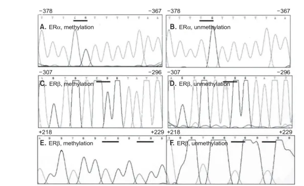
A total of 50 breast cancers and 25 normal tissues were analyzed for the methylation status of ERα and ERβ gene by bisulfite modification DNA sequencing (Fig. 2A-F). Hyperme- thylation of the ERα and ERβ was found in 33 (66.0%) and 25 (50.0%) of 50 breast cancer tissues, respectively. Eleven of
ꠏꠏꠏꠏꠏꠏꠏꠏꠏꠏꠏꠏꠏꠏꠏꠏꠏꠏꠏꠏꠏꠏꠏꠏꠏꠏꠏꠏꠏꠏꠏꠏꠏꠏꠏꠏꠏꠏꠏꠏꠏꠏꠏꠏꠏꠏꠏꠏꠏꠏꠏꠏꠏꠏꠏꠏꠏꠏꠏꠏꠏꠏꠏꠏꠏꠏꠏꠏꠏꠏꠏꠏꠏꠏꠏꠏꠏꠏꠏꠏꠏꠏꠏꠏꠏꠏꠏꠏꠏꠏꠏꠏꠏꠏꠏꠏꠏꠏꠏꠏꠏꠏꠏꠏꠏꠏꠏꠏ
50 samples (22.0%) did not show any methylation of either ERα or ERβ, whereas 19 samples (38.0%) showed methylations of both ERα and ERβ. In contrast to methylation pattern found
in the cancer tissue, no methylation in the CpG site was detected in the normal tissues except one normal tissue adjacent to the cancer. Two CpG sites (-142, -139) in the ERβ gene Fig. 2. The methylated (A, C, E) and unmethylated (B, D, F) samples of ERα and ERβ by use of bisulfite genomic sequencing. cDNA extracted from the microdissected tissues was digested with EcoRI and then subjected to bisulfite treatment. After treatment with bisulfite, DNA was PCR amplified and the resulting products were sequenced on an ABI automated sequencer (Perkin- Elmer Corp., Foster City, CA). The original sequences are a follows; A, B-cctcgttcccaat, C, D-agtgtcggcatc, E, F-aggtggcggcgg. All the unmethylated cytosines are changed to T by the bisulfite treatment but not the methylated cytosines. The CpG sites are indicated by underlines. Upper numbers correspond to the numbering of the genes as in Fig. 1.
-378
-307
+218 +218
-367
-296
+229 +229
-378
-307
-367
-296
A. ER , methylationα
C. ER , methylationβ
E. ER , methylationβ
B. ER , unmethylationα
D. ER , unmethylationβ
F. ER , unmethylationβ
Fig. 3. The methylation profiles of ERβ gene in the cancer (No 44, Table 1, A) and the adjacent normal tissue (B). The methylation pattern in the normal tissue is different from that in the cancer tissue. Upper numbers correspond to the numbering of the genes as in Fig. 1.
-217
-217
-205
-205 -142
-145
-133
-135
A
B
Table 1. Methylation profiles of CpG sites in the promoter regions of ERα and ERβ
ꠚꠚꠚꠚꠚꠚꠚꠚꠚꠚꠚꠚꠚꠚꠚꠚꠚꠚꠚꠚꠚꠚꠚꠚꠚꠚꠚꠚꠚꠚꠚꠚꠚꠚꠚꠚꠚꠚꠚꠚꠚꠚꠚꠚꠚꠚꠚꠚꠚꠚꠚꠚꠚꠚꠚꠚꠚꠚꠚꠚꠚꠚꠚꠚꠚꠚꠚꠚꠚꠚꠚꠚꠚꠚꠚꠚꠚꠚꠚꠚꠚꠚꠚꠚꠚꠚꠚꠚꠚꠚꠚꠚꠚꠚꠚꠚꠚꠚꠚꠚꠚꠚꠚꠚꠚꠚꠚꠚꠚꠚꠚꠚꠚꠚꠚ
No. Histology ERα methylation ERα mRNA ERα protein levela ERβ methylation ERβ mRNA
ꠏꠏꠏꠏꠏꠏꠏꠏꠏꠏꠏꠏꠏꠏꠏꠏꠏꠏꠏꠏꠏꠏꠏꠏꠏꠏꠏꠏꠏꠏꠏꠏꠏꠏꠏꠏꠏꠏꠏꠏꠏꠏꠏꠏꠏꠏꠏꠏꠏꠏꠏꠏꠏꠏꠏꠏꠏꠏꠏꠏꠏꠏꠏꠏꠏꠏꠏꠏꠏꠏꠏꠏꠏꠏꠏꠏꠏꠏꠏꠏꠏꠏꠏꠏꠏꠏꠏꠏꠏꠏꠏꠏꠏꠏꠏꠏꠏꠏꠏꠏꠏꠏꠏꠏꠏꠏꠏꠏꠏꠏꠏꠏꠏꠏꠏ
1 Invasive ductal 1,2,3,4,6b -c 0 1,3b -
2 Invasive ductal 1,2,3,5,7 - 0 1,2,3,23,24,25,27,32,33,34 -
3 Cribriform 3,8 - 0 3,38 -
4 Invasive ductal 1,2,3,4,5,7,8 - 0 3,32,37 -
5 Invasive ductal Noned ++ 60 23 +
6 Invasive lobular None ++ 21 None ++
7 Invasive ductal None ++ 84 None ++
8 Invasive ductal 1,3,4,5,7 - 0 3,28 -
9 Papillary 1,2,6,7,8 - 0 None ++
10 Invasive lobular 4 + 18 None ++
11 Invasive ductal 1,2,3,4,8 - 0 25 -
12 Invasive ductal 1,2,3,4,5,6,7,8 - 0 None ++
13 Invasive ductal None ++ 45 30,37,38,39,40,41 -
14 Invasive ductal 1,2,3,4,7 - 0 38 -
15 Invasive ductal 1,2,3,5 - 15 None ++
16 Invasive ductal 1,2,3,4,6,8 - 0 1 -
17 Invasive ductal None ++ 30 1,3,37 -
18 Invasive ductal None + 0 None +
19 Invasive ductal 1,2,3,5 - 0 23,28 -
20 Invasive ductal 1,2,3,4,7,8 - 0 None ++
21 Papillary None ++ 15 3,32 -
22 Invasive lobular None ++ 69 None ++
23 Invasive ductal None + 6 1 -
24 Invasive ductal 8 + 15 1,2,3,4 -
25 Invasive ductal 1,2,3,4,5,6,7,8 - 0 None ++
26 Invasive ductal 1,2,3,4,5,6,7,8 - 0 None ++
27 Invasive ductal 1,2,3,5 - 0 None +
28 Invasive ductal 1,2,3,4,5,7 - 0 None ++
29 Invasive ductal 1,2,3,6 - 3 None +
30 Invasive ductal 1,3,6,8 - 0 20,36 +
31 Invasive ductal 3,8 NA 0 None NA
32 Invasive ductal 5 NA 3 1,5,23 NA
33 Medullary 6,7 NA 9 4,32 NA
34 Invasive ductal 1,2,3,4,5 NA 0 None NA
35 Invasive ductal None NA 72 None NA
36 Invasive ductal 1,2,3,4,6,7,8 NA 6 4,6 NA
37 Invasive ductal None NA 54 None NA
38 Invasive ductal 1,3,5,6 NA 0 None NA
39 Invasive ductal None NA 0 None NA
40 Papillary 1,2,3,5,6,8 NA 6 1,3 NA
41 Invasive ductal 1,2,3,5 NA 0 37,38,39,40 NA
42 Invasive ductal None NA 30 None NA
43 Invasive ductal None NA 60 None NA
44 Invasive ductal 6, 7 NA 24 1,3,4,10,11 NA
45 Invasive ductal 1,2,3,4,5,6,8 NA 0 32, 37, 38 NA
46 Invasive ductal None NA 72 1,3,32 NA
47 Invasive ductal None NA 150 None NA
48 Invasive ductal 1,2,3,4,5,6,7,8 NA 0 None NA
49 Medullary None NA 180 None NA
50 Invasive ductal 1,2,3,5 NA 0 1,2,3,4 NA
ꠏꠏꠏꠏꠏꠏꠏꠏꠏꠏꠏꠏꠏꠏꠏꠏꠏꠏꠏꠏꠏꠏꠏꠏꠏꠏꠏꠏꠏꠏꠏꠏꠏꠏꠏꠏꠏꠏꠏꠏꠏꠏꠏꠏꠏꠏꠏꠏꠏꠏꠏꠏꠏꠏꠏꠏꠏꠏꠏꠏꠏꠏꠏꠏꠏꠏꠏꠏꠏꠏꠏꠏꠏꠏꠏꠏꠏꠏꠏꠏꠏꠏꠏꠏꠏꠏꠏꠏꠏꠏꠏꠏꠏꠏꠏꠏꠏꠏꠏꠏꠏꠏꠏꠏꠏꠏꠏꠏꠏꠏꠏꠏꠏꠏꠏ
aThe ERα protein level was expressed as the product of the staining intensity and the percentage of staining cells. b indicates the numerical order of CpG out of total CpG sites located in the promoter regions of ERα (8 CpG's) and ERβ (45 CpG's) as in Fig. 1A and Fig.
1B. c: -, negative; +, weak expression; ++ = positive expression. ddenotes for absence of the methylated CpG.
ꠏꠏꠏꠏꠏꠏꠏꠏꠏꠏꠏꠏꠏꠏꠏꠏꠏꠏꠏꠏꠏꠏꠏꠏꠏꠏꠏꠏꠏꠏꠏꠏꠏꠏꠏꠏꠏꠏꠏꠏꠏꠏꠏꠏꠏꠏꠏꠏꠏꠏꠏꠏꠏꠏꠏꠏꠏꠏꠏꠏꠏꠏꠏꠏꠏꠏꠏꠏꠏꠏꠏꠏꠏꠏꠏꠏꠏꠏꠏꠏꠏꠏꠏꠏꠏꠏꠏꠏꠏꠏꠏꠏꠏꠏꠏꠏꠏꠏ
ꠏꠏꠏꠏꠏꠏꠏꠏꠏꠏꠏꠏꠏꠏꠏꠏꠏꠏꠏꠏꠏꠏꠏꠏꠏꠏꠏꠏꠏꠏꠏꠏꠏꠏꠏꠏꠏꠏꠏꠏꠏꠏꠏꠏꠏꠏꠏꠏꠏꠏꠏꠏꠏꠏꠏꠏꠏꠏꠏꠏꠏꠏꠏꠏꠏꠏꠏꠏꠏꠏꠏꠏꠏꠏꠏꠏꠏꠏꠏꠏꠏꠏꠏꠏꠏꠏꠏꠏꠏꠏꠏꠏꠏꠏꠏꠏꠏꠏꠏꠏꠏꠏꠏꠏꠏꠏꠏꠏ were shown to be methylated in that normal tissue, which was
different from the methylation pattern found in the cancer tissue (Fig. 3, Table 1, No 44).
Among all methylation-positive cancers analyzed, -363 site out of total 8 CpG sites in the ERα gene was the most frequent site of methylation followed by -398 and -375 sites (Fig.
4A). The methylation in the ERβ gene was identified as follows in the order of frequency: -217, -302, +160, +224, and +227 (Fig. 4B). Surprisingly, -375 and -363 CpG sites in the ERα gene are located in close proximity to the CCAAT box, while the frequent methylated CpG sites in the ERβ gene are corresponding to the site at or adjacent to the binding sites of GATA (-217, -302) and Sp1 (+160, +224, +227) (Fig.
4A and Fig. 4B). The methylations at or adjacent to the potential binding sites were observed in 29 cases (87.9%) of 33 ERα methylated cancers, and twenty-one (84.0%) out of tumors with ERβ methylation, respectively (Table 1).
2) Correlation between hypermethylation and differ- ential expression of ERα and ERβ
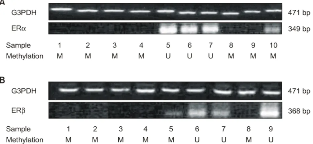
To study whether the aberrant methylation actually correlates with gene silencing, the expression status of ERα and ERβ was determined by RT-PCR for 30 tumors with (ERα, 21 cases; ERβ, 16 cases) or without methylation of these loci (ERα, 9 cases; ERβ, 14 cases). Representative results for mRNA expression of ERα and ERβ are shown in Fig. 5. The expression level of ERα protein was also measured for all the 50 cases by immunohistochemical staining, as described in the section of Materials and Methods (Table 1). As a result, the tumors that showed aberrant methylation of ERα and ERβ did not express mRNA, compared with unmethylated cases (Fig.
5A and 5B, Table 2: P<0.01). In particular, the tumors with methylation at position outside the binding sites of the transcriptional factors (ERα, #10, #24; ERβ, #5, #30) showed a low mRNA band, whereas those at or near the binding sites
Fig. 4. The percentage of methylation at individual CpG site in ERα (A) and ERβ (B) genes. The CpG sites at or near the putative binding sites of transcriptional factors are noted by arrows and their matching transcriptional factors. Numbers on the X-axis correspond to the numbering in Fig. 1A and Fig. 1B.
A
B
-398 -375 -363 -349 -275 -205 -118 -14
-302 -224 -217 -206 -176 -171 -139 -136 +31 +61 +94 +105 +112 +120 +142 +160 +174 +178 +205 +224 +227 +232 +234 +236
60
30
Methylation percentage (%)Methylation percentage (%)
0
0 50
25 30
15 40
20 20
10 10
5
CAAT
GATA GATA SP-1 SP-1
TATA
ꠏꠏꠏꠏꠏꠏꠏꠏꠏꠏꠏꠏꠏꠏꠏꠏꠏꠏꠏꠏꠏꠏꠏꠏꠏꠏꠏꠏꠏꠏꠏꠏꠏꠏꠏꠏꠏꠏꠏꠏꠏꠏꠏꠏꠏꠏꠏꠏꠏꠏꠏꠏꠏꠏꠏꠏꠏꠏꠏꠏꠏꠏꠏꠏꠏꠏꠏꠏꠏꠏꠏꠏꠏꠏꠏꠏꠏꠏꠏꠏꠏꠏꠏꠏꠏꠏꠏꠏꠏꠏꠏꠏꠏꠏꠏꠏꠏꠏ
did not. The incidence of methylated case was significantly correlated with negative expression of ERα protein (24.2% vs 88.2% P<0.01). The expression level of ERα protein was lower among patients with ERα methylation than without methylation (2.82±1.08 vs 55.76±11.95, P<0.01), and also four tumors with ERα methylation outside the binding sites showed higher level of expression than those at or near the sites (16.50±3.12 vs 0.93±0.56, P=0.01).
DISCUSSION
Bisulfite genomic sequencing is the method of choice for the determination of methylation status with single-base resolution and eliminates the possibility of incomplete digestion by the methylation sensitive restriction enzymes, which would yield an inaccurate picture of DNA methylation.(20) And yet, there have
been few reports published that the presence of CpG site methylation in ERα and ERβ gene was confirmed by bisulfite genomic sequencing.(10,18) The studies presented indicate that the promoter regions of ERα and ERβ are methylated in the breast cancer tissues and CpG methylation within the promoter region of the genes induces the transcriptional inactivation by use of bisulfite genomic sequencing and RT-PCR. In particular, we established an in vivo evidence that among the methylation positive cancers of ER gene, the methylation at or adjacent to the potential binding sites of transcriptional factors occurs in most breast cancers (ERα 87.9%; ERβ 84.0%). Exploring the previous results on the ERα and ERβ methylation in the prostatic cancer by the same technique,(10,18) the most frequent sites of methylation in ERα gene was found near the CCAAT box (-375 and -363), whereas methylation of ERβ detected at the putative binding site of Sp1 (+224) although a part of the transcriptional factor binding sites in promoter region of ERβ was not included in that study.(18) We previously reported that p53 gene is methylated in the breast cancer tissue and the CpG sites with methylation are located at or in vicinity of the potential binding sites of transcriptional factors such as AP1 or YY-1. Mancini et al. demonstrated that the methylated CpG in the breast cancer tissues occurs at a putative CREB (cAMP-responsive element binding) trans- cription factor binding site in the BRCA1 promoter and this site is sensitive to the site-specific CpG methylation.(16) Similar methylation interference studies directed at other genes have shown that CpG methylation of CREB abolishes CREB binding.(21) Moreover, the most frequently methylated CpG of Fig. 5. Representative figures of RT-PCR products for ERα (A) and ERβ (B) mRNA in the breast cancer tissue. Total cellular RNA (5g) was reverse transcribed, and the resulting cDNA was amplified by PCR using specific primers for each gene. G3PDH expression demonstrates relatively equal amounts of initial mRNA. The results are summarized in Table 1. The letters M and U in the row of methylation indicate the presence and the absence methylation, respectively. In addition, M* means methylation outside the transcriptional factor binding sites.
G3PDH
G3PDH
471 bp
471 bp 349 bp
368 bp Methylation
Methylation M
M M
M M
M
U
M M
M
U
U U
U M
M M
U M Sample
Sample
1
1 2
2 3
3
5
5 4
4
6
6 7
7 8
8 9
9 10 ERα
ERβ
A
B
Table 2. The correlation of methylation and transcriptional expression in ERα and ERβ gene
ꠚꠚꠚꠚꠚꠚꠚꠚꠚꠚꠚꠚꠚꠚꠚꠚꠚꠚꠚꠚꠚꠚꠚꠚꠚꠚꠚꠚꠚꠚꠚꠚꠚꠚꠚꠚꠚꠚꠚꠚꠚꠚꠚꠚꠚꠚꠚꠚꠚꠚꠚꠚꠚꠚꠚ RNA expression P-value ꠏꠏꠏꠏꠏꠏꠏꠏꠏꠏꠏꠏꠏꠏꠏꠏꠏꠏꠏꠏꠏꠏꠏꠏꠏꠏꠏꠏꠏꠏꠏꠏꠏꠏꠏꠏꠏꠏꠏꠏꠏꠏꠏꠏꠏꠏꠏꠏꠏꠏꠏꠏꠏꠏꠏ ERα methylation(+)(N=21) 2 (9.5%)
<0.01a ERα methylation(-)(N=9) 9 (100%)
ERβ methylation(+)(N=16) 2 (12.5%)
<0.01b ERβ methylation(-)(N=14) 14 (100%)
ꠏꠏꠏꠏꠏꠏꠏꠏꠏꠏꠏꠏꠏꠏꠏꠏꠏꠏꠏꠏꠏꠏꠏꠏꠏꠏꠏꠏꠏꠏꠏꠏꠏꠏꠏꠏꠏꠏꠏꠏꠏꠏꠏꠏꠏꠏꠏꠏꠏꠏꠏꠏꠏꠏꠏ Total number presented in this table was not 50 but 30 because RT-RCR was performed in 30 frozen tissues. Statistical methods:
aFisher's exact test, bchi-squared test
ꠏꠏꠏꠏꠏꠏꠏꠏꠏꠏꠏꠏꠏꠏꠏꠏꠏꠏꠏꠏꠏꠏꠏꠏꠏꠏꠏꠏꠏꠏꠏꠏꠏꠏꠏꠏꠏꠏꠏꠏꠏꠏꠏꠏꠏꠏꠏꠏꠏꠏꠏꠏꠏꠏꠏꠏꠏꠏꠏꠏꠏꠏꠏꠏꠏꠏꠏꠏꠏꠏꠏꠏꠏꠏꠏꠏꠏꠏꠏꠏꠏꠏꠏꠏꠏꠏꠏꠏꠏꠏꠏꠏꠏꠏꠏꠏꠏꠏꠏꠏꠏꠏꠏꠏꠏꠏꠏꠏ connexin gene, a putative tumor suppressor gene, was reported
to be in an Sp1 site known to be important for connexin 26 gene expression in the breast cancer tissue.(22)
Two major hypotheses have been proposed to explain the finding that DNA methylation is responsible for gene silencing.
First, the binding between the methylated DNA and a kind of methyl-CpG binding proteins induces the transcriptional sup- pression, leading to the alteration of the chromatin structure.(7) Second, DNA methylation at or in vicinity to the putative binding sites of transcriptional factors play a role by impeding specific interactions between transcription factors and their matching DNA control elements.(23) Since not many tran- scription factor binding sites contain CpG, the second hypo- thesis has been considered to be infrequently involved.
Recently, the methylation at a CpG site two base pairs upstream of CCAAT box was reported to hinder the binding of the transcriptional factor CBF to CCAAT box, leading to sup- pression of hMLH1 expression.(15) This mechanism may be mediated by the induction of the changes in the DNA double helix configuration, or by interfering with the binding of cofac- tors. Furthermore, the methylation seems to be stabilized when the promoter region is confined by the spread of DNA methylation near or at its transcription factor binding sites and to occur in the organized and sequential, but not the chaotic, manner.(24) The potential role of the hypermethylation at the putative binding sites needs further investigation, as this might be a clue to the initiation or spread pattern of CpG methylation in the human cancer.
Our second concern is that CpG methylation at specific sites might exert an effect on the transcriptional silencing. In this study, the tumors with methylation outside the binding sites in ERα and ERβ gene showed a mRNA expression, whereas those at or near the binding sites did not. And also the tumors with methylation of ERα at other than putative binding sites had higher level of protein expression than those at or near the sites (16.50±3.12 vs. 0.93±0.56, P=0.01). In spite of the small number of samples, this finding implies that the DNA me- thylation present at or in close proximity to putative binding sites of the transcriptional factors may more strongly exert its effect on transcriptional silencing. The transcriptional silencing seems to be diverse according to the location of methylated CpG. Deng et al. demonstrated that the extent of gene silencing by methylation in hMLH1 promoter region shows region- specific pattern in the colorectal cell line, and CpG methylation at the certain part of the promoter region is not critical in silencing the gene expression,(15) supporting our results.
In addition as illustrated in the experiment on the expression
of ERβ in the rodent mammary gland, a considerable numbers of proliferating cells contain ERβ protein.(25) The mechanism of the supposed effect of ER on cell proliferation is unknown.
Functional studies in cancer cell lines have shown differences in stimulating the transcriptional activating function (AF1) of the receptor and activator protein (AP1) cross talk(26);
however, the master genes involved in the mitogenic activity of estrogens are still being debated.
In conclusion, aberrant DNA methylation at regulatory regions in ERα and ERβ genes plays a role in the transcriptional silencing of the genes within subsets of breast carcinomas. Although the number of samples was so limited that our results might take on a sort of hypothesis at this time, it is suggested that the DNA methylation in the ERα and ERβ appears to be most often present in the CpG sites at or near the putative binding sites of the transcriptional factors, and this type of methylation may influence more significantly on transcriptional silencing. This finding is believed to be a clue to the initiation or spread pattern of CpG methylation in the human breast cancer.
ACKNOWLEDGEMENTS
This work is supported by the National Cancer Center Grant.
REFERENCES
1) Laidlaw IJ, Clarke RB, Howell A, Owen AW, Potten CS, Anderson E. The proliferation of normal human breast tissue implanted into athymic nude mice is stimulated by estrogen but not progesterone. Endocrinology 1995;136:164-71.
2) Nishihara E, Nagayama Y, Inoue S, Hiroi H, Muramatsu M, Yamashita S, et al. Ontogenetic changes in the expression of estrogen receptor alpha and beta in rat pituitary gland detected by immunohistochemistry. Endocrinology 2000;141:615-20.
3) Onoe Y, Miyaura C, Ohta H, Nozawa S, Suda T. Expression of estrogen receptor beta in rat bone. Endocrinology 1997;
138:4509-12.
4) Jefferson WN, Couse JF, Banks EP, Korach KS, Newbold RR.
Expression of estrogen receptor beta is developmentally regulated in reproductive tissues of male and female mice. Biol Reprod 2000;62:310-7.
5) Enmark E, Pelto-Huikko M, Grandien K, Lagercrantz S, Lagercrantz J, Fried G, et al. Human estrogen receptor beta gene structure, chromosomal localization, and expression pattern. J Clin Endocrinol Metab 1997;82:4258-65.
6) Petersen DN, Tkalcevic GT, Koza-Taylor PH, Turi TG, Brown TA. Identification of estrogen receptor beta2, a functional variant of estrogen receptor beta expressed in normal rat
ꠏꠏꠏꠏꠏꠏꠏꠏꠏꠏꠏꠏꠏꠏꠏꠏꠏꠏꠏꠏꠏꠏꠏꠏꠏꠏꠏꠏꠏꠏꠏꠏꠏꠏꠏꠏꠏꠏꠏꠏꠏꠏꠏꠏꠏꠏꠏꠏꠏꠏꠏꠏꠏꠏꠏꠏꠏꠏꠏꠏꠏꠏꠏꠏꠏꠏꠏꠏꠏꠏꠏꠏꠏꠏꠏꠏꠏꠏꠏꠏꠏꠏꠏꠏꠏꠏꠏꠏꠏꠏꠏꠏꠏꠏꠏꠏꠏꠏ tissues. Endocrinology 1998;139:1082-92.
7) Jones PA, Laird PW. Cancer epigenetics comes of age. Nat Genet 2000;21:163-7.
8) Ottaviano YL, Issa JP, Parl FF, Smith HS, Baylin SB, Davidson NE. Methylation of the estrogen receptor gene CpG island marks loss of estrogen receptor expression in human breast cancer cells. Cancer Res 1994;54:2552-5.
9) Issa J-P, Ottaviano YL, Celano P, Hamilton SR, Davidson NE, Baylin SB. Methylation of the oestrogen receptor CpG island links ageing and neoplasia in human colon. Nat Genet 1994;7:536-40.
10) Nojima D, Li LC, Dharia A, Perinchery G, Ribeiro-Filho L, Yen TS, et al. CpG hypermethylation of the promoter region inactivates the estrogen receptor-beta gene in patients with prostate carcinoma. Cancer 2001;92:2076-83.
11) Li LC, Yeh CC, Nojima D, Dahiya R. Cloning and char- acterization of human estrogen receptor beta promoter.
Biochem Biophys Res Commun 2000;275:682-9.
12) Hayashi S, Imai K, Suga K, Kurihara T, Higashi Y, Nakachi K. Two promoters in expression of estrogen receptor messen- ger RNA in human breast cancer. Carcinogenesis 1997;18:
459-64.
13) Yoshida T, Eguchi H, Nakachi K, Tanimoto K, Higashi Y, Suemasu K, et al. Distinct mechanisms of loss of estrogen receptor alpha gene expression in human breast cancer:
methylation of the gene and alteration of trans-acting factors.
Carcinogenesis 2000;21:2193-201.
14) Kang JH, Kim SJ, Noh DY, Park IA, Choe KJ, Yoo OJ, et al. Methylation in the p53 promoter is a supplementary route to breast carcinogenesis: correlation between CpG methylation in the p53 promoter and the mutation of the p53 gene in the progression from ductal carcinoma in situ to invasive ductal carcinoma. Lab Invest 2001;81:573-79.
15) Deng G, Chen A, Pong E, Kim YS. Methylation in hMLH1 promoter interferes with its binding to transcription factor CBF and inhibits gene expression. Oncogene 2001;20:7120-7.
16) Mancini D N, Rodenhiser DI, Ainsworth PJ, O'Malley FP,
Singh SM, Xing W, et al. CpG methylation within the 5' regulatory region of the BRCA1 gene is tumor specific and includes a putative CREB binding site. Oncogene 1998;
16:1161-9
17) Zhuang Z, Bertheau P, Emmert-Buck MR, Liotta LA, Gnarra J, Linehan WM, et al. A microdissection technique for archival DNA analysis of specific cell populations in lesions <1 mm in size. Am J Pathol 1995;146:620-5.
18) Li LC, Chui R, Nakajima K, Oh BR, Au HC, Dahiya R.
Frequent methylation of estrogen receptor in prostate cancer:
correlation with tumor progression. Cancer Res 2000;60:702-6.
19) Geisler S, Lonning PE, Aas T, Johnsen H, Fluge O, Haugen DF, et al. Influence of TP53 gene alterations and c-erbB-2 expression on the response to treatment with doxorubicin in locally advanced breast cancer. Cancer Res 2001;61:2505-12.
20) Grunau C, Clark SJ, Rosenthal A. Bisulfite genomic se- quencing: systematic investigation of critical experimental parameters. Nucleic Acids Res 2001;29:E65.
21) Iguchi-Ariga SM, Schaffner W. CpG methylation of the cAMP-responsive enhancer/promoter sequence TGACGTCA abolishes specific factor binding as well as transcriptional activation. Genes Dev 1989;3:612-9.
22) Tan LW, Bianco T, Dobrovic A. Variable promoter region CpG island methylation of the putative tumor suppressor gene Connexin 26 in breast cancer. Carcinogenesis 2002;23:231-6.
23) Tate PH, Bird AP. Effects of DNA methylation on DNA- binding proteins and gene expression. Curr Opin Genet Dev 1993;3:226-31.
24) Turker MS. Gene silencing in mammalian cells and the spread of DNA methylation. Oncogene 2002;21:5388-93.
25) Saji S, Jensen EV, Nilsson S, Rylander T, Warner M, Gusta- fsson JA. Estrogen receptors α and β in the rodent mammary gland. Proc Natl Acad Sci USA 2000;97:337-42.
26) Paech K, Webb P, Kuiper GG, Nilsson S, Gustafsson JA, Kushner PJ, et al. Differential ligand activation of estrogen receptors ERα and ERβ at AP1 sites. Science 1997;277:
1508-10.