www.jkfas.org 대해 알아보고자 한다.
본 론
1. 발목관절염의 특징
체중부하 시 발목관절은 무릎이나 엉덩관절에 비하여 면적은 작 지만 같은 하중을 받게 된다. 500 N의 힘이 가해질 때 발목관절의 접촉면은 350 mm2인 데 비하여 무릎관절은 1,120 mm2, 엉덩관절 은 1,100 mm2로 가해지는 최대 스트레스가 무릎이나 엉덩관절에 비해 약 3배 가량 발목관절에서 높게 나타난다.4,5) 하지만 발목에는 다른 하지 관절에 비해 관절염의 발생 빈도가 낮다.
발목 연골의 두께는 1∼1.7 mm로 얇으면서 일정한 데 비하여 무릎이나 엉덩관절에서는 최소한 3∼6 mm로 두꺼우면서 편차가 있다.6) 발목 연골은 정합적(congruency)으로 구성되어 있고, 얇지 만, 스트레스를 균등하게 받게 되는 것이다. 이러한 특성으로 인해 거골의 연골이 반복적인 부하에 잘 견디고 주기적 압박을 가할 때 발생하는 연골의 표재층 변형이 적다. 나이가 들면서 인장성이 많 이 감소하는 무릎과 엉덩관절에 비해 발목관절 연골의 인장성은 크게 감소하지 않기 때문에 상대적으로 일차성 관절염이 적게 나 타나는 것으로 알려져 있다.7) 발목 연골에서는 neutrophil collage-
서 론
발목에서 관절염의 발생 빈도는 다른 관절에 비해 상대적으로 낮은 편이다. 하지만, 체중부하 시 발목관절은 무릎이나 엉덩관절 과 같은 하중을 받지만 면적은 1/3에 불과하여 단위 면적당 많은 압력을 받게 된다. 또한 다른 관절에 비해 손상에 취약하여 외상후 관절염이 많이 발생하게 된다.1,2) 관절염을 초기, 중기, 말기 관절 염으로 분류한다면 발목에서 초기 관절염 환자가 많이 발생하고, 환자가 호소하는 불편함도 단순방사선 영상에 보이는 소견에 비해 높게 나타나는 경우가 있다.3) 초기 발목관절염에 대한 이해와 치 료가 중요한 이유이다. 이에 본 종설에서는 발목에서 발생하는 관 절염의 특징을 살펴보고, 초기 발목관절염의 양상과 치료 방법에
This is an Open Access article distributed under the terms of the Creative Commons Attribution Non-Commercial License (http://creativecommons.org/licenses/CC
by-nc/4.0) which permits unrestricted non-commercial use, distribution, and reproduction in any medium, provided the original work is properly cited.
Copyright 2017 Korean Foot and Ankle Society. All rights reserved.ⓒ
The incidence of arthritis in the ankle is relatively low compared to other joints. On the other hand, it receives a lot of pressure per unit area, is vulnerable to damage, and arthritis can arise after trauma. Early ankle arthritis can be considered a case of osteophyte subchon- dral sclerosis without narrowing of the joint space. Conservative treatment, such as weight control, insole use, drug use, and injection therapy for early ankle arthritis, is effective and can be considered before surgical treatment. Nevertheless, if pain is persistent, surgical treatment to remove bony spurs is effective. Ensuring that there is no other cause of pain when deciding whether to perform an opera- tion is very important.
Key Words: Osteoarthritis, Ankle, Osteophyte, Debridement
초기 발목관절염의 진단과 치료
서상교
울산대학교 의과대학 서울아산병원 정형외과학교실
Diagnosis and Treatment of Early Ankle Osteoarthritis
Sang Gyo Seo
Department of Orthopedic Surgery, Asan Medical Center, University of Ulsan College of Medicine, Seoul, Korea
Received November 20, 2017 Revised December 11, 2017 Accepted December 11, 2017 Corresponding Author: Sang Gyo Seo
Department of Orthopedic Surgery, Asan Medical Center, University of Ulsan College of Medicine, 88 Olympic-ro 43-gil, Songpa-gu, Seoul 05505, Korea Tel: 82-2-3010-3530, Fax: 82-2-488-7877, E-mail: sgseo@amc.seoul.kr The contents of this paper are presented at the 2017 Spring Congress of the Korean Orthopaedic Association.
Financial support: None.
Conflict of interest: None.
3. 초기 발목관절염의 영상학적 검사
발목관절염의 확인을 위해 단순방사선 영상의 촬영은 가장 기본 적이면서도 중요한 검사로 반드시 체중부하 상태에서 확인을 하여 야 관절의 상태를 명확히 파악할 수 있다. 이를 확인하기 위해 발 목의 전후방, 측면 사진, 격자(mortise) 사진 촬영을 시행하고, 후족 부 정렬(hindfoot alignment)을 확인하여 내반 또는 외반이 없는지 를 확인하는 것도 매우 중요하다.
초기 관절염을 골극이 있는 관절염이라고 정의를 한다면 이를 확인하기 위해 컴퓨터 단층촬영(computed tomography, CT)이 필 요한 경우도 있다. 발목 내측부의 압통, 통증을 동반한 환자의 경 우 압통이 임상적으로 명확히 내측부에 확인되나 단순방사선 영상 에서는 특이 소견이 명확히 확인되지 않는 경우가 있다(Fig. 2). 이 런 경우 압통이나 통증이 임상적으로 명확하다면 CT를 통해 통증 을 유발하는 골극 부위를 확인할 수 있다(Fig. 3). 최근에는 체중부 하 CT가 보급되어 환자의 체중부하 시 관절염의 상태를 정확히 파 악하는 데 도움을 주고 있다.12)
관절염이 심하지 않은 경우에도 오랜 기간 지속되는 통증이 있 는 경우 연골이나 연부조직의 동반 이상 여부를 확인하기 위해 자 기공명영상(magnetic resonance imaging)의 촬영이 필요한 경우도 있다. 또한 SPECT-CT (single-photon emission CT/CT)를 통하여 발목관절염이나 연골의 이상을 확인하는 연구도 보고되고 있다.13)
무엇보다는 발목의 다른 질환과 마찬가지로 영상학적 검사를 시 행하기 전에 환자의 병력을 잘 파악하고, 증상의 위치나 발생 기전 을 먼저 유추하는 것이 필수적이라 할 것이다.
4. 보존적 치료
초기 발목관절염은 기본적으로 수술적 치료보다는 보존적 치료 를 우선적으로 시행하게 된다.
먼저 환자에게 발목관절염에 대하여 이해시키는 것이 필요하다.
nase (MMP8)에 대한 mRNA가 존재하지 않고, interleukin-1 (IL-1) receptor antagonist에 의한 IL-1의 억제 효과가 크다는 것도 발목에 서 관절염이 적게 발생하는 만드는 특성이다.8,9)
발목관절염은 무릎관절염에 비해 1/8∼1/10에서 발생하는 것으 로 알려져 있으며, 발목관절염으로 인한 인공관절치환술은 무릎관 절염으로 인한 인공관절치환술의 1/24에 불과한 것으로 보고되었 다.10) 하지만 외상으로 인하여 정합성이 무너지거나 만성적인 불 안정성이 있는 경우에는 관절염의 발생이 증가하며, 발목 골절 이 후에는 14%에서 70% 정도까지 관절염이 발생하는 것으로 보고되 어 손상에는 매우 취약한 편이다.2,11)
2. 초기 발목관절염의 정의
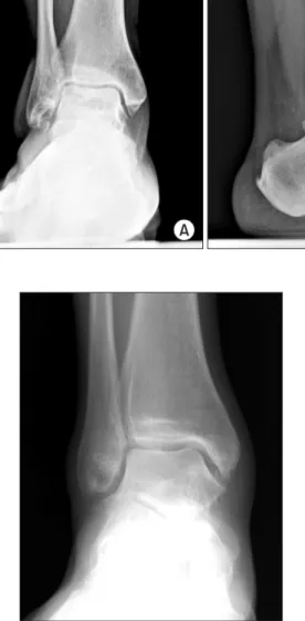
어디까지가 초기 발목관절염인가에 대한 명확한 정의는 내려지 지 않았다. 하지만 이전의 관절염 분류 체계를 참고로 이를 유추해 볼 수 있다. Kellgren-Lawrence의 관절염 분류는 골극의 유무, 관절 간격의 변화를 중심으로 분류하였다(Table 1). 반면, Takakura의 관절염 분류는 관절 간격의 변화를 중심으로 하면서 내측 관절 간 격의 변화를 분류 기준으로 삼았다는 점이 특징이다(Table 2). 이 외에도 발목관절염의 분류는 여러 종류가 있으나 대표적으로 사 용되는 위 두 가지 방법을 참조하자면 골극의 존재 여부, 관절 간 격의 축소 여부가 기준이 된다고 할 수 있다. 초기 관절염이 분명 하게 정의되어 있지는 않으나, 관절 간격이 일부라도 좁아진 경우 가 있다면 통증에 대한 수술 결정시 관절 교정술이나 인공관절치 환술, 관절고정술 등을 고려해야 하기 때문에 중기 또는 말기 관 절염이라 분류할 수 있을 것으로 생각된다. 따라서 초기 관절염은 관절 간격 차체에는 큰 이상이 없으면서 골극이 있거나 연골화 경 화(subchondral sclerosis)가 있는 경우로 간주할 수 있으며, 이는 Kellgren-Lawrence 분류의 Grade 1 또는 2, Takakura 분류의 Stage I에 해당하는 경우라 할 수 있다(Fig. 1).
Table 2. Osteoarthritis Classification of Takakura
Stage Definition
I No joint space narrowing but early sclerosis and osteophyte formation II Narrowing of the joint space medially
IIIa Obliteration of the joint space limited to the facet of medial malleolus with subchondral bone contact IIIb Obliteration of the joint space advanced to the roof of the talar dome with subchondral bone contact
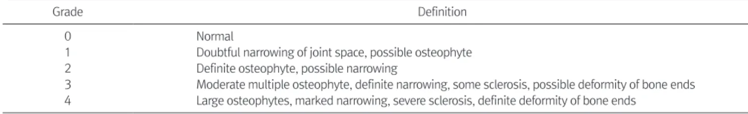
IV Obliteration of the joint space with complete bone contact Table 1. Osteoarthritis Classification of Kellgren-Lawence
Grade Definition
0 Normal
1 Doubtful narrowing of joint space, possible osteophyte 2 Definite osteophyte, possible narrowing
3 Moderate multiple osteophyte, definite narrowing, some sclerosis, possible deformity of bone ends 4 Large osteophytes, marked narrowing, severe sclerosis, definite deformity of bone ends
www.jkfas.org 관절 간격이 좁아져 있지 않은 관절염의 경우 비교적 보존적 치료
에 효과적이며, 추후 관절염의 진행에 대한 추시관찰이 필요하다 는 점에 대해 주지시킬 필요가 있다.
체중 조절을 통하여 발목으로 가는 압력을 줄이고, 특정 부위 통 증이 심할 경우 깔창이나 신발 등을 사용하여 압력을 분산시키는 방법을 찾는 것도 중요한 보존적 치료 중의 하나이다. 발목관절염 에 의한 통증을 줄이기 위해 압력을 분산시키는 보조기 등을 사용 하는 경우도 보고되고 있으나,14) 실내에서 맨발 생활을 하는 국내 에서는 적용하기에 무리가 있고, 환자들의 불편도가 높아 자주 사 용하기는 어려운 실정이다.
골극의 위치에 따라 등산이나 기타 특정 운동 시 통증이 유발되 는 경우 스트레칭 또는 다른 운동으로 전환시키는 것으로 관절염 을 조절하기도 한다. 하지만 여가 활동의 중요성이 강조되는 최근 의 경향을 생각한다면 환자 개개인의 운동 방식을 바꾸는 방식보 다는 보다 적극적인 치료를 고려해 볼 필요도 있을 것이다.
가장 많이 사용하는 치료는 약물 치료이며, nonsteroidal anti- inflammatory drug (NSAID)을 주로 사용하며, NSAID는 30% 정도 의 통증 감소와 15%의 관절 기능 개선을 유도할 수 있다고 보고되 었다. 아세트아미노펜이나 opioid도 통증의 정도에 따라 사용할 수 있으며, 통증의 양상이 화끈거리고 저린 양상의 신경병성 통증 이 발생하는 경우 tricyclic antidepressants (TCA), selective norepi- nephrine reuptake inhibitors (SNRI), selective serotonin reuptake inhibitors (SSRI) 등의 항우울제 계통의 약을 사용하여 통증을 조절 하기도 한다.
관절강 내 주사 치료도 보고되고 있는데, steroid (dexametha- sone, triamcinolone)를 주사하는 것에 대한 보고가 있으나15) 연부 조직에 대한 catabolic risk가 있어 피부가 위축되거나 피부 색깔이 변화되는 부작용이 있을 수 있고, 뚜렷한 효과를 입증한 논문이 많 지 않아 자주 사용하기보다는 특별한 사용 목적(결혼식, 휴가 등)
B A
Figure 1. Simple radiographs of ankle anteroposterior (A) and lateral (B) show osteopyhte and early sclerosis.
Figure 2. Simple radiographs of early osteoarthritis. Figure 3. Computed tomography (CT) of early ankle osteoarthritis. CT shows the medial osteophyte.
결 론
발목관절은 다른 관절에 비해 관절염의 발생 빈도는 낮지만 외 상에 취약하고, 관절염 발생 시 지속적인 통증을 유발한다. 초기 발목관절염은 관절 간격 자체의 변화는 없이 골극 등으로 인해 통 증이 유발되는 관절염으로 비교적 보존적 치료에 효과적이나 지속 적인 통증이 유발되는 경우 골극을 제거하는 수술적 치료가 효과 적이다. 하지만 지속적인 발목 통증을 호소하는 경우 이것이 관절 염으로 인한 것인지 아니면 다른 동반된 변형, 연부조직 이상이나 신경병성 통증이 없는 것인지를 다시 한번 확인하는 것도 매우 중 요하다.
REFERENCES
11 Saltzman CL, Salamon ML, Blanchard GM, Huff T, Hayes A, Buckwalter JA, et al. Epidemiology of ankle arthritis: report of a consecutive series of 639 patients from a tertiary orthopaedic center1 Iowa Orthop J1 2005;25:44-61
21 Valderrabano V, Horisberger M, Russell I, Dougall H, Hinter- mann B. Etiology of ankle osteoarthritis1 Clin Orthop Relat Res1 2009;467:1800-61
31 Lateef S, Golightly YM, Renner JB, Jordan JM, Nelson AE. A cross-sectional analysis of radiographic ankle osteoarthritis fre- quency and associated factors: the Johnston county osteoarthri- tis project1 J Rheumatol1 2017;44:499-5041
41 Beaudoin AJ, Fiore SM, Krause WR, Adelaar RS. Effect of isolated talocalcaneal fusion on contact in the ankle and talonavicular joints1 Foot Ankle1 1991;12:19-251
51 Ihn JC, Kim SJ, Park IH. In vitro study of contact area and pres- sure distribution in the human knee after partial and total men- iscectomy1 Int Orthop1 1993;17:214-81
61 Athanasiou KA, Niederauer GG, Schenck RC Jr. Biomechani- cal topography of human ankle cartilage1 Ann Biomed Eng1 1995;23:697-7041
71 Kempson GE. Age-related changes in the tensile properties of human articular cartilage: a comparative study between the femoral head of the hip joint and the talus of the ankle joint1 Biochim Biophys Acta1 1991;1075:223-301
81 Chubinskaya S, Huch K, Mikecz K, Cs-Szabo G, Hasty KA, Kuettner KE, et al. Chondrocyte matrix metalloproteinase-8:
up-regulation of neutrophil collagenase by interleukin-1 beta in human cartilage from knee and ankle joints1 Lab Invest1 1996;74:232-401
91 Huch K, Wilbrink B, Flechtenmacher J, Koepp HE, Aydelotte MB, Sampath TK, et al. Effects of recombinant human osteogenic protein 1 on the production of proteoglycan, prostaglandin E2, and interleukin-1 receptor antagonist by human articular chon- drocytes cultured in the presence of interleukin-1beta1 Arthritis Rheum1 1997;40:2157-611
101 Cushnaghan J, Dieppe P. Study of 500 patients with limb joint osteoarthritis1 I1 Analysis by age, sex, and distribution of symp- tomatic joint sites1 Ann Rheum Dis1 1991;50:8-131
에 일시적으로 사용해 볼 수 있는 방법이다. Platelet-rich plasma (PRP) 주사는 최근 Repetto 등16)이 효과적인 결과를 보고한 바 있 으나 국내에서는 의료법 관련하여 사용이 어려운 상황이다. 최근 히알루론산(hyaluronic acid) 주사의 좋은 결과에 대한 보고들도 많 이 나오고 있다.17,18) 하지만 2015년 발표된 Cochrane review19)에서 는 발목관절염에 대해 히알루론산 주사를 사용하는 것이 placebo 에 비해 이득도 위해도 없는 것으로 알려져 효과 자체는 불분명한 편이다. 반복적인 steroid injection을 요구하는 환자에 대해 대안으 로 고려해 볼 수는 있을 것이다.
5. 수술적 치료
비수술적 치료에도 통증이 지속되는 경우 수술적인 치료를 고려 한다. 하지만 과도한 수술적 치료는 원치 않았던 여러 합병증을 유 발하기도 한다. 따라서 초기 발목관절염의 수술적 치료를 시행하 기에 앞서 지속되는 환자의 통증이 관절염으로 인한 것인지, 다른 원인이 있는 것이 아닌지에 대해 반드시 확인할 필요가 있다.
초기 관절염에서 발생한 연부조직이나 골극이 유발하는 통증을 충돌 증후군이라 표현하기도 한다. 충돌 증후군은 활액막과 관절 막, 인대 부위의 손상에 의한 상흔(scarring), 섬유화(fibrosis)로 나 타나는 연부조직 충돌 증후군과 골극이 보행 시 직접 충돌하면서 통증을 유발하는 골성 충돌 증후군으로 구분하는데, 이러한 충돌 증후군은 초기 관절염에서 수술적 치료를 시행하는 주요 적응증이 된다. 충돌증후군은 특정한 동작에서 연부조직이나 골성조직의 압 박으로 통증이 유발되고, 운동각도의 제한을 가져오기도 한다.20)
초기 관절염에 대한 수술적 치료는 관절경을 이용한 변연절제술 과 절개 후 골극을 제거하는 골절제술(cheilectomy)로 나눌 수 있 다. Tol 등21)은 57명의 초기 관절염 환자에 대해 관절경적 변연절 제술 및 골절제술을 시행하여 성공적인 결과를 보고하였고, Parma 등,22) Choi 등23)도 각각 105명, 71명에 대해 관절경적 변연절제술을 시행하여 성공적인 결과를 보고하였다. 반면 Hassouna 등24)은 60 명에 대해 관절경적 변연절제술을 시행하였으나 28%는 추후 재수 술을 시행하였고, 기존의 관절염이 중기 이상 진행된 경우는 관절 경적 치료가 부족할 수 있음을 상기시켰다.
골극이 뚜렷하고 통증 부위와 일치되는 경우 개방적 골절제술 (open cheilectomy)이 효과적이다. 특히 원위 경골의 내측부에 지 붕(roof)처럼 발생하는 골극(Fig. 3)의 경우 단순방사선 영상에서 확인되지 않는 경우가 많으며, 이 경우 임상 증상을 확인하고 CT를 시행하여 골극이 확인되면, 절개 골절제술을 시행하는 것이 효과 적이다.
최근에는 골수유래세포(bone marrow derived cell)를 조기 발목 관절염에 적용한 보고25)도 나오고 있으나 실제 환자들에게 보다 많 이 적용하기 위해서는 추가 연구가 더 필요한 실정이다.
www.jkfas.org 111 Saltzman C. Editorial: why ankle replacement? Clin Orthop Relat
Res1 2004;(424):21
121 Tuominen EK, Kankare J, Koskinen SK, Mattila KT. Weight- bearing CT imaging of the lower extremity1 AJR Am J Roent- genol1 2013;200:146-81
131 Hassink G, Testa EA, Leumann A, Hügle T, Rasch H, Hirschmann MT. Intra- and inter-observer reliability of a new standardized diagnostic method using SPECT/CT in patients with osteochon- dral lesions of the ankle joint1 BMC Med Imaging1 2016;16:671 141 Saltzman CL, Shurr D, Kamp J, Cook TA. The leather ankle lacer1
Iowa Orthop J1 1995;15:204-81
151 Pekarek B, Osher L, Buck S, Bowen M. Intra-articular cortico- steroid injections: a critical literature review with up-to-date findings1 Foot (Edinb)1 2011;21:66-701
161 Repetto I, Biti B, Cerruti P, Trentini R, Felli L. Conservative treat- ment of ankle osteoarthritis: can platelet-rich plasma effectively postpone surgery? J Foot Ankle Surg1 2017;56:362-51
171 DeGroot H 3rd, Uzunishvili S, Weir R, Al-omari A, Gomes B.
Intra-articular injection of hyaluronic acid is not superior to saline solution injection for ankle arthritis: a randomized, double-blind, placebo-controlled study1 J Bone Joint Surg Am1 2012;94:2-81
181 Witteveen AG, Sierevelt IN, Blankevoort L, Kerkhoffs GM, van Dijk CN. Intra-articular sodium hyaluronate injections in the osteoarthritic ankle joint: effects, safety and dose dependency1
Foot Ankle Surg1 2010;16:159-631
191 Witteveen AG, Hofstad CJ, Kerkhoffs GM. Hyaluronic acid and other conservative treatment options for osteoarthritis of the ankle1 Cochrane Database Syst Rev1 2015;(10):CD0106431 201 Ogilvie-Harris DJ, Gilbart MK, Chorney K. Chronic pain follow-
ing ankle sprains in athletes: the role of arthroscopic surgery1 Arthroscopy1 1997;13:564-741
211 Tol JL, Verheyen CP, van Dijk CN. Arthroscopic treatment of anterior impingement in the ankle1 J Bone Joint Surg Br1 2001;83:9-131
221 Parma A, Buda R, Vannini F, Ruffilli A, Cavallo M, Ferruzzi A, et al. Arthroscopic treatment of ankle anterior bony impingement:
the long-term clinical outcome1 Foot Ankle Int1 2014;35:148-551 231 Choi WJ, Choi GW, Kwon HM, Lee JW. Arthroscopic treatment in mild to moderate osteoarthritis of the ankle1 Knee Surg Sports Traumatol Arthrosc1 2013;21:1338-441
241 Hassouna H, Kumar S, Bendall S. Arthroscopic ankle debride- ment: 5-year survival analysis1 Acta Orthop Belg1 2007;73:737- 401
251 Buda R, Castagnini F, Cavallo M, Ramponi L, Vannini F, Giannini S. "One-step" bone marrow-derived cells transplantation and joint debridement for osteochondral lesions of the talus in ankle osteoarthritis: clinical and radiological outcomes at 36 months1 Arch Orthop Trauma Surg1 2016;136:107-161