DOI : 10.3341/jkos.2009.50.1.27
= 증례보고 =
건성안이 있는 이식편대숙주질환에서
사이클로스포린 A 점안치료 후 장기간의 경과관찰
이승현1⋅임성규1⋅우제문2⋅윤경철1
전남대학교 의과대학 안과학교실1, 울산대학교 울산병원 안과2
목적: 건성안을 동반한 이식편대숙주질환 환자에서 0.05% 사이클로스포린 A 점안액(Restasis, Allergan Inc. USA.)의 장기간 치료효과 에 대해 알아보고자 하였다.
대상과 방법: 이식편대숙주질환 환자 8명 16안을 대상으로 치료 전과 0.05% 사이클로스포린 A를 1일 2회 점안하여 치료한 후 1, 3, 6, 12개월째 눈물표면인자를 조사하였다.
결과: 눈물막파괴시간은 치료 전(3.88±1.78초)에 비해 치료 1개월 후에(4.17±1.90초, P=0.02), 증상점수와 기본눈물분비는 각각 치료 전(3.00±0.38, 4.44±1.59 mm)과 비교하여 치료 3개월 후에(2.33±0.52 P=0.04, 5.42±2.50 mm P=0.04) 호전되었다. 각막상피병증 의 정도는 치료 전(2.38±1.31)에 비해 치료 12개월 후에(1.13±0.35, P=0.02) 호전을 보였다.
결론: 이식편대숙주질환 환자에서 0.05% 사이클로스포린 A 점안액의 사용은 수 개월내에 기본눈물분비량과 눈물막의 상태 및 안구건 조증상을 호전시키는 효과가 있었으며, 각막상태의 호전을 위해서는 1년 이상의 장기간 치료가 필요할 것으로 생각된다.
<대한안과학회지 2009:50(1):27-33>
■ 접 수 일: 2008년 4월 14일 ■ 심사통과일: 2008년 9월 24일
■ 통 신 저 자: 윤 경 철 광주시 동구 학동 8 전남대학교병원 안과
Tel: 062-220-6742, Fax: 062-227-1642 E-mail: [email protected]
혈액종양환자의 치료에 있어서 조혈모세포이식술은 가 장 먼저 선택되는 치료법 중의 하나이다.1,2조혈모세포이식 술 후에 발생하는 급성 혹은 만성이식편대숙주질환 환자의 45~60%에서 안 증상이 발생하고 이중 건성안은 가장 흔 히 발생하는 합병증이다.1-4 건성안은 점모양각막염, 지속 성 각막상피결손, 각막의 각질화, 각막궤양, 각막천공 등을 야기한다.5-7이식편대숙주질환 환자에서 발생한 건성안의 치료로 인공누액점안, 치료용콘택트렌즈 착용, 보호안경 착용, 누점폐쇄, retinoic acid 점안, 스테로이드 점안 혹은 전신투여, FK 506 등의 면역억제제가 이용되고 있으며, 중 등도 이상의 건성안 환자에서는 양막이식술과 전층각막이 식술 등의 수술적 치료도 시행되고 있다.3,8-11하지만 이와 같은 고식적인 방법으로 이식편대숙주질환 환자에서 발생 한 건성안을 효과적으로 치료하는 데는 한계가 있다.
사이클로스포린 A는 면역반응에 의해 활성화된 T 림프 구를 억제하고 결막의 전염증성 사이토카인을 하향조절한
다.12-16또한 사이클로스포린 A의 점안으로 결막 내의 술
잔세포가 증가하고 상피세포의 증식이 억제된다. 건성안 환 자에서 사이클로스포린 A 점안액을 사용하는 경우 기본눈
물분비 증가, 각결막상피병증의 호전, 표층점상각막염의 호 전, 결막술잔세포의 밀도 및 Transforming growth factor- β2의 생산이 증가되며, 시력개선, 자극감 감소, 인공누액 사용 횟수 감소 등의 주관적인 증상에 효과가 있음이 보고
되었다.17-26 최근에는 이식편대숙주질환 환자에서 발생하
는 건성안의 치료에 사이클로스포린 A를 이용하는 일부 시 도가 있었으나, 아직 효용성과 안정성에 대한 장기간 추적 관찰은 부족한 실정이다.27,28이에 저자들은 건성안이 있는 이식편대숙주질환 환자에서 0.05% 사이클로스포린 A 점 안액(Restasis, Allergan Inc., Irvine, CA, USA)을 사용한 후 1년 이상 안구표면과 눈물막의 변화를 관찰하여 그 효과 를 알아보고자 하였다.
대상과 방법
2006년 3월부터 2007년 3월까지 본원에서 이식편대숙 주질환에 동반된 건성안이 있으면서 6개월 이상의 인공누 액 점안과 눈물점폐쇄술 등 고식적인 치료에 반응하지 않 는 환자 중 사이클로스포린 A 점안액 사용 후 최소 12개월 이상 추적관찰이 가능하고 사용 중 통증으로 인한 점안중 단이 없었던 8명 16안을 대상으로 조사하였다.
건성안의 진단은 눈물막파괴시간이 10초 이내, 기본눈물 분비검사가 5 mm 이하, 눈부심, 가려움, 이물감, 작열감, 통증 등의 안구자극증상, 각막노출로 인한 각막상피병증이 있어 형광색소염색을 보이는 경우로 하였다. 급성 감염 또
는 염증이 건성안에 동반된 경우, 약물독성반응이 있는 경 우, 눈꺼풀이나 눈썹의 이상이 있는 경우, 외상에 의한 손상 이 있는 경우에서처럼 이식편대숙주질환 외의 건성안의 원 인이 될 수 있는 경우는 연구 대상에서 제외하였다.29-32 모 든 환자에서 개개인의 동의를 얻어 임상시험윤리위원회의 승인을 받아 연구를 시행하였다. 0.05% 사이클로스포린 A 점안액(Restasis)을 매 12시간마다 점안하고 보존제가 없 는 인공누액제를 병행하여 사용하도록 하였다.
안구건조증상, 각막감각, 눈물막파괴시간, 쉬르머검사를 통한 기본눈물분비, 눈물청소율, 형광색소염색을 통한 각막 상피병증의 정도에 대한 검사를 사이클로스포린 A 점안액 사용 전과 사용 후 1개월, 3개월, 6개월, 12개월째에 각각 시행하여 비교하였다.
안구건조증상은 0에서 4단계로 나누었으며, 이 때 0은 증상이 없는 경우로 4는 심한 안구불편감으로 인해 일상생 활에 지장을 주는 경우로 하였고 각막감각검사는 Cochet- Bonnet 촉각계(Luneau Optalmologie, Chartres Cedex, France)를 이용하였는데 나일론 세사를 최대한으로 늘린 후에 그 끝을 각막 중심부와 수직으로 접촉시켰다. 세사의 길이를 5 mm 단위로 단계적으로 줄여가면서 피검자가 처 음으로 감각을 느낄 때의 촉각계의 길이를 밀리미터로 기 록하였다.
눈물막파괴시간 측정은 형광검사지를 결막낭에 접촉시 킨 후 피검자가 수 초간 몇 번 동안 눈을 깜박이게 한 후 염색된 눈물막 층에서 검은 점, 구멍, 또는 줄의 형태로 형 광 색소 염색의 결손이 관찰될 때까지 시간을 세극등현미 경의 코발트블루 광원을 이용하여 측정하였다. 0.5%로 희석시킨 형광색소(Fluorescite, Fort Worth, Tx,Alcon, USA) 10 ㎕와 0.5% proparacain hydrochloride (Alcaine, Fort Worth, Tx Alcon, USA)를 하측 구결막에 넣고 5분 후 쉬르머 검사지(Eagle Vision, Memphis TN, USA)를 아 래 눈꺼풀의 외측 1/3부분의 구결막에 5분간 접촉시킨 다 음 젖은 부위의 길이를 재어 기본눈물분비량을 밀리미터 단위로 측정하고, 검사지의 끝부분의 염색된 정도를 표준 검사지와 비교하여 눈물청소율을 측정하였다. 표준 검사지 는 점안된 형광색소에 대하여 각각 1, 1/2, 1/4, 1/8, 1/16, 1/32, 1/64, 1/128. 1/256으로 희석한 것으로 형광색소의 농도가 연해짐에 따라 9단계로 구분하였다.33,34염색정도의 수치적인 비교를 위하여 log2-1
값으로 나타내었다.
각막상피병증의 정도는 형광색소(Haag-Streit AG, CH- 3098 Koniz, Switzerland) 염색으로 평가하였으며, 염색된 면적과 밀도를 점수화하여 각각을 0부터 3까지로 구분하고 이를 곱한 수치로 나타내었다. 형광 염색은 각막이 염색되 지 않은 경우를 0, 각막의 1/3 미만인 경우를 1, 1/3과 2/3
미만 사이를 2, 2/3 이상을 3으로 하였고, 밀도 점수는 염색 이 되지 않은 경우를 0, 경도를 1, 중증도를 2, 밀도가 높으 면서 병변이 서로 겹치는 경우를 3으로 나타내었다.
통계는 SPSS 14.0에서 Wilcoxon signed rank 검사를 이 용하였으며, P값이 0.05 이하인 경우를 유의하다고 하였다.
결 과
총 8명의 환자 모두 남자였고 평균 나이는 34.38±11.53 (19~50)세였다. 대상자 중 급성 골수구성 림프종 6명, 맨 틀 세포 림프종 1명, 비호즈킨성 림프종으로 진단받은 환자 가 1명이었으며, 이 중 골수이식을 시행받은 환자가 4명, 말초혈 조혈모세포이식술을 시행받은 환자가 3명이었고 골 수이식과 말초혈 조혈모세포이식술 모두를 시행받은 환자 가 1명 이었다. 조혈모세포이식술 후에 건성안으로 진단받 는 데까지 소요되는 평균 기간은 21.00±19.71개월(3~55 개월)이었고, 총 8명의 환자 중 7명(87.5%)은 피부, 폐, 간, 구강점막 등의 다른 장기로의 침범이 있었다. 대부분의 환자들은 사이클로스포린과 스테로이드를 전신으로 투여받 고 있었으며, 건성안에 대하여 인공누액점안, 눈물점 폐쇄 술, 치료용콘택트렌즈 사용 등의 고식적인 치료를 받고 있 었다(Table 1).
이식편대숙주질환에서 발생한 건성안 환자에게0.05% 사 이클로스포린 A를 점안 후 시간에 따른 증상의 변화, 안구 표면 및 눈물막의 변화는 Table 2에 나타내었다. 0.05% 사 이클로스포린 A 점안액 사용 전 증상점수는 3.00±0.38이 었고, 점안액 사용 1개월, 3개월, 6개월, 12개월 후에 각각 2.67±0.52 (P=0.157), 2.33±0.52 (P=0.046), 1.80±0.33 (P=0.044), 1.80±0.83 (P=0.043)로 점안 3개월 후부터 증상이 호전되어 최종 관찰시까지 유지되었다. 각막감각검 사는 치료 전 58.13±5.12 mm이었고, 치료 1개월, 3개월, 6개월, 12개월 후에 각각 57.50±5.84 mm (P=0.317), 58.33±3.26 mm (P=0.317), 58.00±2.58 mm (P=0.157), 58.50±2.42 mm (P=0.357)로 의미있는 변화를 보이지 않 았다. 눈물막파괴시간은 치료 전 3.88±1.78초였고, 치료 1개 월, 3개월, 6개월, 12개월 후에 각각 4.17±1.90초(P=0.020), 5.25±1.82초(P=0.007), 8.80±4.13초(P=0.011), 6.50±
2.80초(P=0.011)로 점안 1개월 후부터 의미있는 호전을 보여 최종관찰시까지 유지되었다. 쉬르머검사는 치료 1개 월, 3개월, 6개월, 12개월 후에 각각 4.67±1.72 mm (P=0.180), 5.42±2.50 mm (P=0.035), 5.80±1.81 mm (P=0.040), 6.50±2.50 mm (P=0.017)로 치료 전 4.44±
1.19 mm과 비교하여 치료 3개월 후부터 의미있는 증가를 보였고 최종관찰시까지 유지되었다. 눈물청소율검사는 치
No. Age
(years) Sex Original disease
Stem cell Transplan -tation
Duration between SCT* and dry eye (months)
GVHD† other than the
eyes
Systemic
immunosuppression Conventional ocular treatment
1 34 M AML‡ (M3) BMT§ 12 Steroid,
cyclosporine A
Punctal plug, artificial tears, therapeutic contact lens, amniotic membrane transplantation
2 34 M AML (M5) PBSCT∏ 14 Skin, lung Steroid Punctal plug, artificial tears 3 41 M Mantle cell
lymphoma
PBSCT 3 Liver, lung
oral mucosa
Steroid, cyclosporine A
Punctal plug, artificial tears
4 42 M NHL#(diffuse large B cell)
PBSCT 7 Liver,
intestine
Steroid Punctal plug, artificial tears
5 19 M AML (M2) BMT/PBS
CT
55 Skin, lung, bone
Steroid Punctal plug, artificial tears
6 30 M AML (M2) BMT 4 Skin,
oral mucosa
Steroid, cyclosporine A
Punctal plug, artificial tears Therapeutic contact lens
7 43 M AML (M3) BMT 28 Skin, lung,
oral mucosa
Steroid, cyclosporine A
Punctal plug, artificial tears, therapeutic contact lens,
amniotic membrane transplantation scleral patch graft
8 16 M AML (M2) BMT 45 Lung,
intestine
Steroid Punctal plug, artificial tears therapeutic contact lens
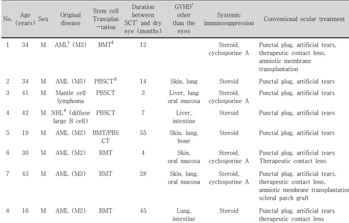
*SCT=stem cell transplantation; †GVHD=graft-versus-host-disease; ‡AML=acute myelogenous leukemia; §BMT=bone marrow transplantation; ∏PBSCT=peripheral blood stem cell transplantation; #NHL=non-Hodgkin’s lymphoma.
Table 1. Characteristics of patients with severe dry eye associated with GVHD
Before treatment 1 month after treatment
3 months after treatment
6 months after treatment
12 months after treatment Symptom score 3.00±0.38 2.67±0.52 2.33±0.52* 1.80±0.33* 1.80±0.83* Corneal sensitivity (mm) 58.13±5.12 57.50±5.84 58.33±3.26 58.00±2.58 58.50±2.42 Tear film break up time (s) 3.88±1.78 4.17±1.90* 5.25±1.82* 8.80±4.13* 6.50±2.80* Basal secretion test (mm) 4.44±1.59 4.67±1.72 5.42±2.50* 5.80±1.81* 6.50±2.50* Tear clearance rate ((log2)-1) 2.81±0.83 2.92±0.67 3.00±0.60 2.90±0.57 2.90±0.57 Keratoepitheliopathy score 2.38±1.31 2.25±1.14 2.50±1.62 1.70±1.57 1.13±0.35*
*P<0.05 compared with baseline.
Table 2. Tear film and ocular surface changes after treatment with topical 0.05% cyclosporineA in severe dry eye associated with graft-versus-host disease
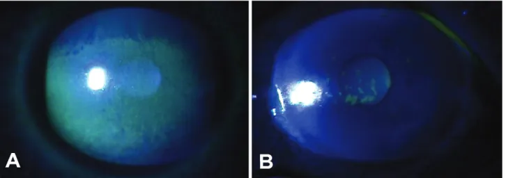
료 전 2.81±0.83이었고, 치료 1개월, 3개월, 6개월, 12개월 후에 각각 2.92±0.67 (P=1.000), 3.00±0.60 (P=0.564), 2.90±0.57 (P=0.083), 2.90±0.57 (P=0.083)로 변화가 없었다. 각막상피병증검사는 치료 전 2.38±1.31이었고, 치료 1개월, 3개월, 6개월, 12개월 후에 각각 2.25±1.14 (P=0.414), 2.50±1.62 (P=0.877), 1.70±1.57 (P=0.131), 1.13±0.35 (P=0.021)로 사용 전과 비교할 때 점안 12개 월 후에 각막상피병증이 호전되었다(Fig. 1). 점안액 사용
12개월 동안 의미있는 합병증은 발생하지 않았다.
고 찰
안구건조증 환자에서 관찰되는 안표면 및 눈물부속기의 만성 염증이 건성안의 중요한 병인으로 받아들여지고 있다.
면역조직화학연구에서 이러한 조직 내에 CD4 보조 T 세포 의 침윤과, IL-6, IL-8, IL-10 그리고 TNF-α와 같은 사
Figure 1. Slit lamp photographs with fluorescein staining in GVHD patient with dry eye syndrome before (A) and 1 year after (B) 0.05% cyclosporine A therapy. Note the marked improvement in the corneal epithelium.
이토카인이 증가하고 면역활성의 지표인 HLA DR, ICAM-1 등이 발현된다고 보고되어 안구건조증과 면역계와의 연관 성이 밝혀졌다.7,35-37 만성 이식편대숙주질환 환자에서 건 성안은 가장 흔히 발견되는 안과적 합병증이며, 조혈모세포 이식술 후 약 6개월 후에 발병하고, 건성안의 증상은 이식 편대숙주질환의 중증도와 마이봄샘기능부전과 관련이 있다 고 알려져 있다.7,38-41
이식편대숙주질환 환자에서 건성안이 발생하는 기전은 명확히 밝혀지지 않았지만 건성안 환자의 결막과 눈물샘에 서 염증반응과 섬유화가 관찰된다.11,42이식편대숙주반응으 로 발생한 건성안 환자의 결막에서는 T 세포 위주의 림프 구가 발견되고 눈물샘을 포함한 눈물관 주위, 눈물관 상피 에 T 세포의 침윤이 관찰되며 눈물관과 혈관 주위에는 섬 유화가 나타난다.11이와 같은 소견은 건성안으로 진행하는 일련의 과정에 혈관의 치밀판 손상으로 인한 T 세포의 결 막과 눈물샘으로 이동이 주요한 역할을 한다는 것을 의미 한다. 활성화된 T 세포는 눈물관 주위와 혈관 주변으로 이 동하여 눈물관과 결막상피를 손상시킨다. 동시에 CD34 양 성 간질섬유모세포는 T 세포와 같은 염증세포들이 분비하 는 사이토카인에 의해 쉽게 활성화된다.11,42-43이러한 반응 으로 발생한 안구 표면의 만성적인 염증은 술잔세포의 감 소, 결막편평상피화생의 증가와 같은 결막표면의 변화를 야 기한다.3,43
이식편대숙주질환에서 발생한 건성안의 치료로 인공누 액점안이나 누점폐쇄와 같은 기존의 고식적인 방법은 한계 가 있다. Ogawa et al11은 이식편대숙주질환 환자 2명에게 전신적으로 FK506과 스테로이드를 투여하여 건성안을 치 료하여 보고한 바 있으며 Yoon et al44은 12명의 이식편대 숙주질환 환자에서 발생한 중등도 이상의 건성안에서 제대 혈청 점안액의 사용은 안전하고 효과적인 치료라고 하였다.
또한 만성 이식편대숙주질환 환자에서 건성안의 증상이 심 각하거나 석회각막변성이 있는 경우 양막이식술을 고려해 볼 수 있다.
사이클로스포린 A는 cyclophilin으로 알려진 세포내 펩 타이드에 결합하여 안표면 조직에서 면역 매개 염증반응의 주된 사이토카인인 IL-2의 생성을 억제시킨다. 감소된 IL-2는 보조 T 세포의 비활성화를 초래하여 눈물샘의 림 프구 침윤을 줄여 눈물 생성을 호전시킨다고 알려져 있다.
현재까지 일반적인 건성안에서 0.05%와 0.1% 사이클로스 포린 점안액을 사용하는 경우 수개월 후에 기본눈물분비량 증가 및 눈물막파괴시간 연장, 표층점상각막염의 호전을 보 이고 시력의 개선, 인공누액 사용 횟수 감소, 자극감 감소 등의 주관적인 증상에 효과가 있음이 보고되었다.3,45 안과 적 영역에서는 안구표면의 염증에 의해 눈물분비가 감소되 어 발생한 건성안에서 눈물분비를 증가시키기 위해 사용되 고 있으며, 알레르기성결막염, 타이거슨 각막염, 상윤부결 막염, 안검의 마이봄샘 기능부전 등의 다양한 염증성 및 비 염증성 질환에서 사이클로스포린 점안액의 사용이 시도되 고 있다.24,46-53또한 사이클로스포린 A는 항 염증효과가 있 는 스테로이드에 비하여 안압 상승, 수정체 혼탁 등의 부작 용이 없다는 장점이 있다.
Lelli et al27은 0.05% 사이클로스포린 A 점안액을 3개월 동안 건성안이 심한 이식편대숙주질환 환자16명에게 사용 하여 증상 및 눈물분비기능, 각막표면의 변화 등을 관찰한 결과 10명의 환자에서 건성안의 증상이 호전되었고 16명 모두에서 각막상피병증이 호전되었다고 하였다. Wang et al28은 이식편대숙주질환으로 인한 심한 건성안을 갖고 있 는 15명을 대상으로 0.05% 사이클로스포린 점안액을 사용 하고 1개월 후에 증상점수, 각막감각, 눈물증발율, 눈물막 파괴시간, 각막상피병증의 정도, 술잔세포의 밀도, 결막편
평상피화생 정도, 염증세포의 수 및 MUC5AC 발현이 의미 있게 호전되는 것을 관찰하여 발표하였다.
본 연구에서는 중증도 이상의 건성안이 있는 이식편대숙 주질환 환자에서 0.05% 사이클로스포린 점안액을 사용하 여 1개월, 3개월, 6개월, 12개월 후에 증상 및 눈물막의 기 능, 각막표면의 변화를 관찰하였다. 점안액 사용 1개월 후 에 눈물막파괴시간이 의미있는 증가를 보였고 점안액 사용 3개월 후에는 증상점수와 기본눈물분비가 유의한 호전을 보였으며, 사용 12개월 후 최종관찰시까지 눈물분비기능이 지속적으로 증가하였다. 이는 이식편대숙주질환에서 발생 한 건성안에서 0.05% 사이클로스포린 A 점안액의 사용은 여러 연구의 발표결과와 같이 사이토카인과 염증세포의 발 현을 억제하여 초기에 술잔세포의 밀도를 증가시키고 눈물 막파괴시간을연장하며, 시간이 경과할수록 눈물분비기능을 호전시켜 건성안의 증상 및 안구표면인자의 호전에 기여했 을 것으로 생각된다.25,27,28,31
그러나 형광색소를 이용한 각막상피병증의 정도는 0.05% 사이클로스포린 A점안액 사용 12개월 후에 호전되었고 각막지각검사는 최종관찰 시까지 의미있는 변화를 보이지 않았다. 그 이유는 이식편 대숙주질환에서 발생한 건성안은 눈물기능단위(lacriaml functional unit, LFU)의 손상의 정도가 심하므로, 사이클 로스포린 점안액 사용 후 눈물막파괴시간과 기본눈물분비 등이 호전되었지만 치료 1년이 지나도 정상범위에는 도달 하지 못했기 때문으로 생각되며 각막지각검사 결과가 향상 되기 위해서는 각막상피병증 호전 후에도 더 장기간의 치 료가 필요할 것이다.
눈물청소율은 눈물교체 정도를 간단히 알 수 있고 눈물 분비 및 눈물배출과 강한 연관성을 갖는다.34 점안액 사용 전과 후에 눈물청소율의 의미 있는 변화가 없었던 이유는 대상자 8명 모두에서 눈물점 폐쇄 후에 발생한 눈물배출 지 연이 눈물청소율에 영향을 주었기 때문에 기본눈물분비량 의 증가에도 불구하고 눈물청소율의 의미있는 변화가 없었 던 것으로 보인다. 0.05% 사이클로스포린 점안액 사용이 눈물청소율에 미치는 영향을 정확히 평가하기 위해서는 눈 물점 폐쇄를 시행하지 않은 대상군을 통한 연구가 필요할 것이다. 또한 향후 안구표면질환의 정도를 나타내는 가장 민감한 지표인 술잔세포의 밀도와 병이 진행함에 따라 안 구표면에 나타나는 결막상피세포의 형태학적 변화에 대한 연구가 필요할 것으로 생각된다.
Barber et al29의 연구에 따르면 0.1% 사이클로스포린 점 안액을 1년 이상 사용한 일반적인 건성안 환자에서 약 10%가 작열감을 호소하였고 결막 자극 등을 호소한 경우 도 4%정도였으며, 약 5%에서 치료 중단을 원하였다. 하지 만 본 연구에서는 사이클로스포린 점안액을 사용한 8명 모
두에서 통증 등으로 인한 사용중단은 없었다. 그 이유는 첫 째, 본 연구의 대상군이 적고 둘째, 본 연구의 대상자 모두 증상이 심하고 인공누액이나 눈물점폐쇄 등의 고식적인 치 료에 잘 반응하지 않아 사이클로스포린 점안액에 대한 호 응도가 좋았을 것으로 생각된다.
본 연구의 제한점으로는 이식편대숙주질환자의 수가 많 지 않아 대조군을 설정할 수 없었다는 것이다. 하지만 본 연구는 건성안의 치료 효과가 사이클로스포린 점안액에 의 한 것인지 아니면 다른 치료에 의한 것인지 구별하기 위해 인공누액과 눈물점폐쇄술 등의 고식적인 건성안 치료에 최 소 6개월 이상 반응하지 않는 환자를 대상으로 하였고, 눈 물점폐쇄술의 경우 시간이 경과할수록 효과가 감소되는 양 상을 보이므로 건성안의 호전은 사이클로스포린 점안액에 의한 것이라고 판단할 수 있다.22
결론적으로 이식편대숙주질환으로 인한 건성안 환자에 서 0.05% 사이클로스포린 점안액은 사용전에 비하여 시간 이 지남에 따라 안구건조증상, 눈물분비 및 눈물막의 성상 을 호전시키는 효과가 있었으며 각막상태의 호전을 위해서 는 1년 이상의 장기간의 치료가 필요할 것으로 보인다.
참고문헌
1) Horowitz M. Uses and growth of hematopoietic cell trans- plantation immunology. In : Blume KG, Forman SJ, Applebaum FR, eds. Thomas’ Hematopoietic Cell Transplantation, 3rd ed.
Blackwell: Malden, MA, 2004: v. 1 chap. 2.
2) Martin PJ. Overview of hematopoietic cell transplantation. In : Blume KG, Forman SJ, Applebaum FR, eds. Thomas’
Hematopoietic Cell Transplantation, 3rd ed. Blackwell:
Malden, MA, 2004: v. 1 chap. 3.
3) Ogawa Y, Kuwana M. Dry eye as a major complication associated with chronic graft-versus-host disease after hematopoietic stem cell transplantation. Cornea 2003;22:19-27.
4) Franklin RM, Kenyon KR, Tutschka PJ, et al. Ocular mani- festations of graft-vs-host disease. Ophthalmology 1983;90:4-13.
5) Anderson NG, Regillo C. Ocular manifestations of graft versus host disease. Curr Opin Ophthalmol 2004;15:503-7.
6) Johnson DA, Jabs DA. The ocular manifestations of graft- versus-host diseases. Int Ophthalmol Clin 1997;37:119-33.
7) Ogawa Y, Okamoto S, Wakui M, et al. Dry eye after hematopoietic stem cell transplantation. Br J Ophthalmol 1999;83:1125-30.
8) Ogawa Y, Okamoto S, Mori T, et al. Autologous serum eye drops for the treatment of severe dry eye in patients with chronic graft-versus-host disease. Bone Marrow Transplant 2003;31:579-83.
9) Murphy PT, Sivakumaran M, Fahy G, Hutchinson RM.
Successful use of topical retinoic acid in severe dry eye due to chronic graft-versus-host disease. Bone Marrow Transplant 1996;18:641-2.
10) RochaEM, Pelegrino FS, de Paiva CS, et al. GVHD dry eyes
treated with autologous serum tears. Bone Marrow Transplant 2000;25:1101-3.
11) Ogawa Y, Okamoto S, Kuwana M, et al. Successful treatment of dry eye in two patients with chronic graft versus host disease with systemic administration of FK506 and corticos- teroids. Cornea 2001;20:430-4.
12) Pflugfelder SC, Solomon A, Stern ME. The diagnosis and management of dry eye: a twenty-five-year review. Cornea 2000;19:644-9.
13) Nussenblatt RB, Palestine AG. Cyclosporine: immunology, pharmacology and therapeutic uses. Surv Ophthalmol 1986;31:
159-69.
14) Hemady R, Tauber J, Foster SC. Immunosuppressive drugs in immune and inflammatory ocular disease. Surv Ophthalmol 1991;35:369-85.
15) Jabs DA, Wingard J, Green R, et al. The eye in bone marrow transplantation. Arch Ophthalmol 1989;107:1343-8.
16) Bhan AK, Fujikawa LS, Foster CS. T-cell subsets and Langer- hans cells in normal and diseased conjunctiva. Am J Ophthal- mol 1982;94:205-12.
17) Gilbard JP, Rossi SR, Azar DT, Heyda KG. Effect of punctual occlusion by Freeman silicone plug insertion on tear osmolarity in dry eye disorder. CLAO J 1989;15:216-8.
18) Lee EH, Jang JW, Lew HM. The changes of tear osmolarity and protein after silicone punctal plug insertion in dry eye. J Korean Ophthalmol Soc 2001;42:1509-14.
19) Yen MT, Pflucfelder SC, Feuer WJ. The effect of punctual occlusion on tear production, tear clearance and ocular surface sensation in normal subjects. Am J Ophthalmol 2001;131:314 -23.
20) Pflugfelder SC, De Paiva CS, Villarreal AL, Stern ME. Effects of sequential artificial tear and cyclosporine emulsion therapy on conjunctival goblet cell density and transforming growth factor-β2 production. Cornea 2008;27:64-9.
21) Wilson SE, Perry HD. Long-term resolution of chronic dry eye symptoms and signs after topical cyclosporine treatment.
Ophthalmology 2007;114:76-9.
22) Roberts CW, Carniglia PE, Brazzo BG. Comparison of topical cyclosporine, punctal occlusion, and a combination for the treatment of dry eye. Cornea 2007;26:805-9.
23) Salib GM, McDonald MB, Smolek M. Safety and efficacy of cyclosporine 0.05% drops versus unpreserved artificial tears in dry-eye patients having laser in situ keratomileusis. J Cataract Refract Surg 2006;32:772-8.
24) Tatlipinar S, Akpek EK. Topical ciclosporin in the treatment of ocular surface disorders. Br J Ophthalmol 2005;89:1363-7.
25) Sall K, Stevenson OD, Mundorf TK, Reis BL. Two multicenter, randomized studies of the efficacy and safety of cyclosporine ophthalmic emulsion in moderate to severe dry eye disease. CsA Phase 3 Study Group. Ophthalmology 2000;107:631-9.
26) Stevenson D, Tauber J, Reis BL. Efficacy and safety of cyclosporine A ophthalmic emulsion in the treatment of moderate-to-severe dry eye disease: a dose-ranging, randomized trial. The cyclosporine A phase 2 study group. Ophthal- mology 2000;107:967-74
27) Lelli GJ Jr, Musch DC, Gupta A, et al. Ophthalmic cyclosporine use in ocular GVHD. Cornea 2006;25:635-8.
28) Wang Y, Ogawa Y, Dogru M,et al. Ocular surface and tear functions after topical cyclosporine treatment in dry eye patients with chronic graft-versus-host disease. Bone Marrow Transplant 2008;41:293-302.
29) Barber LD, Pflugfelder SC, Tauber J, Foulks GN. Phase III safety evaluation of cyclosporine 0.1% ophthalmic emulsion administered twice daily to dry eye disease patients for up to 3 years. Ophthalmology 2005;112:1790-4.
30) Sall K, Stevenson OD, Mundorf TK, Reis BL. CsA Phase 3 Study Group. Two multicenter, randomized studies of the efficacy and safety of cyclosporine ophthalmic emulsion in moderate to severe dry eye disease. Ophthalmology 2000;107:
631-9.
31) Stevenson D, Tauber J, Reis BL. The Cyclosporine A Phase 2 Study Group. Efficacy and safety of cyclosporine A ophthalmic emulsion in the treatment of moderate to severe dry eye syndrome: a dose-ranging, randomized trial. Ophthal- mology 2000;107:967-74.
32) Djalilian AR, Nussenblatt RB, Holland EJ. Immuno- suppressive therapy in ocular surface transplantation. In : Holland EJ, Mannis MJ, eds. Ocular surface disease Medical and surgical management, 1st ed. New York: Springer, 2002;
v. 1. chap. 22.
33) Tsubota K. Tear dynamics and dry eye. Prog Retin Eye Res 1998;17:565-96.
34) Kaido M, GotoE, Dogru M, Tsubota K. Punctal occlusion for Stevens-Johnson syndrome. Ophthalmology 2004;111:895-900.
35) Stern ME, Geo J, Siemasko KT, et al. Role of the lacrimal gland functional unit in the pathophysiology of dry eye. Exp Eye Res 2004;78:409-16.
36) Pepose JS, Akata RF, Pflugfelder SC, Voigt W. Mononuclear cell phenotypes and immunoglobulin gene rearrangements in lacrimal gland biopsies from patients with Sjogren's syndrome.
Ophthalmology 1990;97:1599-605.
37) Poon AC, Geerling G, Dart JK, et al. Autologous serum eyedrops for dry eye and epithelial defect: clinical and in vitro toxicity studies. Br J Ophthalmol 2001;85:1188-97.
38) Hirst LW, Jabs DA, Tutschka PJ, et al. The eye in bone marrow transplantation. I. Clinical study. Arch Ophthalmol 1983;101:580-4.
39) Shulman HM, Sullivan KM, Weiden PL, et al. Chronic graft- versus-host syndrome in man. A long-term clinicopathologic study of 20 Seattle patients. Am J Med 1980;69:204-17.
40) Mencucci R, Rossi Ferrini C, Bosi A, et al. Ophthalmological aspects in allogenic bone marrow transplantation: Sjogren-like syndrome in graft-versus-host disease. Eur J Ophthalmol 1997;7:13-8.
41) Tichelli A, Duell T, Weiss M, et al. Late-onset keratoconjunc- tivitis sicca syndrome after bone marrow transplantation:
incidence and risk factors. European group for blood and marrow transplantation (EBMT) Working party on late effects. Bone Marrow Transplant 1996;17:1105-11.
42) Ogawa Y, Kuwana M, Yamazaki K, et al. Periductal area as the primary site for T-cell activation in lacrimal gland chronic graft-versushost disease. Invest Ophthalmol Vis Sci 2003;44:
1888-96.
43) Ogawa Y, Yamazaki K, Kuwana M, et al. A significant role of stromal fibroblasts in rapidly progressive dry eye in patients
=ABSTRACT=
Long-term Evaluation After Topical Cyclosporine Treatment in Dry Eye Patients With Graft-Versus-Host Disease
Seung Hyun Lee, MD1, Seoung Kyu Im, MD1, Je-Moon Woo, MD, PhD2, Kyung Chul Yoon, MD. PhD1
Department of Ophthalmology, Chonnam National University Medical School1, Gwangju, Korea Department of Ophthalmology, Ulsan Hospital, Ulsan University Medical School2, Ulsan, Korea
Purpose: This study was performed to evaluate the long-term effect of 0.05% cyclosporine A emulsion (Restasis, Allergan Inc.
USA.) on dry eye associated with graft-versus-host disease (GVHD).
Methods: Sixteen eyes of 8 patients with severe dry eye associated with GVHD were treated with 0.05% cyclosporine A emulsion twice a day. Tear film parameters were evaluated before treatment and after 1, 3, 6, and 12 months of treatment.
Results: One month after treatment, tear break-up time improved from 3.88±1.78 s to 4.17±1.90 s (P=0.02). Three months after treatment, symptom score and basal secretion improved from 3.00±0.38 and 4.44±1.59 mm to 2.33±0.52 (P=0.04) and 5.42±2.50 mm (P=0.04), respectively. Twelve months after treatment, the keratoepitheliopathy score improved from 2.38±1.31 to 1.13±0.35 (P=0.02).
Conclusions: Use of a 0.05% cyclosporine A emulsion is effective for dry eye associated with GVHD as it helps to increase tear secretion and to relieve symptoms. Treatment with 0.05% cyclosporine A emulsion for longer than 1 year may be necessary to improve keratoepitheliopathy.
J Korean Ophthalmol Soc 2009;50(1):27-33
Key Words: Cyclosporine, Dry eye syndrome, Graft-versus-host disease
Address reprint requests to Kyung Chul Yoon, MD, PhD
Department of Ophthalmology, Chonnam National University Medical School & Hospital
#8 Hak-dong, Dong-gu, Gwang-ju 501-757, Korea
Tel: 82-62-220-6742, Fax: 82-62-227-1642, E-mail: [email protected] with chronic GVHD. Invest Ophthalmol Vis Sci 2001;42:111-9.
44) Yoon KC, Jeong IY, Im SK, et al. Therapeutic effect of umbilical cord serum eyedrops for the treatment of dry eye associated with graft-versus-host disease. Bone Marrow Transplant 2007;3:231-5.
45) Vajpayee RB, Mukerji N, Tandon R, et al. Evaluation of umbilical cord serum therapy for persistent corneal epithelial defects. Br J Ophthalmol 2003;87:1312-6.
46) Ozcan AA, Ersoz TR, Dulger E. Management of severe allergic conjunctivitis with topical cyclosporine A 0.05%
eyedrops. Cornea 2007;26:1035-8.
47) Pucci N, Massi C, Bernardini R, et al. Eyelash length in children with verna keratoconjunctivitis: effect of treatment with cyclosporine eye drops. Int J Immunopathol Pharmacol 2007;20:595-9.
48) Doan S, Gabison E, Abitbol O, et al. Efficacy of topical 2%
cyclosporine A as a steroid-sparing agent in steroid-dependent vernal keratoconjunctivitis. J Fr Ophtalmol 2007;30:697-701.
49) Kiliç A, Gürler B. Topical 2% cyclosporine A in preservative-
free artificial tears for the treatment of vernal keratoconjunc- tivitis. Can J Ophthalmol 2006;41:693-8.
50) Reinhard T, Sundmacher R. Local cyclosporin A therapy in Thygeson superficial punctate keratitis--a pilot study. Klin Monatsbl Augenheilkd 1996;209:224-7.
51) Rubin M, Rao SN. Efficacy of topical cyclosporin 0.05% in the treatment of posterior blepharitis. J Ocul Pharmacol Ther 2006;22:47-53.
52) Perry HD, Doshi-Carnevale S, Donnenfeld ED, et al. Efficacy of commercially available topical cyclosporine A 0.05% in the treatment of meibomian gland dysfunction. Cornea 2006;25:
171-5.
53) Sahin A, Bozkurt B, Irkec M. Topical cyclosporine a in the treatment of superior limbic keratoconjunctivitis: a long-term follow-up. Cornea 2008;27:193-5.
54) Perry HD, Doshi-Carnevale S, Donnenfeld ED, Kornstein HS.
Topical cyclosporine A 0.5% as a possible new treatment for superior limbic keratoconjunctivitis Ophthalmology 2003;110:
1578-81.