www.cmj.ac.kr
Long-Term Outcome of Treatment with Topical Corticosteroids for Severe Dry Eye Associated with Sjögren’s Syndrome
Hyun Ho Jung1, Yong Sok Ji1, Mi Sun Sung1, Kyung Keun Kim2 and Kyung Chul Yoon1,*
1Department of Ophthalmology, Chonnam National University Medical School and Hospital, Gwangju, 2Medical Research Center of Gene Regulation and Center for Creative Biomedical Scientists, Chonnam National University Medical School, Gwangju, Korea
This retrospective study was performed to analyze the long-term outcome of topical corticosteroid treatment for severe dry eye associated with Sjögren’s syndrome (SS).
Patients who had severe dry eye associated with SS were topically treated with lote- prednol etabonate 0.5% (group A, n=66) or fluorometholone 0.1% (group B, n=67) twice daily and were followed up for 2 years. Visual acuity (VA), intraocular pressure (IOP), Schirmer test, tear film breakup time (BUT), keratoepitheliopathy, and symptom scores were measured at baseline and 6, 12, 18, and 24 months after treatment. VA and IOP were not changed significantly during follow-up in either group. Schirmer test results, keratoepitheliopathy, and symptom scores at 6, 12, 18, and 24 months (p<0.05) and tear film BUT at 12, 18, and 24 months (p<0.05) significantly improved after treat- ment compared with baseline in both groups. No significant differences between the groups were found in any parameter during follow-up. At 24 months, the number of patients with IOP elevation of more than 2 mmHg compared with baseline was 4 in group A (6.1%) and 9 in group B (13.4%). The mean IOP in these patients was lower in group A than in group B (15.00±0.82 mmHg versus 16.50±1.12 mmHg; p=0.04).
Long-term application of low-dose topical corticosteroids is effective for controlling signs and symptoms of chronic, severe dry eye associated with SS. Loteprednol eta- bonate 0.5% may have a lower risk for IOP elevation than fluorometholone 0.1%.
Key Words: Dry eye syndromes; Sjögren’s syndrome; Fluorometholone; Loteprednol etabonate
This is an Open Access article distributed under the terms of the Creative Commons Attribution Non-Commercial License (http://creativecommons.org/licenses/by-nc/3.0) which permits unrestricted non-commercial use, distribution, and reproduction in any medium, provided the original work is properly cited.
Article History:
received 20 December, 2014 revised 10 January, 2015 accepted 13 January, 2015
Corresponding Author:
Kyung Chul Yoon
Department of Ophthalmology, Chonnam National University Medical School and Hospital, 8 Hak-dong, Dong-gu, Gwangju 501-757, Korea TEL: +82-62-220-6741
FAX: +82-62-227-1642 E-mail: [email protected]
INTRODUCTION
Dry eye is one of the most common problems encountered by ophthalmologists. About 14% to 33% of the worldwide population suffers from dry eye.1 Dry eye disease is defined as “a multifactorial disease of the tears and ocular surface that results in symptoms of discomfort, visual disturbance, and tear film instability with potential damage to the ocu- lar surface, accompanied by increased osmolarity of the tear film and inflammation of the ocular surface.”2 Ocular surface inflammation is known to play a key role in the pathogenesis of dry eye disease. Therefore, topical anti-in- flammatory medications such as corticosteroids, cyclo- sporine A, and nonsteroidal anti-inflammatory drugs (NSAIDs) in addition to artificial tears have been used in
patients with dry eye disease.
Corticosteroids induce several anti-inflammatory effects.3 For instance, they suppress inflammatory cytokines and chemokines, decrease the expression of cell adhesion mole- cules, and stimulate apoptosis of lymphocytes. Topical cor- ticosteroids are useful in a variety of inflammatory ocular surface diseases, including dry eye and allergic conjunc- tivitis. However, adverse events have been reported with their use, such as increased intraocular pressure (IOP), de- velopment of cataract, increased risk of infection, and de- layed wound healing.4 On the other hand, topical cyclo- sporine A has been shown to suppress activated T cells and proinflammatory cytokines in the conjunctiva and to in- crease the numbers of goblet cells and the proliferative ac- tivity of the epithelium, leading to an increase in tear
production.5-7 However, cyclosporine A may need to be used for several months before any therapeutic effect is ob- served, and some patients may discontinue medications owing to ocular discomforts associated with either the pH of the emulsion or a direct toxic effect of the cyclosporine.8 Sjögren’s syndrome (SS) is a systemic autoimmune dis- ease, extending from sicca symptoms, such as xeroph- thalmia and xerostomia, to systemic manifestations (extraglandular involvement).9 Dry eye with SS is repre- sentative of aqueous-deficient dry eye associated with lac- rimal gland inflammation. Patients with SS have more se- vere clinical features and chronic inflammation than do pa- tients without SS.10,11 Therefore, anti-inflammatory medi- cations are often needed in these patients.
To date, several studies have been conducted of the effec- tiveness of anti-inflammatory treatment for dry eye asso- ciated with SS. In those studies, short-term improvements were observed in subjective and objective parameters of dry eye after the use of topical corticosteroids, cyclosporine A, or NSAIDs.12-15 Although topical cyclosporine A is com- monly used for the long-term treatment of dry eye disease, clinicians frequently encounter patients with SS-dry eye who do not improve with treatment. Currently, no studies involving the long-term outcome of treatment with topical corticosteroids in dry eye associated with SS have been reported. Accordingly, the purpose of this study was to ana- lyze the long-term efficacy and safety of topical low-dose corticosteroid treatment for chronic dry eye associated with SS in patients with severe inflammation with central corneal staining whose symptoms were refractory to top- ical cyclosporine A or artificial tears.
MATERIALS AND METHODS 1. Participants
In this retrospective, clinical comparative analysis, par- ticipants who met the eligibility criteria were enrolled from a clinical database of Korean patients who were treated for severe SS-dry eye between January 2009 and September 2014 at the Department of Ophthalmology, Chonnam National University Medical School and Hospital. The study was conducted in accordance with the Declaration of Helsinki. Institutional review board/ethics committee ap- proval was obtained from the Institutional Review Board of the Chonnam National University Hospital.
Dry eye was diagnosed according to the patients’ report of ocular discomfort (soreness, scratchiness, grittiness, dryness, and/or burning sensation), tear film breakup time (BUT), Schirmer test, and corneal staining with fluo- rescein as well as other general ophthalmic examinations.
To be included in the study, all patients had to meet the fol- lowing criteria: chronic dry eye disease with International Dry Eye Workshop (DEWS) level III (marked, central cor- neal staining and Schirmer test results <5 mm/5 minutes or tear film BUT <5 s) or higher, Sjögren’s syndrome (primary or secondary) according to the criteria proposed by the American-European Consensus Group,16 and 18
years of age or older. Patients who did not respond to topical cyclosporine A and artificial tears and who were treated with topical corticosteroids and sodium hyaluronate 0.1%
artificial tears for at least 24 months were enrolled.
Exclusion criteria included active ocular infection, in- flammation not associated with dry eye, recent use of con- tact lenses (within 1 month), punctal plug insertion, his- tory of ocular surgery (within the past 3 months), use of sys- temic medication potentially interfering with tear pro- duction or evaporation (such as dopaminergics, benzodia- zepines), history of glaucoma or suspicion of glaucoma, or history of systemic diseases such as thyroid disease, dia- betes mellitus, hypertension, peptic ulcer, and osteoporosis.
When both eyes satisfied the inclusion criteria, the right eye from each patient was selected for analysis in the study.
Of a total of 612 patients with SS-dry eye, this study in- cluded 133 eyes of 133 patients. Group A consisted of 66 pa- tients treated with topical loteprednol etabonate 0.5%
(Lotemax, Bausch & Lomb, Rochester, NY, USA), in addi- tion to sodium hyaluronate 0.1% artificial tears (Kynex, Alcon Korea, Seoul, Korea). Group B consisted of 67 pa- tients treated with topical fluorometholone 0.1%
(Ocumetholone, Samil Pharm. Co., Korea) in addition to so- dium hyaluronate 0.1% artificial tears. Topical cortico- steroids were applied as 1 drop twice daily, and artificial tears were applied as 1 drop six times per day. All patients were followed up bimonthly.
2. Clinical assessments
All patients underwent ophthalmic examinations in- cluding uncorrected visual acuity (UCVA), manifest re- fraction, IOP measurements with Goldmann applanation tonometry, slit-lamp biomicroscopy, fundus examination, Schirmer test, tear film BUT measurements, keratoepi- theliopathy score assignment with fluorescein, and the grading of dry eye symptoms before treatment and after 6, 12, 18, and 24 months of treatment. UCVA measured with the Snellen chart was expressed as logMAR. Considering diurnal variation in IOP, an IOP elevation of more than 2 mmHg compared to baseline was regarded as a significant IOP elevation in this study.17
The tear film and ocular surface parameters were meas- ured as follows: Schirmer test, tear film BUT, and keratoe- pitheliopathy score. All tear film and ocular surface param- eters were measured by the same investigator (Y.K.C.).
Schirmer test was performed without anesthetic, measur- ing the total tear secretion (basic and reflex) in each eye.
The eyes were gently dried with tissue paper over closed lids to mop up any excess secretion. The standard strip was folded 5 mm from one end and inserted at the junction of the middle and outer third of the lower lids. After 5 minutes, the filter papers were removed. The length of the wet strip was measured by using a millimeter scale, and the average of 2 measurements for each eye was calculated.18 Tear film BUT was evaluated 2 minutes after instillation of 2 l of 0.5% fluorescein. The subjects were asked to blink several times and then to keep their eyes open. The time in seconds
TABLE 1. Baseline characteristics of patients who were treated with topical corticosteroids and sodium hyaluronate (SH) 0.1% artificial tears for severe dry eye associated with Sjögren’s syndrome
Group A Group B p valuea
Number of patients 66 67
Age (years) 53.17±11.82 52.02±12.23 0.96
Sex (male/female) 2/64 3/64 0.99
UCVA (logMAR) 0.38±0.12 0.37±0.13 0.75
Refractive errors (diopter) −2.49±3.60 −2.04±1.85 0.68
Intraocular pressure (mmHg) 13.80±1.21 13.74±1.20 0.39
Tear film break-up time (seconds) 3.66±1.00 3.57±0.97 0.87
Schirmer test (mm) 4.38±1.04 4.37±1.38 0.90
Keratoepitheliopathy score (0-9) 5.75±1.19 5.72±1.35 0.78
Symptom score (0-4) 3.04±0.85 2.99±0.89 0.38
UCVA: Uncorrected visual acuity, logMAR: log of the minimum angle of resolution, Group A: Loteprednol etabonate 0.5% group, Group B: Fluorometholone 0.1% group. Data are expressed as the mean±standard deviation. aChi-square test (sex distribution) and in- dependent t-test or Mann-Whitney U test (other variables).
between the last complete blink and the appearance of the first corneal black spot was measured and the mean value of the measurements was recorded.19 Keratoepitheliop- athy was scored by multiplying the area score (0-3) by the density score (0-3) after staining with 1% unpreserved fluorescein. The staining area was graded on a numerical scale of 0 to 3, with 0 representing no punctate staining, 1 representing less than one-third, 2 representing one-third to two-thirds, and 3 representing more than two-thirds staining. The staining density was graded according to the number of punctate epithelial erosions (PEEs) as follows:
0, no PEE; 1, less than 5; 2, 5-30; 3, more than 30 PEEs.20 Subjective dry eye symptoms such as burning, foreign body sensation, and dryness were graded on a numerical score of 0 to 4, with 0 representing no symptoms, 1 representing mild symptoms that did not constitute discomfort, 2 repre- senting moderate symptoms causing discomfort for less than half a day, 3 representing severe symptoms causing discomfort for at least half a day, and 4 representing very severe symptoms causing discomfort and interfering with normal activities.20 Clinical signs and symptoms were re- corded by using standard clinical medical records over a pe- riod of at least 24 months after treatment.
3. Statistical analysis
The Statistical Package for the Social Sciences (SPSS, ver- sion 17.0, Chicago, IL, USA) was used for statistical analysis. The normality of distribution was verified by us- ing the Shapiro-Wilk normality test. Results are presented as mean±standard deviation. Patients’ demographic data were analyzed by using descriptive statistics. Differences in various parameters between groups were evaluated by using the chi-square test and the independent t-test or Mann-Whitney U test. To compare changes in parameters in both groups, repeated-measures analysis of variance (ANOVA) was used. A post hoc test with Bonferroni adjust- ment was then used to determine significance between pairs of relevant groups. The probability level of p<0.05
was considered to be statistically significant.
RESULTS
1. Clinical characteristics of participants
Table 1 summarizes the demographics and baseline characteristics of the patients. In group A, the mean age of the 2 men and 64 women was 53.17±11.82 years. In group B, the mean age of the 3 men and 64 women was 52.02±12.23 years. There were no statistically significant differences in age, sex, UCVA, refractive errors, or IOP between the groups. In addition, the tear film and ocular surface param- eters showed no significant differences (p>0.05).
Group A included 48 patients (72.7%) with primary SS and 18 patients (27.3%) with secondary SS, including 10 with rheumatoid arthritis, 6 with systemic lupus eryth- ematosus, and 2 with systemic sclerosis. In group B, 50 pa- tients (74.6%) had primary SS and 17 patients (25.4%) had secondary SS, including 11 with rheumatoid arthritis, 4 with systemic lupus erythematosus, and 2 with ankylosing spondylitis. All patients were treated with oral hydroxy- chloroquine by rheumatologists. The patients with pri- mary SS were additionally treated with oral pilocarpine.
2. Tear film and ocular surface parameters
Schirmer test results at baseline and 6, 12, 18, and 24 months after treatment are shown in Fig. 1A. The mean scores in group A were 4.38±1.04 mm at baseline, 4.88±1.15 mm at 6 months, 5.50±1.20 mm at 12 months, 5.88±1.10 mm at 18 months, and 5.90±1.08 mm at 24 months after treatment, and respective scores in group B were 4.36±1.50 mm at baseline, 4.80±1.35 mm at 6 months, 5.20±1.24 mm at 12 months, 5.35±1.43 mm at 18 months, and 5.48±1.30 mm at 24 months. The mean Schirmer’s scores at all fol- low-up periods were significantly increased compared with baseline in both groups (all p<0.05).
Fig. 1B illustrates tear film BUTs at baseline and 6, 12, 18, and 24 months after treatment. The mean tear film
FIG. 1. Changes of Schirmer test (A), Tear film break-up time (BUT), (B) Keratoepitheliopathy, (C) and symptom score (D) in the lote- prednol etabonate 0.5% and fluorometholone 0.1% groups. Group A: loteprednol etabonate group, Group B: fluorometholone group, Tear film-BUT: tear film break-up time, *p<0.05 compared to the baseline.
BUTs in group A were 3.66±1.00 s, 3.74±0.95 s, 3.84±1.05 s, 4.00±1.05 s, and 4.35±1.19 s at baseline, 6, 12, 18, and 24 months, respectively. The respective values in group B were 3.57±0.97 s, 3.75±1.10 s, 3.90±1.10 s, 4.20±1.24 s, and 4.21±1.29 s. In both groups, the mean tear film BUT at 12, 18, and 24 months after treatment showed a significant in- crease compared with baseline (all p<0.05).
The mean keratoepitheliopathy scores at baseline, 6, 12, 18, and 24 months were 5.75±1.19, 5.06±1.70, 4.76±1.30, 4.45±1.30, and 4.41±1.04 in group A, and 5.72±1.35, 5.10±1.25, 4.85±1.66, 4.70±1.83, and 4.45±1.40 in group B, respectively (Fig. 1C). The mean keratoepitheliopathy score in all follow-up periods showed a statistically sig- nificant decrease (all p<0.05) compared with baseline in both groups.
The mean symptom scores in group A were 3.04±0.85, 2.58±0.90, 2.45±0.94, 2.20±1.18, and 2.17±1.20 at base- line, 6, 12, 18, and 24 months, respectively, and the re- spective mean scores in group B were 2.99±0.89, 2.60±0.80, 2.40±0.99, 2.25±1.00, and 2.21±1.11 (Fig. 1D). Similar to the Schirmer test and keratoepitheliopathy score, sig- nificant improvements in the dry eye symptom were ob- served at all follow-up periods in both groups (all p<0.05).
There were no significant differences in the Schirmer test results, tear film BUT, keratoepitheliopathy score, or symptom score between group A and group B at any time point after treatment.
3. UCVA and IOP
The mean UCVA at 6 months was 0.38±0.12 in group A and 0.38±0.14 in group B. At 12, 18, and 24 months, the mean UCVA was, respectively, 0.38±0.15, 0.40±0.12, and 0.40±0.18 in group A and 0.40±0.13, 0.41±0.15, and 0.40±0.15 in group B. There were no significant differences at any follow-up visit compared with baseline in either group. In addition, no significant differences between groups A and B were found during follow-up.
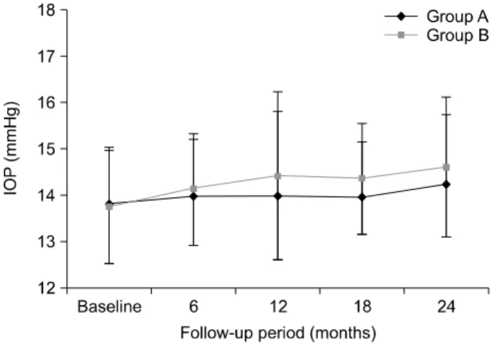
The mean IOP values were 13.80±1.21 mmHg at baseline, 13.99±1.20 mmHg at 6 months, 14.00±1.80 mmHg at 12 months, 14.06±1.18 mmHg at 18 months, and 14.23±1.50 mmHg at 24 months in group A, and 13.74±1.20 mmHg, 14.12±1.20 mmHg, 14.40±1.21 mmHg, 14.34±1.32 mmHg, and 14.60±1.25 mmHg at baseline, 6, 12, 18, and 24 months, respectively, in group B (Fig. 2). No significant ele- vations in the mean IOP value were found in either group at any follow-up visit compared with baseline. In addition,
FIG. 2. Changes of intraocular pressure (IOP) in the loteprednol etabonate 0.5% and fluorometholone 0.1% groups. IOP: intra- ocular pressure, Group A: loteprednol etabonate group, Group B: flu- orometholone group.
FIG. 3. (A) Proportion of patients with intraocular pressure (IOP) elevation by more than 2 mmHg compared to the baseline at 6, 12, 18 and 24 months in the loteprednol etabonate 0.5% and fluorometholone 0.1% groups. (B) Mean IOP value in patients with IOP elevation by more than 2 mmHg compared to the baseline at 24 months. IOP: intraocular pressure, Group A: loteprednol etabonate group, Group B: fluorometholone group, *p values (comparison between group A and group B)<0.05.
there were also no significant differences between groups A and B. No patients with IOP elevation required an- ti-glaucoma medications.
IOP elevations of more than 2 mmHg compared with baseline were analyzed at 6, 12, 18, and 24 months after treatment in both groups: one patient at 6 months (1.5%), one at 12 months (1.5%), three at 18 months (4.5%), and four at 24 months (6.1%) in group A, and two patients at 6 months (3.0%), three at 12 months (4.5%), six at 18 months (9.0%), and nine at 24 months (13.4%) in group B (Fig. 3A).
Fig. 3B illustrates the changes in the mean IOP value in patients with an IOP elevation of more than 2 mmHg at 24 months compared with baseline. A significant difference in the mean IOP value between the groups was found at 24 months after treatment (15.00±0.82 mmHg in group A ver- sus 16.50±1.12 mmHg in group B; p=0.04).
DISCUSSION
According to the DEWS guideline, anti-inflammatory treatment in dry eye disease is recommended when the se- verity level reaches level II or higher.2 Dry eye in SS is caused by lacrimal hypo-secretion with inflammatory changes in the lacrimal gland, leading to the presence of inflammatory mediators in the tears. Several studies have reported that dry eye associated with SS is more severe than non-SS dry eye.10,11 Therefore, topical anti-inflam- matory medications such as corticosteroids, NSAIDs, and cyclosporine A 0.05% are needed to treat SS-dry eye. The anti-inflammatory mechanism of cyclosporine A 0.05% is limited to the inhibition of T-cell activation, whereas corti- costeroids have multifaceted effects on anti-inflammatory mechanisms as well as immunomodulation.21 Corticoste- roids exert their biological effects through nuclear gluco- corticoid receptors. Corticosteroids inhibit interleukin-2 (IL-2) production by T cells.22 Furthermore, corticosteroids down-regulate the production of the pro-inflammatory cy- tokines IL-1, IL-6, and tumor necrosis factor- (TNF-) by monocytes and macrophages.23 Avunduk et al.24 noted that topical corticosteroids were superior for improving the clinical parameters of dry eye compared with NSAIDs in both patients with and patients without SS. Some case studies have been carried out with the short-term use of topical corticosteroids to treat SS-dry eye, showing im- provements of signs and symptoms.12,14 Marsh et al.,12 in a retrospective, noncomparative case study, reported that application of topical nonpreserved methylprednisolone 3 to 4 times per day for 2 weeks was an effective treatment option for patients with SS-associated severe kerato- conjunctivitis sicca, leading to relief of irritation symptoms and improvement of the corneal staining score. Aragona et al., in a prospective, placebo-controlled study, evaluated 40 patients with SS.14 The treatment group was treated with
topical 0.1% clobetasone butyrate twice daily for 30 days.
Corneal and conjunctival stain significantly improved at day 15, and the symptoms score improved at day 30. No changes in IOP or fundus examination were observed in the placebo group or the treatment groups at either time point.
In the present study, the Schirmer test results, tear film BUT, keratoepitheliopathy, and subjective symptoms im- proved during the follow-up period after treatment with both loteprednol etabonate 0.5% and fluorometholone 0.1%. Topical corticosteroids can decrease ocular surface inflammation and promote a healthy tear composition, leading to the recovery of epithelial damage. Interestingly, the tear film BUT did not significantly improve at 6 months after treatment. This may be because all patients in the present study had severe dry eye with marked corneal staining.
Generally, the long-term use of corticosteroids has been known to have a risk of adverse events, such as increased IOP, development of cataract, and infection. In the present study, patients with chronic, severe dry eye with SS, which was refractory to topical cyclosporine A or NSAIDs, were treated with long-term, low-frequency topical corticosteroids.
No infectious lesions were observed during the follow-up periods in either group. UCVA and IOP at 6, 12, 18, and 24 months after corticosteroid treatment had no statistically significant changes compared with baseline in either the loteprednol etabonate or fluorometholone group. We sup- pose that several factors may influence the absence of sig- nificant changes in mean IOP values in our study. First, both corticosteroids have a lower tendency to induce IOP elevation compared with other corticosteroids such as dex- amethasone or methylprednisolone. Second, we adminis- trated topical corticosteroids at a low frequency. Third, we excluded patients with glaucoma or glaucoma suspect and known steroid responders.
Diurnal variations in IOP in the normal eye range be- tween 2 and 10 mmHg.17 To minimize the diurnal variation in IOP, we made an effort to measure IOP at the same time of day during follow-up visits for each patient, and analysis was performed to identify the patients with an IOP ele- vation of more than 2 mmHg compared with baseline. The number of these patients at each follow-up visit differed be- tween the groups. At 24 months after treatment, the pro- portions of patients were 6.9% in the loteprednol etabonate group and 15.3% in the fluorometholone group. The mean IOP value was significantly lower in the loteprednol eta- bonate group than in the fluorometholone group.
Loteprednol etabonate is a novel ester-based cortico- steroid, the 17-chloromethyl ester of Δ1-cortienic acid etabonate, a derivative of the prednisolone metabolite, Δ1-cortienic acid.25 Loteprednol etabonate is highly lip- ophilic and has strong binding to the glucocorticoid receptor. Also, it is rapidly metabolized after action by tis- sue esterases, thereby limiting any potential adverse effects.26 A number of studies have demonstrated that lote- prednol etabonate has potent anti-inflammatory efficacy with less impact on IOP elevation than other corticosteroids.27
Consistent with previous reports, in the present study, the risk of IOP elevation was lower with the use of loteprednol etabonate than with fluorometholone.
Although some patients experienced IOP elevation, no elevation to values higher than 21 mmHg was observed in either group. In addition, no abnormalities of the optic nerve head, retinal nerve fiber layer, or visual field were found in the patients with IOP elevation. Nevertheless, it is important to recognize that there is a possibility of IOP elevation, which could lead to glaucomatous optic nerve damage.
Our study had several limitations. The study design was retrospective, and the sample size was relatively small. A further prospective, randomized, large-scale clinical trial could confirm and better address our results. Second, this study had no control group. We included patients who had chronic dry eye disease with DEWS level III or higher.
Inflammation of the ocular surface such as severe central corneal staining was not controlled with lubricants such as topical cyclosporine A in all patients. Therefore, in this ret- rospective study, patients who were treated with lu- bricants only could not be recruited. Third, although we performed slit lamp biomicroscopy on every patient over the course of the study, cataract grading by the Lens Opacity Classification System (LOCS) was not evaluated.
However, there was no remarkable development or pro- gression of cataract in the patients of this study. In addi- tion, no significant differences in UCVA or visual symp- toms associated with cataract such as clouded vision, glare, and sensitivity to light were found during the follow-up pe- riod compared with baseline. Further studies on assess- ment of cataract grading are warranted. Finally, although effort was made to measure IOP at the same time point dur- ing the follow-up visits for each patient, it is possible that IOP spikes in between the follow-up visits were missed.
In conclusion, long-term application of low-dose topical corticosteroids, such as loteprednol etabonate 0.5% or fluo- rometholone 0.1%, can be effective to control signs and symptoms of chronic, severe dry eye associated with SS.
Loteprednol etabonate 0.5% may have a lower risk for IOP elevation than fluorometholone 0.1%. Although long-term use of topical corticosteroids seems to be an effective treat- ment option, it is important to monitor possible side effects, including IOP elevation.
CONFLICT OF INTEREST STATEMENT None declared.
REFERENCES
1. Schaumberg DA, Sullivan DA, Buring JE, Dana MR. Prevalence of dry eye syndrome among US women. Am J Ophthalmol 2003;
136:318-26.
2. The definition and classification of dry eye disease: report of the Definition and Classification Subcommittee of the International Dry Eye WorkShop (2007). Ocul Surf 2007;5:75-92.
3. Rhen T, Cidlowski JA. Antiinflammatory action of glucocorti- coids--new mechanisms for old drugs. N Engl J Med 2005;353:
1711-23.
4. Carnahan MC, Goldstein DA. Ocular complications of topical, peri-ocular, and systemic corticosteroids. Curr Opin Ophthalmol 2000;11:478-83.
5. Nussenblatt RB, Palestine AG. Cyclosporine: immunology, phar- macology and therapeutic uses. Surv Ophthalmol 1986;31:159-69.
6. Hemady R, Tauber J, Foster CS. Immunosuppressive drugs in im- mune and inflammatory ocular disease. Surv Ophthalmol 1991;35:369-85.
7. Tatlipinar S, Akpek EK. Topical ciclosporin in the treatment of ocular surface disorders. Br J Ophthalmol 2005;89:1363-7.
8. Sall K, Stevenson OD, Mundorf TK, Reis BL. Two multicenter, randomized studies of the efficacy and safety of cyclosporine oph- thalmic emulsion in moderate to severe dry eye disease. CsA Phase 3 Study Group. Ophthalmology 2000;107:631-9.
9. Liew MS, Zhang M, Kim E, Akpek EK. Prevalence and predictors of Sjogren's syndrome in a prospective cohort of patients with aqueous-deficient dry eye. Br J Ophthalmol 2012;96:1498-503.
10. Goto E, Matsumoto Y, Kamoi M, Endo K, Ishida R, Dogru M, et al. Tear evaporation rates in Sjögren syndrome and non-Sjögren dry eye patients. Am J Ophthalmol 2007;144:81-5.
11. Horwath-Winter J, Berghold A, Schmut O, Floegel I, Solhdju V, Bodner E, et al. Evaluation of the clinical course of dry eye syndrome. Arch Ophthalmol 2003;121:1364-8.
12. Marsh P, Pflugfelder SC. Topical nonpreserved methylpredni- solone therapy for keratoconjunctivitis sicca in Sjögren syndrome.
Ophthalmology 1999;106:811-6.
13. Gündüz K, Ozdemir O. Topical cyclosporin treatment of kerato- conjunctivitis sicca in secondary Sjögren's syndrome. Acta Ophthalmol (Copenh) 1994;72:438-42.
14. Aragona P, Spinella R, Rania L, Postorino E, Sommario MS, Roszkowska AM, et al. Safety and efficacy of 0.1% clobetasone bu- tyrate eyedrops in the treatment of dry eye in Sjögren syndrome.
Eur J Ophthalmol 2013;23:368-76.
15. Aragona P, Stilo A, Ferreri F, Mobrici M. Effects of the topical treatment with NSAIDs on corneal sensitivity and ocular surface of Sjögren's syndrome patients. Eye (Lond) 2005;19:535-9.
16. Shiboski SC, Shiboski CH, Criswell L, Baer A, Challacombe S, Lanfranchi H, et al; Sjögren's International Collaborative
Clinical Alliance (SICCA) Research Groups. American College of Rheumatology classification criteria for Sjögren's syndrome: a data-driven, expert consensus approach in the Sjögren's International Collaborative Clinical Alliance cohort. Arthritis Care Res (Hoboken) 2012;64:475-87.
17. de Venecia G, Davis MD. Diurnal variation of intraocular pres- sure in the normal eye. Arch Ophthalmol 1963;69:752-7.
18. Yoon KC, Heo H, Im SK, You IC, Kim YH, Park YG. Comparison of autologous serum and umbilical cord serum eye drops for dry eye syndrome. Am J Ophthalmol 2007;144:86-92.
19. Dogru M, Katakami C, Inoue M. Tear function and ocular surface changes in noninsulin-dependent diabetes mellitus. Ophthalmology 2001;108:586-92.
20. Kaido M, Goto E, Dogru M, Tsubota K. Punctal occlusion in the management of chronic Stevens-Johnson syndrome. Ophthal- mology 2004;111:895-900.
21. Samudre SS, Lattanzio FA Jr, Williams PB, Sheppard JD Jr.
Comparison of topical steroids for acute anterior uveitis. J Ocul Pharmacol Ther 2004;20:533-47.
22. Daynes RA, Araneo BA. Contrasting effects of glucocorticoids on the capacity of T cells to produce the growth factors interleukin 2 and interleukin 4. Eur J Immunol 1989;19:2319-25.
23. Linden M, Brattsand R. Effects of a corticosteroid, budesonide, on alveolar macrophage and blood monocyte secretion of cyto- kines: differential sensitivity of GM-CSF, IL-1 beta, and IL-6.
Pulm Pharmacol 1994;7:43-7.
24. Avunduk AM, Avunduk MC, Varnell ED, Kaufman HE. The com- parison of efficacies of topical corticosteroids and nonsteroidal an- ti-inflammatory drops on dry eye patients: a clinical and im- munocytochemical study. Am J Ophthalmol 2003;136:593-602.
25. Bodor N, Buchwald P. Soft drug design: general principles and recent applications. Med Res Rev 2000;20:58-101.
26. Druzgala P, Hochhaus G, Bodor N. Soft drugs--10. Blanching ac- tivity and receptor binding affinity of a new type of glucocorticoid:
loteprednol etabonate. J Steroid Biochem Mol Biol 1991;38:
149-54.
27. Controlled evaluation of loteprednol etabonate and prednisolone acetate in the treatment of acute anterior uveitis. Loteprednol Etabonate US Uveitis Study Group. Am J Ophthalmol 1999;
127:537-44.