ORIGINAL ARTICLE
헬리코박터 음성 혹은 제균에 반응이 없는 위 점막 연관 림프조직 림프종에서 방사선 치료의 임상적 유용성
박병삼, 이시형
영남대학교 의과대학 내과학교실
Clinical Efficacy of Radiotherapy in Helicobacter pylori Negative or Unresponsive to Eradication Therapy Primary Gastric Mucosa-Associated Lymphoid Tissue Lymphoma
Byung Sam Park and Si Hyung Lee
Department of Internal Medicine, Yeungnam University College of Medicine, Daegu, Korea
Background/Aims: The eradication of Helicobacter pylori (H. pylori) is an effective treatment in gastric mucosa-associated lymphoid tissue (MALT) lymphoma associated with H. pylori infection. However, the treatment strategy in gastric MALT lymphoma patients who are H. pylori-negative or unresponsive to H. pylori eradication therapy remains controversial. In this study, we investigated the clinical efficacy of treatments other than H. pylori eradication therapy in these groups of patients.
Methods: This was a retrospective single-center study based on the medical records of patients diagnosed with gastric MALT lympho- ma at Yeungnam University Medical Center between January 2005 and December 2016. Patients were treated with H. pylori erad- ication therapy, chemotherapy, or radiotherapy according to their H. pylori infection status and stage of gastric MALT lymphoma.
Results: Of the 68 eligible patients, 50 were enrolled in the study. Of the 42 patients with H. pylori-positive gastric MALT lymphoma, 36 (81.7%) were treated with H. pylori eradication therapy as primary treatment and 25 (69.4%) achieved a complete response (CR).
Patients without a CR after H. pylori eradication therapy (n=11, 30.6%) received radiotherapy as a secondary treatment. Two patients with H. pylori-positive gastric MALT lymphoma and eight with H. pylori-negative gastric MALT lymphoma received radiotherapy as the primary treatment. CR was achieved in all 21 patients treated with radiotherapy as primary or secondary treatment. The 5-year pro- gression-free survival rate after radiotherapy was 92.9%.
Conclusions: Radiotherapy may be a worthwhile treatment option in patients with H. pylori-negative MALT lymphoma or H. pylori-pos- itive MALT lymphoma that is not responsive to H. pylori eradication therapy. (Korean J Gastroenterol 2019;73:19-25)
Key Words: Lymphoma, B-cell, marginal zone; Stomach neoplasms; Helicobacter Pylori; Radiotherapy
Received July 30, 2018. Revised November 17, 2018. Accepted November 23, 2018.
CC This is an open access article distributed under the terms of the Creative Commons Attribution Non-Commercial License (http://creativecommons.org/licenses/
by-nc/4.0) which permits unrestricted non-commercial use, distribution, and reproduction in any medium, provided the original work is properly cited.
Copyright © 2019. Korean Society of Gastroenterology.
교신저자: 이시형, 42415, 대구시 남구 현충로 170, 영남대학교 의과대학 내과학교실
Correspondence to: Si Hyung Lee, Department of Internal Medicine, Yeungnam University College of Medicine, 170 Hyeonchung-ro, Nam-gu, Daegu 42415, Korea.
Tel: +82-53-620-3830, Fax: +82-53-623-8038, E-mail: [email protected], ORCID: https://orcid.org/0000-0001-7221-7506 Financial support: This work was supported by the 2017 Yeungnam University Research Grant (217A480003).
Conflict of interest: None.
서 론
점막 연관 림프조직(mucosa-associated lymphoid tis- sue, MALT) 림프종은 모든 B세포 기원 림프종의 5-8%를 차 지하며,1,2 위장관, 특히 위에서 가장 흔하게 발생한다. 위
MALT 림프종은 헬리코박터 파일로리(
Helicobacter pylori
,H. pylori
) 감염과 밀접한 관련이 있다. 위 MALT 림프종의 90%에서H. pylori
감염이 동반되어 있고,3,4 이러한 환자의 80%에서H. pylori
제균 치료로 관해를 유도할 수 있다.3,5 또 한 위 MALT 림프종에서H. pylori
제균 치료 후의 장기 예후도 양호하게 보고되고 있다.6,7 이에
H. pylori
제균 치료는 국 소 위 MALT 림프종의 첫 번째 치료 방안으로 제시되고 있다.하지만
H. pylori
양성 위 MALT 림프종의 일부에서는H. pylori
제균 치료에 반응을 보이지 않으며, 위 MALT 림프 종의 약 5-10%는H. pylori
감염의 증거가 없다. 이러한 경우 에 기다리고 지켜보는 전략(watch and wait), 항암화학요법, 방사선 치료 등 여러 치료 방안이 제시되고 있다.3 위와 위 주변 림프절을 포함한 중선량(24-30 Gray)의 방사선 치료의 경우 좋은 효과가 보고되고 있으나, 국소적 위 MALT 림프종 의 경우 아직까지 대규모 무작위 연구는 없는 실정이다.8,9 항 암화학요법은 가장 효과적인 요법(regimen)에 대한 증거가 아직은 부족하며, t (11;18) 유무가 항암화학요법의 효과에 영 향을 미칠 수 있다.10 또한 항암화학요법을 통한 전신 치료는 국소 질환에서 적절하지 않아 방사선 치료와 같은 국소 치료 에 실패한 경우에 고려될 수 있는 치료 방안이다.11 현재까지H. pylori
음성이거나H. pylori
제균 치료에 반응을 보이지 않는 위 MALT 림프종의 치료에 있어서 일치된 합의나 권고 는 아직 없으며 치료 방안에 대하여 논란이 있는 실정이다.이에
H. pylori
제균 치료에 반응이 없거나H. pylori
감염의 증거가 없는 환자에서 방사선 치료와 같은H. pylori
제균 치 료 외 치료 방안의 치료적 효과 및 임상 양상을 알아보기 위하 여 본 연구를 진행하였다.대상 및 방법
1. 방법
본 연구는 후향적 연구로 2005년 3월 1일부터 2016년 12월 31일까지 영남대학교 의료원에서 위 MALT 림프종을 진단받 은 환자들을 대상으로 진행하였다. 내시경을 통한 조직 검사 로 위 MALT 림프종을 진단하였으며, 급성 요소분해효소 검 사(rapid urase test)로 진단시
H. pylori
감염 유무를 평가하 였다. 질환의 병기 평가를 위하여 혈액학적 검사, 전산화단층 촬영(CT), 골수 생검 등을 실시하였다. 병기 평가는 위장관 림프종의 병기 평가에 사용되는 Lugano staging system12을 사용하였다. 추적 관찰 기간이 3개월 미만인 환자들은 본 연 구에서 제외하였다.2. 치료 방법 및 반응 평가
각각의 환자들은 질환의 병기 및
H. pylori
감염 상태 등에 따라H. pylori
제균 치료, 항암화학요법, 방사선 치료 중 한 가지 방법으로 치료를 하였다. 치료 종료 2-3개월 후 내시경 및 내시경 조직 검사를 통하여 치료 반응을 평가하였으며, 필 요시 CT 등 영상학적 검사를 추가적으로 시행하였다. 치료 종료 후 시행한 추적 내시경 및 내시경 조직 검사에서 MALT림프종의 증거가 없고, 이러한 상태가 한 차례 더 시행한 내시경 조직 검사에서 지속되는 경우 완전 관해(complete response) 로 정의하였고, 그 외의 경우에는 비관해(no complete re- sponse)로 정의하였다.
H. pylori
제균 치료는 아목시실린(1000 mg, 1일 2회), 클 래리스로마이신(500 mg, 1일 2회), 양성자펌프억제제(proton pump inhibitor)의 표준 3제 요법을 1-2주간 실시하였다.H.
pylori
제균 치료 성공 여부는 급성 요소분해효소 검사, Giemsa 염색을 이용한 내시경 조직 검사, 요소호기 검사 (urea breath test) 등으로 평가하였다. 항암화학요법은 cy- clophosphamide, doxorubicin, vincristine, prednisolone 의 약제를 병용하여 시행하였으며, 매 3주마다 실시하여 총 6회 실시하였다. 방사선 치료 시작 전 CT simulator를 이용하 여 시뮬레이션 시행 후 방사선 치료를 시행하였으며, 총 방사 선 용량은 평균 31.4±2.4 Gy/16.6±1.7 fraction이었다. 방사 선 치료를 시행하는 도중에 발생하는 이상반응 유무를 정도에 따라 경도, 중등도, 중증, 생명을 위협하는 등의 4단계로 평가 하였다.3. 통계분석
각 환자들의 인구통계학적 인자들과 의학적 자료들은 본원 의 전자 의무기록 차트 리뷰를 통하여 수집하고 분석하였다.
연속적인 변수들은 평균과 표준편차를 이용하여 값을 제시하 였고, 비연속적인 변수들은 각각의 수치와 백분율을 이용하여 제시하였다. 카플란-마이어 방법을 통하여 무진행 생존율 (progression free survival)을 측정하였다. 모든 통계적 분석은 마이크로소프트 윈도우용으로 나온 SPSS version 20.0 (IBM Inc., Chicago, IL, USA)을 이용하여 분석하였다.
결 과
1. 대상 환자의 특징
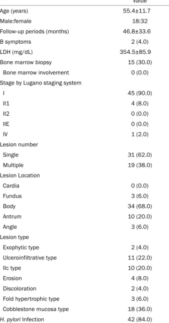
연구 기간 동안 총 68명의 환자들이 위 MALT 림프종으로 진단되었다. 이 중 18명은 추적 관찰에서 탈락되어 최종적으로 50명이 본 연구에 포함되었다. 연구 대상자의 임상 특징은 Table 1과 같다. 평균 연령은 55.4세, 남녀비는 18:32로 상대적 으로 여성에서 유병률이 높았다. 연구 대상자 중 B 증상이 있는 경우는 드물어 단지 2예(4%)에 불과하였으며, 혈청 LDH 의 평균값도 354.5 mg/dL로 정상 범위 이내였다. 골수 생검은 15예(30%)에서 시행하였으며, 질환의 골수 침범이 존재하는 경우는 없었다. 진단 시점에서 대부분(90.0%) Lugano 병기 I기로 국소 질환의 양상이었으며, 1예(2%)에서 진단 시점에 복막 파종의 원격 전이를 동반하고 있었다.
Table 1. Baseline Characteristics of Study Subjects Value
Age (years) 55.4±11.7
Male:female 18:32 Follow-up periods (months) 46.8±33.6
B symptoms 2 (4.0)
LDH (mg/dL) 354.5±85.9
Bone marrow biopsy 15 (30.0)
Bone marrow involvement 0 (0.0)
Stage by Lugano staging system
I 45 (90.0)
II1 4 (8.0)
II2 0 (0.0)
IIE 0 (0.0)
IV 1 (2.0)
Lesion number
Single 31 (62.0)
Multiple 19 (38.0)
Lesion Location
Cardia 0 (0.0)
Fundus 3 (6.0)
Body 34 (68.0)
Antrum 10 (20.0)
Angle 3 (6.0)
Lesion type
Exophytic type 2 (4.0)
Ulceroinfiltrative type 11 (22.0)
IIc type 10 (20.0)
Erosion 4 (8.0)
Discoloration 2 (4.0)
Fold hypertrophic type 3 (6.0)
Cobblestone mucosa type 18 (36.0)
H. pylori Infection 42 (84.0)
Values are presented as mean±standard deviation or n (%).
LDH, lactic dehydrogenase; H. pylori, Helicobacter pylori.
Table 2. Treatment Details of Study Subjects
Value
H. pylori eradication therapy 42 (84.0)
Eradication therapy alone 36 (87.5)
Eradication therapy with radiotherapy 2 (4.8) Eradication therapy with chemotherapy 4 (9.5) Duration of H. pylori eradication therapy
7 days 11 (26.2)
10 days 3 (7.1)
14 days 26 (61.9)
Unknown 2 (4.8)
H. pylori eradication rate 39/41 (95.1)
Test for H. pylori eradication success
Rapid urase test and biopsy with Giemsa stain 14 (34.1) Rapid urase test and UBT test 1 (2.4)
Rapid urase teat alone 19 (46.3)
Biopsy with Giemsa stain alone 5 (12.2)
UBT test alone 2 (4.9)
Radiotherapy 21 (42.0)
Total radiation dose (Gy) 31.3±2.4
Fraction 16.6±1.7
Adverse events
Grade 1 11 (52.4)
Grade 2 3 (14.3)
Grade 3-4 0 (0.0)
Values are presented as mean±standard deviation or n (%).
H. pylori, Helicobacter pylori; UBT, urea breath test; Gy, Gray.
2. 치료 반응
1) H. pylori 양성 환자
연구 대상자 전체 중 42명(84.0%)에서
H. pylori
감염이 확 인되었고, 42예 모두에서 표준 3제 요법으로H. pylori
제균 치료를 시행하였다(Table 2).H. pylori
제균 치료를 시행한 42예 중 1예는H. pylori
제균 성공 여부를 확인할 수 없었고, 39예에서 제균이 성공하여 95.1%의 제균 성공률을 보였다.표준 3제 요법으로
H. pylori
제균에 실패한 2예 중 1예는 추가 표준 3제의 14일 요법으로H. pylori
제균에 성공하였으 며, 다른 1예는 bismuth를 포함한 4제의 14일 요법으로H.
pylori
제균에 성공하였다.H. pylori
감염이 확인된 42예 중 36예(85.7%)에서 위 MALT 림프종에 대한 초치료로H. pylori
제균 치료 단독 요법 을 시행하였다. 치료 반응 평가를 위한 추적 내시경 및 조직 검사는H. pylori
제균 치료 종료 2.0±0.8개월 및 6.4±2.3개월 후에 2차례 시행하였으며,H. pylori
제균 치료를 시행한 36예 중 25예(69.4%)에서 치료 후 완전 관해를 보였다.H. pylori
제균 치료로 완전 관해를 이룬 25명의 환자 중 1예(4.0%)에서 추적 관찰 기간 동안 위 MALT 림프종이 진행 하였다. H. pylori
제균 치료 8개월 추적 내시경 조직 검사에 서 위 MALT 림프종 재발이 확인되었으며, 재발 시점에서H.
pylori
재감염 소견은 보이지 않았다. 재발 후 방사선 치료를 추가로 시행하였으며, 이후 완전 관해를 이루었다(Fig. 1).H. pylori
제균 치료로 완전 관해를 보이지 못한 11예 (30.6%)는 2차 치료로 방사선 치료를 시행하였다. 제균 치료 종료 3.4±1.7개월 후 방사선 치료를 시작하였으며, 방사선 치 료를 받은 모든 환자(100%)가 완전 관해를 이루었다.H. pylori
양성인 42예에서H. pylori
제균 치료를 초치료Fig. 1. Treatment pathway and outcomes for patients. In total 50 patients, 42 patients (84.0%) had H. pylori infection. Eleven patients with no-CR after H. pylori eradication therapy received radiotherapy as secondary treatment and all achieved CR without relapse. Eight patients with H. pylori-negative gastric MALT lymphoma were treated with radiotherapy as the primary treatment and the CR rate after radiotherapy was 100%. MALT, mucosa-associated lymphoid tissue; H. pylori, Helicobacter pylori; RTx, radiotherapy; CTx, chemotherapy; CR, complete response.
로 시행한 36예를 제외한 6명의 환자 중 2예(4.8%)는
H. py- lori
제균 치료와 동시에 방사선 치료를 시행하였고, 4예 (9.5%)는H. pylori
제균 치료와 동시에 항암화학요법을 실시 하였다.H. pylori
제균 치료와 동시에 방사선 치료를 시행한 2예 모두에서 위 MALT 림프종은 완전 관해를 이루었고,H.
pylori
제균도 성공하였다. 2예 중 1예는 방사선 치료 종료 17.4개월 후 위 MALT 림프종이 재발하여 2차 치료로 방사선 치료를 시행하였고, 추가적인 완전 관해를 달성하였다.H.
pylori
제균 치료와 동시에 항암화학요법을 시행한 4예 중 3예(75%)에서 완전 관해를 확인할 수 있었으며, 3예 모두 추 적 관찰 기간 동안 재발은 없었다. 하지만 1예는 진단 시점에 서 복막 파종의 원격 전이를 동반한 상태였고(병기 IV), 항암 화학요법에도 불구하고 질환이 진행하여 진단 6.8개월 후 사 망하였다(Fig. 1).2) H. pylori 음성 환자
위 MALT 림프종의 진단 시
H. pylori
감염의 증거가 없 는 8예는 초치료로 방사선 치료를 시행하였다. 방사선 치료 를 실시한 8예 모두(100%)에서 치료 후 완전 관해에 도달 하였으며, 추적 관찰 기간 동안 위 MALT 림프종의 재발은 없었다(Fig. 1).3. 무진행 생존율
평균 추적 관찰 기간은 42.5개월(범위, 3.0-133.6개월)이었 다. 추적 관찰 기간 동안 1예에서 치료 중 질환이 악화되었고, 2예에서 완전 관해 후 재발하였다. 전체 환자에서 치료 후 질 환의 진행까지 중앙값 7.9개월(범위, 6.8-19.3개월)이 걸렸으 며, 5년 무진행 생존율은 93.1%였다(Fig. 2). 1차 또는 2차 치료로 방사선 치료를 받은 21예 중 1예에서 추적 관찰 기간 동안 위 MALT 림프종이 진행하였고, 질환의 진행까지 19.3개 월이 걸렸다. 위 MALT 림프종 치료로 방사선 치료를 받은 환자의 5년 무진행 생존율은 92.9%였다(Fig. 2). 전제 환자 중 치료 후 질환이 진행한 3예 중 2예에서 추가 방사선 치료 를 시행하였으며, 2예 모두에서 추가적인 완전 관해에 도달할 수 있었다(Table 3). 하지만 1예는 항암화학요법 시작 6.8개 월 후 질환이 진행하여 사망에 이르렀다.
고 찰
H. pylori
감염은 위 MALT 림프종의 발병에 있어서 중요 한 역할을 한다. 위 MALT 림프종은H. pylori
제균 치료로 대개 60-90%의 완전 관해율을 달성할 수 있다고 알려져 있으며,13-15 최근 연구들에서는 80-86% 정도의 완전 관해율이 보고
Table 3. Patients Whose Gastric MALT Lymphoma Had Progressed during Follow-up Periods
Age (years) Sex Stage H. pylori
infection
H. pylori
eradication 1st Tx TTP (months) 2nd Tx Final result
Case 1 64 F I Positive Yes Era 7.9 RTx CR
Case 2 63 M I Positive Yes RTx 19.3 RTx CR
Case 3 34 M IV Positive Yes CTx 6.8 - Death
MALT, mucosa-associated lymphoid tissue; H. pylori, Helicobacter pylori; Tx, treatment; TTP, time to progression; F, female; Era, eradication therapy; RTx, radiotherapy; CR, complete response; M, male; CTx, chemotherapy.
Fig. 2. Progression-free survival rates after treatment. 5-year progression-free survival rate of all patients was 91.1%. Median survival period after all treatments was 7.4 months (range, 4.3-19.3 months). 5-year progression-free survival rate after radiotherapy was 92.3%. Median survival period after radiotherapy was 34.6 months (range, 9.8-133.6 months). RTx, radiotherapy.
되고 있다.16,17이에 National Comprehensive Cancer Network 가이드라인에서
H. pylori
양성 위 MALT 림프종 치료에H.
pylori
제균 치료를 첫 번째 치료 방안으로 제시하고 있다.18 위 MALT 림프종에서H. pylori
양성률은 대개 높은 편이며 한 연구에서는H. pylori
감염 유병률이 거의 90%까지 보고 되기도 한다.19본 연구에서H. pylori
유병률은 84.0%였으며,H. pylori
양성 위 MALT 림프종 환자 중H. pylori
제균 치료 로 69.4%에서 완전 관해에 도달할 수 있었다. 더욱이H. py- lori
양성 위 MALT 림프종의 경우H. pylori
제균 치료 1년 이후에 조직학적 완전 관해에 도달하는 경우도 있어,20 당장H. pylori
제균 치료에 반응을 보이지 않더라도 추가 치료 없 이 기다리며 관찰하는 치료 전략(wait and see)을 취하여 볼 수 있다.21 하지만 wait and see의 치료 전략은 아직까지는 완전하게 정립된 치료 방안이 아니며, 더욱이 환자의 불안감 등 기타 다른 요인으로 이러한 치료 전략이 제한될 수 있다.H. pylori
감염의 증거가 없는 위 MALT 림프종의 경우에 도H. pylori
제균 치료는 1차 치료로 고려할 수 있는 치료적옵션이다.21
H. pylori
음성 위 MALT 림프종에서H. pylori
제균 치료의 반응률은 적게는 11.1%에서 많게는 57.1%에 이 르기까지 다양하게 보고되고 있으며, 긍정적인 치료 반응을 보고한 연구들이 있으나 전반적인H. pylori
음성 위 MALT 림프종에 대한H. pylori
제균 치료의 치료 반응률은H. pylo- ri
양성 위 MALT 림프종에 비하여 낮게 보고되고 있는 실정 이다.22-24위 MALT 림프종에 대한 방사선 치료의 반응률은 높은 편 으로 90% 이상 완전 관해율을 보이며, 5년 생존율 95% 이상 및 10년 생존율 70%로 장기간 예후 역시 매우 훌륭하다.8,25-30 더욱이 National Comprehensive Cancer Network 가이드 라인에서는
H. pylori
제균 치료에 반응을 보이지 않는H. py- lori
양성 위 MALT 림프종의 경우 방사선 치료를 수술이나 항암화학요법보다 우선적으로 제시하고 있다.18 반면, 방사선 치료에 따른 독성은 적어, 천공이나 출혈과 같이 방사선 치료 에 따른 심각한 이상반응이 발생할 위험은 4% 미만으로 알려 져 있다.31본 연구에서 1차 치료로 방사선 치료를 시행한 10예 (H. pylori
양성 2예,H. pylori
음성 8예) 및 구제 요법으로 방사선 치료를 시행한,H. pylori
제균 치료에 반응하지 않는H. pylori
양성 위 MALT 림프종 11예 등 방사선 치료를 시행 한 21예 모두에서 치료 후 완전 관해를 달성하였다. 또한, 방사 선 치료에 따른 독성으로 치료를 중단한 경우는 없었다. 1차 치료로 방사선 치료를 시행한 10예 중 추적 관찰 기간 동안 1예에서 위 MALT 림프종이 재발하였으며, 재발 후 2차 치료 로 방사선 치료를 시행하여 완전 관해를 재달성 할 수 있었다.본 방사선 치료를 한 환자의 5년 무진행 생존율은 92.9%로 이전의 연구들과 비슷한 양상으로 좋은 예후를 보였다.
본 연구에서는 표준 3제 요법에 대한 95.1%의
H. pylori
제균 성공률을 보였다. 95.1%의 제균 성공률은 최근의 경향 에 비하여 매우 높은 편이며, 본 연구와 동일 지역에서 시행된 표준 3제 요법에 대한H. pylori
제균 성공률에 관한 연구와 비교하였을 때도 다소 높은 수준이다.32,33 반면, 높은 수준의 제균 성공률에 비하여H. pylori
제균 치료에 대한 위 MALT 림프종의 관해율은 69.4%로 다소 떨어지는 양상이다.6,7 급성 요소분해효소 검사와 조직 검사의 경우H. pylori
제균 치료 후에는 박멸 여부와 관계없이H. pylori
집락의 감소와 불균등 분포 등으로 인하여 위음성의 가능성이 있어 검사 시 전정 부와 체부에서 적어도 2개 이상의 조직을 얻어 검사를 시행할 것을 권고하고 있다.34 본 연구에서
H. pylori
제균 치료 후 제균 여부를 판정하기 위하여 사용된 급성 요소분해효소 검 사, Giemsa 염색을 이용한 조직 검사, 요소호기 검사 등의 3가지 검사 중 2가지 검사를 동시에 사용한 경우는 42예 중 15예(급성 요소분해효소 검사와 조직 검사를 동시에 시행한 14예와 급성 요소분해효소 검사와 요소호기 검사를 동시에 시행한 1예, 35.7%)인 반면, 3가지 검사 중 한 가지 검사만으 로H. pylori
제균 여부를 평가한 경우는 27예(64.3%)로 특히 급성 요소분해효소 검사 혹은 조직 검사 단독만 사용한 경우 가 25예로 대부분을 차지하였다. 또한 급성 요소분해효소 검 사 및 조직 검사 시 전정부와 체부에서 2개 이상의 조직을 얻어 검사를 시행하였는지 명확히 평가할 수 없었다. 이러한 요인들은H. pylori
제균 여부를 평가하기 위하여 시행된 급 성 요소분해효소 검사 또는 조직 검사 결과의 위음성을 가능 성을 높이고, 최종적으로H. pylori
제균율이 과대평가되었을 가능성을 배제할 수 없을 것이다.본 연구는 몇몇 제한점이 있다. 첫째, 본 연구는 후향적 연 구로 연구 대상자 선정, 치료 방법 선정, 치료 반응 평가에 제한이 있다. 특히 연구 대상자의 선정 및 치료 방법을 선택하 는데 있어서 높은 수준의 일관성을 유지하는데 한계가 있었 다. 둘째, 본 연구에 선정된 연구 대상자 수는 비교적 적으며, 특히
H. pylori
음성 위 MALT 림프종 환자의 수는 10명에 불과할 정도로 적다. 끝으로, 분자생물학적 유전학적 분석을 시행하지 못하였으며, 특히 t(11;18)/API2-MALT1 상태에 대 한 평가를 시행하지 못하였다. 하지만 본 연구는 이러한 제한 점에도 불구하고 위 MALT 림프종 환자의 치료 계획을 수립 하는데 도움이 되는 연구라는 점에서 의의가 있다고 할 수 있을 것이다. 결론적으로, 방사선 치료는H. pylori
음성인 위 MALT 림프종과H. pylori
제균 치료에 반응하지 않는H.
pylori
양성 환자의 치료에 매우 효과적인 치료 방안이 될 수 있으며, 양호한 장기적 예후를 기대할 수 있을 것이다.요 약
목적: 헬리코박터 파일로리(
H. pylori
) 제균 치료는 위 MALT 림프종 치료에 효과적인 방법이다. 그러나H. pylori
감염 증 거가 없거나H. pylori
제균 치료에 반응을 보이지 않는 위 MALT 림프종 환자에서 여러 가지 치료 방안이 제시되고 있 으나 아직까지 일치된 방안이나 권고가 없는 실정이다. 이에 저자들은H. pylori
제균 치료에 반응을 보이지 않거나H.
pylori
감염의 증거가 없는 환자에서 방사선 치료와 같은H.
pylori
제균 치료 외 치료 방안의 치료적 효과 및 임상 양상을알아보고자 본 연구를 진행하였다.
대상 및 방법: 2005년 1월부터 2016년 12월까지 영남대학교 의료원에서 위 MALT 림프종을 진단받은 환자들을 대상으로 후향적 연구를 시행하였다. 내시경 및 내시경 조직 검사를 시 행하여 위 MALT 림프종을 진단하였으며, 질환의 병기 및
H.
pylori
감염 상태에 따라H. pylori
제균 치료, 항암화학요법, 방사선 치료 중 한 가지 방법으로 치료를 하고 치료 반응 및 임상 경과를 평가하였다.결과: 전체 68명이 위 MALT 림프종으로 진단되었으며, 최종 50명의 환자가 본 연구의 대상자로 선정되었다. 평균 나이는 55.4±11.7세였고, 평균 추적 관찰 기간은 42.5±31.0개월이었 다. 42예의
H. pylori
양성 위 MALT 림프종 환자 중 36예 (85.7%)에서H. pylori
제균 치료를 1차 치료로 시행하였으 며, 25예(69.4%)에서 완전 관해에 도달하였다.H. pylori
제균 치료 후 완전 관해에 도달하지 못한 11예(30.6%)에서 2차 치 료로 방사선 치료를 시행하였으며, 모든 환자가 완전 관해에 도달할 수 있었다.H. pylori
양성인 위 MALT 림프종 환자 중 2예은 1차 치료로H. pylori
제균 치료와 동시에 방사선 치료를 시행하였고, 2예 모두 완전 관해를 보였다.H. pylori
감염의 증거가 없는 8예는 1차 치료로 방사선 치료를 시행하 였고, 모두 완전 관해를 달성하였다. 1차 또는 2차 치료로 방 사선 치료를 받은 21예는 모두 치료 후 완전 관해에 도달하였 으며, 1예에서 추적 관찰 기간 동안 위 MALT 림프종이 진행 하였고 방사선 치료를 받은 환자의 5년 무진행 생존율은 92.9%였다.결론: 방사선 치료는
H. pylori
음성 위 MALT 림프종과H.
pylori
제균 치료에 반응을 보이지 않는H. pylori
양성 위 MALT 림프종의 치료에 효과적인 치료 방안이 될 수 있다.색인단어: 림프종 B세포 변연대; 위 종양; 헬리코박터 파일로리;
방사선 치료
REFERENCES
1. A clinical evaluation of the international lymphoma study group classification of non-Hodgkin's lymphoma. The non-Hodgkin's lymphoma classification project. Blood 1997;89:3909-3918.
2. Olszewski AJ, Castillo JJ. Survival of patients with marginal zone lymphoma: analysis of the surveillance, epidemiology, and end results database. Cancer 2013;119:629-638.
3. Ruskoné-Fourmestraux A, Fischbach W, Aleman BM, et al. EGILS consensus report. Gastric extranodal marginal zone B-cell lym- phoma of MALT. Gut 2011;60:747-758.
4. Zucca E, Copie-Bergman C, Ricardi U, et al. Gastric marginal zone lymphoma of MALT type: ESMO clinical practice guidelines for di- agnosis, treatment and follow-up. Ann Oncol 2013;24 Suppl 6:vi144-vi148.
5. Zullo A, Hassan C, Cristofari F, et al. Effects of Helicobacter pylori eradication on early stage gastric mucosa-associated lymphoid
tissue lymphoma. Clin Gastroenterol Hepatol 2010;8:105-110.
6. Ono S, Kato M, Takagi K, et al. Long-term treatment of localized gastric marginal zone B-cell mucosa associated lymphoid tissue lymphoma including incidence of metachronous gastric cancer.
J Gastroenterol Hepatol 2010;25:804-809.
7. Nakamura S, Sugiyama T, Matsumoto T, et al. Long-term clinical outcome of gastric MALT lymphoma after eradication of Helicobacter pylori: a multicentre cohort follow-up study of 420 patients in Japan. Gut 2012;61:507-513.
8. Wirth A, Gospodarowicz M, Aleman BM, et al. Long-term outcome for gastric marginal zone lymphoma treated with radiotherapy:
a retrospective, multi-centre, international extranodal lympho- ma study group study. Ann Oncol 2013;24:1344-1351.
9. Ruskoné-Fourmestraux A, Matysiak-Budnik T, Fabiani B, et al.
Exclusive moderate-dose radiotherapy in gastric marginal zone B-cell MALT lymphoma: results of a prospective study with a long term follow-up. Radiother Oncol 2015;117:178-182.
10. Lévy M, Copie-Bergman C, Gameiro C, et al. Prognostic value of translocation t(11;18) in tumoral response of low-grade gastric lymphoma of mucosa-associated lymphoid tissue type to oral chemotherapy. J Clin Oncol 2005;23:5061-5066.
11. Ikoma N, Badgwell BD, Mansfield PF. Multimodality treatment of gastric lymphoma. Surg Clin North Am 2017;97:405-420.
12. Rohatiner A, d'Amore F, Coiffier B, et al. Report on a workshop convened to discuss the pathological and staging classifications of gastrointestinal tract lymphoma. Ann Oncol 1994;5:397-400.
13. Wotherspoon AC, Doglioni C, Diss TC, et al. Regression of primary low-grade B-cell gastric lymphoma of mucosa-associated lym- phoid tissue type after eradication of Helicobacter pylori. Lancet 1993;342:575-577.
14. Kim JS, Chung SJ, Choi YS, et al. Helicobacter pylori eradication for low-grade gastric mucosa-associated lymphoid tissue lym- phoma is more successful in inducing remission in distal com- pared to proximal disease. Br J Cancer 2007;96:1324-1328.
15. Zullo A, Hassan C, Andriani A, et al. Eradication therapy for Helicobacter pylori in patients with gastric MALT lymphoma: a pooled data analysis. Am J Gastroenterol 2009;104:1932-1937;
quiz 1938.
16. Choi YJ, Lee DH, Kim JY, et al. Low grade gastric mucosa-asso- ciated lymphoid tissue lymphoma: clinicopathological factors associated with Helicobacter pylori eradication and tumor regression. Clin Endosc 2011;44:101-108.
17. Ryu KD, Kim GH, Park SO, et al. Treatment outcome for gastric mucosa-associated lymphoid tissue lymphoma according to Helicobacter pylori infection status: a single-center experience.
Gut Liver 2014;8:408-414.
18. NCCN Guidelines. [Internet]. Plymouth Meeting (PA): National Comprehensive Cancer Network; [updated 2018 May 15; cited 2018 Jul 2]. Available from: https://www.nccn.org/professionals/
physician_gls/pdf/b-cell.pdf
19. Asenjo LM, Gisbert JP. Prevalence of Helicobacter pylori infection in gastric MALT lymphoma: a systematic review. Rev Esp Enferm
Dig 2007;99:398-404.
20. Wündisch T, Thiede C, Morgner A, et al. Long-term follow-up of gastric MALT lymphoma after Helicobacter pylori eradication. J Clin Oncol 2005;23:8018-8024.
21. Thieblemont C, Zucca EA. Clinical aspects and therapy of gastro- intestinal MALT lymphoma. Best Pract Res Clin Haematol 2017;
30:109-117.
22. Akamatsu T, Mochizuki T, Okiyama Y, Matsumoto A, Miyabayashi H, Ota H. Comparison of localized gastric mucosa-associated lymphoid tissue (MALT) lymphoma with and without Helicobacter pylori infection. Helicobacter 2006;11:86-95.
23. Choi YJ, Kim N, Paik JH, et al. Characteristics of Helicobacter pylori- positive and Helicobacter pylori-negative gastric mucosa-asso- ciated lymphoid tissue lymphoma and their influence on clinical outcome. Helicobacter 2013;18:197-205.
24. Gong EJ, Ahn JY, Jung HY, et al. Helicobacter pylori eradication therapy is effective as the initial treatment for patients with H. py- lori-negative and disseminated gastric mucosa-associated lym- phoid tissue lymphoma. Gut Liver 2016;10:706-713.
25. Vrieling C, de Jong D, Boot H, de Boer JP, Wegman F, Aleman BM.
Long-term results of stomach-conserving therapy in gastric MALT lymphoma. Radiother Oncol 2008;87:405-411.
26. Tsang RW, Gospodarowicz MK, Pintilie M, et al. Localized muco- sa-associated lymphoid tissue lymphoma treated with radiation therapy has excellent clinical outcome. J Clin Oncol 2003;21:
4157-4164.
27. Goda JS, Gospodarowicz M, Pintilie M, et al. Long-term outcome in localized extranodal mucosa-associated lymphoid tissue lym- phomas treated with radiotherapy. Cancer 2010;116:3815-3824.
28. Abe S, Oda I, Inaba K, et al. A retrospective study of 5-year out- comes of radiotherapy for gastric mucosa-associated lymphoid tissue lymphoma refractory to Helicobacter pylori eradication therapy. Jpn J Clin Oncol 2013;43:917-922.
29. Kim SW, Lim DH, Ahn YC, et al. Clinical outcomes of radiation ther- apy for early-stage gastric mucosa-associated lymphoid tissue lymphoma. World J Gastroenterol 2013;19:6062-6068.
30. Yahalom J. Patients with H pylori-independent MALT lymphoma are curable with radiotherapy. Oncology (Williston Park) 2011;
25:1147-1149.
31. Schechter NR, Yahalom J. Low-grade MALT lymphoma of the stomach: a review of treatment options. Int J Radiat Oncol Biol Phys 2000;46:1093-1103.
32. Heo J, Jeon SW. Changes in the eradication rate of conventional triple therapy for Helicobacter pylori infection in Korea. Korean J Gastroenterol 2014;63:141-145.
33. Jung YS, Lee SH, Park CS, et al. Trends in the eradication rates of Helicobacter pylori infection in Daegu and Gyeongsangbuk-do, Korea: multicenter study over 13 years. Korean J Gastroenterol 2014;63:82-89.
34. Kim SG, Jung HK, Lee HL, et al. Guidelines for the diagnosis and treatment of Helicobacter pylori infection in Korea, 2013 revised edition. J Gastroenterol Hepatol 2014;29:1371-1386.