Mucinous Nevus
Vol. 30, No. 4, 2018 465
Received July 14, 2017, Revised October 20, 2017, Accepted for publication October 27, 2017
Corresponding author: You Won Choi, Department of Dermatology, Ewha Womans University School of Medicine, 1071 Anyangcheon-ro, Yangcheon-gu, Seoul 07985, Korea. Tel: 82-2-2650-2665, Fax: 82-2- 2652-6925, E-mail: [email protected]
ORCID: https://orcid.org/0000-0001-6315-3889
This is an Open Access article distributed under the terms of the Creative Commons Attribution Non-Commercial License (http://creativecommons.
org/licenses/by-nc/4.0) which permits unrestricted non-commercial use, distribution, and reproduction in any medium, provided the original work is properly cited.
Copyright © The Korean Dermatological Association and The Korean Society for Investigative Dermatology
pISSN 1013-9087ㆍeISSN 2005-3894 Ann Dermatol Vol. 30, No. 4, 2018 https://doi.org/10.5021/ad.2018.30.4.465
CASE REPORT
Mucinous Nevus
Min Young Lee, Ji Yeon Byun, Hae Young Choi, You Won Choi
Department of Dermatology, Ewha Womans University College of Medicine, Seoul, Korea
Mucinous nevus is an uncommon entity classified as either a cutaneous mucinosis or a connective tissue nevus. The condition presents as grouped papules and coalescent pla- ques growing in a unilateral or zosteriform manner. The key histopathological feature is a band-like deposition of mucin in the superficial dermis. A 34-year-old male presented with grouped gray-brown papules and confluent plaques exhibit- ing a zosteriform distribution on the right side of the lower back. The lesions had commenced in childhood. Histological examination revealed mucin deposition in the papillary dermis. Thus, we diagnosed a mucinous nevus. To date, only a few reports of such nevi have been reported in the literature. Therefore we report a rare case of mucinous nevus.
(Ann Dermatol 30(4) 465∼467, 2018) -Keywords-
Cutaneous, Mucin, Nevus
INTRODUCTION
Mucinous nevus is a rare disorder classified as either a cu- taneous mucinosis or a connective tissue nevus1,2. The condition was first described by Redondo Bellón et al.1 in 1993. Clinically, asymptomatic grouped papules or pla-
ques grow to form a verrucous or nevoid feature exhibit- ing a unilateral or zosteriform distribution3,4. The nevus usually develops on the trunk at birth or in early adult- hood3,4. Histologically, the nevus is characterized by mu- cin deposits localized to the superficial dermis3,4.
To the best of our knowledge, only 25 cases have been re- ported in the English-language literature3,5-11 and only one case in the Korean literature12. Herein, we report an un- usual case of mucinous nevus.
CASE REPORT
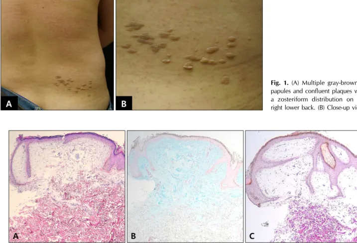
A previously healthy 34-year-old Korean male presented with asymptomatic grouped gray-brown papules and con- fluent plaques exhibiting a zosteriform distribution on his right lower back (Fig. 1). The skin lesions had commenced in childhood, gradually coalesced, and grew slowly. In our patient, pigmentary abnormalities as like freckles ex- cept for skin lesions of the right lower back were not observed. He had neither any past medical problem nor a family history of similar lesions and pigmentary abnor- malities. He denied any trauma.
Histological examination revealed an acanthosis with elongated rete ridges and amorphous materials associated with loosely separated collagen fibers in the papillary der- mis (Fig. 2A). The amorphous materials stained with alcian blue at pH 2.5 (Fig. 2B); this confirmed a mucin deposit limited to the papillary dermis. Verhoeff-van Gieson stain- ing revealed that the numbers of elastic fibers in the papil- lary dermis were reduced in the regions of mucin deposi- tion (Fig. 2C).
This clinicopathological analysis enabled us to diagnose a mucinous nevus. Our patient decided to allow us to ob- serve the lesion; no treatment was performed.
DISCUSSION
Mucinous nevus is an uncommon entity initially described
MY Lee, et al
466 Ann Dermatol
Fig. 2. (A) Amorphous materials and loosely separated collagen fibers in the papillary dermis and acanthosis with elongated rete ridges (H&E, ×40). (B) Positively stained bluish amorphous materials in the papillary dermis (Alcian blue at pH 2.5, ×40). (C) Reduced numbers of elastic fibers in the papillary dermis bearing mucin deposits (Verhoeff-van Gieson, ×40).
Fig. 1. (A) Multiple gray-brownish papules and confluent plaques with a zosteriform distribution on the right lower back. (B) Close-up view.
by Redondo Bellón et al.1 in 1993 and classified as either a cutaneous mucinosis or a connective tissue nevus1,2. The cutaneous mucinoses are a heterogeneous group of dis- eases in which abnormal amounts of mucin are deposited in the skin6. Connective tissue nevi are hamartomas with unusual levels (excesses or deficiencies) of collagen, elas- tin, and/or proteoglycans2.
The term “mucinous nevus” refers to the its nevoid ap- pearance and the characteristic pattern of mucin deposits in the papillary dermis2,6,13. Clinically, mucinous nevi present as asymptomatic, multiple skin-colored to brown- ish papules or plaques; separate lesions coalesce and then grow to form a verrucous or nevoid feature with a unilat- eral or zosteriform pattern3,4,6. It usually develops at birth or in early adulthood6,13. The principal site is the trunk, in- cluding the back3,6. The male:female ratio is 5:1; the rea- son is not clear5. To date, there are two reports of familial mucinous nevus6,13. However, there was no report about the genetic abnormality as like mosaicism. Histologically, mucinous nevus is characterized by diffuse band-like mu- cin deposits in the uppermost portion of the dermis1-4. The
mucin is thought to be composed of hyaluronic acid stain- ing positively with alcian blue at pH 2.5 but not staining at pH 0.514,15. The origin of the mucin remains un- known14, but is presumed to be attributable to a primary metabolic process (such as overproduction) rather than a secondary catabolic process2. Mucinous nevi are divided into two histopathological types depending on whether epidermal changes are present; these are connective tissue nevi of the proteoglycan (CTNP) type and the combined epidermal-CTNP type5. The epidermis is normal, in the CTNP type but, in the combined epidermal-CTNP type, exhibits hyperkeratosis and acanthosis with elongation of the rete ridges2. Our case featured an epidermal change;
thus, we diagnosed the combined epidermal-CTNP type of mucinous nevus.
Both an epidermal nevus and nevus lipomatosus super- ficialis exhibit nevoid features similar to those of a muci- nous nevus. It is difficult, therefore, to clinically dis- tinguish among the conditions. Histological data are necessary. Histologically, an epidermal nevus and nevus lipomatosus superficialis can be distinguished from a mu-
Mucinous Nevus
Vol. 30, No. 4, 2018 467 cinous nevus; only the latter exhibits mucin deposits in
the papillary dermis.
However, such mucin deposition is also observed in cuta- neous mucinosis of infancy, but is very superficial and ap- pears to be hugged by the epidermis5. Clinically, cuta- neous mucinosis of infancy presents as scattered small papules unlike mucinous nevus.
Mucinous nevi do not require treatment (except for cos- metic purposes); the nevi are benign3. Surgical excision, scalpel dermabrasion, and carbon dioxide laser treatment are possible5. Surgical excision is not indicated if several discrete lesions are evident5. Mucinous nevus of the CTNP type was treated via scalpel dermabrasion, but scarring de- veloped 1 year later16. There is one report of mucinous ne- vus of the combined epidermal-CTNP type which did not recur after carbon dioxide laser vaporization5. Our patient did not voice any cosmetic concern; thus, we decided to simply observe the lesions.
To the best of our knowledge, only 25 cases of mucinous nevi have been reported in the English-language liter- ature3,5-11 and only one in the Korean literature12. The principal location is the trunk including the back.
Approximately 80% of all cases were reported in males and about 30% of all cases present at birth.
Herein, we report a rare case of mucinous nevus of the combined epidermal-CTNP type.
CONFLICT OF INTEREST
The authors have nothing to disclose.
REFERENCES
1. Redondo Bellón P, Vázquez-Doval J, Idoate M, Quintanilla E. Mucinous nevus. J Am Acad Dermatol 1993;28:797-798.
2. Rongioletti F, Rebora A. Mucinous nevus. Arch Dermatol 1996 ;132:1522-1523.
3. Cobos G, Braunstein I, Abuabara K, Chu EY, James W.
Mucinous nevus: report of a case and review of the literature. JAMA Dermatol 2014;150:1018-1019.
4. Tardío JC, Granados R. The cellular component of the mucinous nevus consists of CD34-positive fibroblasts. J Cutan Pathol 2010;37:1019-1020.
5. Chi CC, Wang SH, Lin PY. Combined epidermal-connective tissue nevus of proteoglycan (a type of mucinous nevus): a case report and literature review. J Cutan Pathol 2009;
36:808-811.
6. Perez-Crespo M, Lopez-Navarro N, Betlloch I, Herrera E, Niveiro M, Gallego E. Acquired and familial mucinous nevus. Int J Dermatol 2011;50:1283-1285.
7. Vukicevic JS, Milobratovic DJ, Milinkovic MV, Bogdanovic Z. Extensive, adulthood inflammatory linear verrucous epidermal nevus associated with mucinous nevus. Indian J Dermatol Venereol Leprol 2011;77:607-608.
8. Song BH, Park S, Park EJ, Kwon IH, Kim KH, Kim KJ.
Mucinous nevus with fat: an unusual case report and literature review. Am J Dermatopathol 2012;34:e146-e148.
9. Kim EJ, Jo SJ, Cho KH. A case of mucinous nevus clinically mimicking nevus lipomatosus superficialis. Ann Dermatol 2014;26:549-550.
10. Sasaki T, Yoneda K, Yokoi I, Moriue J, Demitsu T, Kubota Y.
Comorbidity of dermatofibromas and mucinous nevi. Int J Dermatol 2016;55:e53-e55.
11. Walter Lepage A, Frouin É, Junca A, Cante V, Monégier du Sorbier C, Hulin-Desquiret MC, et al. [Mucinous nevus of late onset]. Ann Dermatol Venereol 2016;143:547-553.
French.
12. Joo HJ, Yoo HJ, Kim JE, Kang H. A case of congenital mucinous nevus on the back. Korean J Dermatol 2014;52:
892-894.
13. Chen CW, Tsai TF, Chen YF, Hung CM. Familial mucinous nevus. Pediatr Dermatol 2008;25:288-289.
14. Lim JH, Cho SH, Kim HO, Kim CW, Park YM. Mucinous naevus with atypical features. Br J Dermatol 2003;148:
1064-1066.
15. Brakman M, Starink TM, Tafelkruyer J, Bos JD. Linear connective tissue naevus of the proteoglycan type ('naevus mucinosus'). Br J Dermatol 1994;131:368-370.
16. Yokogawa M, Kamakura T, Ishiguro H, Ikeda M, Kodama H. Mucinous nevus. J Dermatol 2005;32:30-33.