Vo1. 14, No. 1, May, 2005
※통신저자: 장 학
서울특별시 종로구 연건동 2 8번지 서울대학교 의과대학 성형외과학교실
Tel: 82-2-2072-3086, Fax: 82-2-3675-7792, E-mail: [email protected]
✽ 본 논문의 요지는 2 0 0 4년 1 1월 2 7일 연세대학교 의과대학 강당에서 개최된 대한미세수술학회 주최 제 2 3차 대한 미세 수술학회 학술대회에서 구연발표 되었음.
두경부 유리피판 수술에 있어서의
비천공성 혈관 클립을 이용한 미세혈관 문합술
서울대학교 의과대학 성형외과학교실, 울산대학교 의과대학 성형외과학교실*
장@ 학・민경원・김우람*・신현우*・고경석*
─ Abstract ─
Microvascular Anastomosis with Non-penetrating Vascular Clips in Head and Neck Free Flap Surgery
Hak Chang, M.D., Kyung Won Minn, M.D., Woo Ram Kim, M.D.*, Hyun Woo Shin, M.D.*, Kyung Suck Koh, M.D.*
Department of Plastic and Reconstructive Surgery Seoul National University College of Medicine, Department of Plastic Surgery Asan Medical Center University of Ulsan College of Medicine*
Microvascular anastomosis with suture technique is a basic skill but there are several problems such as stenosis, thrombosis and long operating time. Recently plastic surgeons have developed non-suturing mechanical coupling devices for microvascular anastomosis. The authors applied non-penetrating vascular clips (VCS clips) in the field of free flap surgery of head and neck area.
Between August of 2004 and January of 2005, we performed 9 free flaps (16 vessels) using small- sized VCS clips. Four stay sutures were applied first and then VCS clips were placed between sutures about 1 mm apart. Vascular pedicle of free flap included the descending branch of lateral circumflex femoral vessel, thoracodorsal vessel, deep inferior epigastric vessel and cephalic vein.
The recipient vessels were the superior thyroid artery, superficial temporal artery, internal jugular vein, external jugular vein, and superficial temporal vein. We performed 13 end-to-end (4 arteries and 9 veins) and 3 venous end-to-side anastomoses. No flap related complication occurred but we applied additional clips or sutures in two cases due to blood leakage after completion of anastomo- sis. Primary patency rates seemed to be good and more rapid anastomosis could be done than con-
Ⅰ. 서 론
유리피판술에 있어서 봉합사를 이용한 미세혈관문 합은 가장 기본적인 술기이나 바늘이 혈관을 관통하 며 혈관내막에 손상을 입히고 혈관내면에 노출된 봉 합사는 혈전현성을 야기하는 등의 문제가 있다. 비 천공성 혈관 클립 (Vascular Closure System Clip Applier, 이하 V C S클립)은 혈관을 관통하지 않고 혈관을 문합시키고 사용이 간편하여 흉부외과 및 혈관외과 영역에서 이미 사용되어 왔으며 최근에 는 성형외과 영역에서도 보고가 있다.1 , 2 , 3 저자들은 두경부 재건에 있어서 V C S클립을 이용한 미세혈관 문합의 사용 경험을 바탕으로 그 문합 방법의 장단 점을 점검하고 유리피판수술에 있어서의 V C S클립 의 유용성을 검토하고자 하였다.
Ⅱ. 재료 및 방법
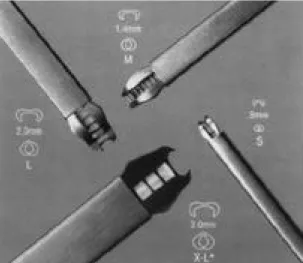
2 0 0 4년8월부터 2 0 0 5년1월까지 9명의 유리피판을 이용한 두경부 재건수술에서 V C S클립을 사용하여 총 1 6곳의 미세혈관문합을 시행하였다. VCS클립은 티타니움( t i t a n i u m )으로 되어있고 클립의 크기에 따라 소형, 중형, 대형, 특대형이 있다.(Fig. 1).
소형(클립 폭 0.9 mm) 은 직경 0.9-2.4 mm, 중
형(클립 폭 1.4 mm)은 직경 2.4-4.0 mm, 대형 (클립 폭 2.0 mm)은 직경 4.0-5.0 mm, 특대형 (클립 폭3.0 mm)은 직경 5.5 mm이상의 혈관에 적용하도록 되어 있다(Fig. 2). 환자는 남성 7명, 여성 2명이었으며, 나이는 3 2 ~ 7 5세(평균 5 0 . 7세) 였다. 원인질환은 두경부암 7례, 동정맥기형 1례, 뇌수술후 감염 1례 등 이였으며, 재건에 사용한 피 판은 전외측 대퇴천공지 피판 4례, 요측전완피판 2 례, 광배근피판 1례, 공장피판 1례, 복직근 피판 1 례 등 이였다 (Table 1). 미세혈관문합에 있어서 혈관은 일반적인 방법으로 준비하고 봉합은 먼저 1 2 시, 3시, 6시, 9시 등 균등한 간격으로 4곳을 9 - 0 혹은 10-0 나일론으로 stay suture를 시행하였다.
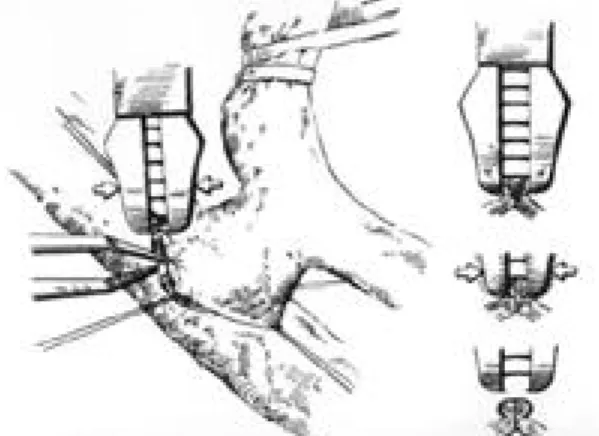
다음에 인접한 stay suture를 조수와 술자가 서로 반대방향으로 당겨서 문합할 부분이 일직선이 되게 한 뒤, VCS클립을 사용하여 stay suture 사이를 약 1 mm간격으로 s t a p l i n g하였다(Fig. 3).
증례 1
5 3세 남자(Case 1)로, 재발된 우측 편도암에 대 하여 우선 이비인후과에서 하악골 정중절골술로 접 ventional suture technique. Advantages of VCS technique are high patency rate, low thrombo- genecity and rapidity. Although the high cost of VCS instrument may be a problem, this clip could be applied safely in microvascular free tissue transfer.
Fig. 1. Four types of vascular closure staple clip (VCS) applier. Small (S), Medium (M), Large (L) and X-Large (XL) are available. The metallic shaft head can be rotated by 360 degree.
Fig. 2. VCS clip dimensions. Approximate size of clips is demonstrated. The number in millimeter means the interval between two tips of the clip.
근하여 광범위절제술 및 경부임파선곽청술을 시행하 였다. 이어서 성형외과에서 경부의 수혜부 혈관을 준비하고 요측전완피판을 거상하였다. 요골동맥은 천경부동맥에, 요골동맥의 두개의 동반정맥 중 굵은 것을 천경부정맥에 9-0 및 10-0 나일론 봉합사를 이
용하여 단단문합하여 우선 피판을 재관류 시켰다. 2 번째 정맥 문합으로 선택한 노쪽피부정맥( c e p h a l i c v e i n )은 V C S클립을 이용하여 외경정맥에 단단문합 하였다. 먼저 9-0 나일론으로 4곳( 1 2 , 3 , 6 , 9시 방 향)를 혈관벽이 외번( e v e r s i o n )이 되도록 s t a y s u t u r e를 두고, 그 사이사이를 소형 V C S클립을 이 용하여 약 1 mm간격으로 4 ~ 5개씩 c l i p p i n g하였 으며 총 1 5분이 소요되었다(Fig. 4). 피판은 합병 증 없이 모두 생착 하였다.
증례 2
7 5세 여자(Case 6)로 좌측 측두 및 두정부에 걸 친 두피의 편평상피암에 대하여 광벙위절제술이 시 행되었다. 15×13 cm크기의 두피 전층결손 및 1 0
×10 cm의 전층두개골 결손이 발생하였다. 좌측 측 두부에서 수혜부 혈관을 준비하고 광배근 유리 피판 을 거상하였다. 피판의 혈관경인 가슴등동맥과 정맥 은 각각 얕은관자동맥과 정맥에 소형 V C S클립을 이 용하여 단단문합하였고, 동맥은 8분, 정맥은 9분이 소요되었다(Fig. 5). 재관류 직후에 문합부위에서의 혈액 누출은 없었으며 피판은 문제없이 생착 되었 다.
Ⅲ. 결 과
Table 1. Patients Characteristics Case Sex
Diagnosis Flap Microvascular
VCS application
No. /Age anastomoses
1 M/53 Tonsil cancer RF 1a/2v 1v (EE)
2 M/32 Tonsil cancer ALT 1a/1v 1v (EE)
3 M/55 AVM, forehead ALT 1a/1v 1v (EEx2*)
4 M/40 Tongue cancer RF 1a/3v 2v (EE, ES)
5 M/44 Buccal mucosa cancer ALT 1a/2v 1v (EE)
6 F/75 SCC, scalp LD 1a/1v 1a/1v(EE/EE)
7 M/69 Hypopharynx cancer Jejunum 1a/1v 1a/1v(EE/EE)
8 F/38 Infection, subdural space RA 1a/1v 1a/1v (EE/EE)
9 M/51 Tonsil cancer ALT 1a/2v 1a/2v (EE/ESx2)
AVM, arteriovenous malformation, SCC, squamous cell carcinoma
RF, radial forearm, ALT, anterolateral thigh, LD, latissimus dorsi, RA, rectus abdominis 1a/2v, one artery & two veins were anastomosed
*Two venous anastomoses were done because arterial graft was used for venous gap.
EE, end-to-end anastomosis, ES, end-to-side anastomosis Fig. 3. Schematic drawing of clip application technique.
Usually four stay sutures are first applied, which aids in obtaining symmetric eversion. If eversion is not enough, aid by a forceps is recommended.
The clips must be applied between stay sutures close together about 1 mm apart. As shown in right side, VCS clips remain outside the lumen after clipping, which means minimal intimal dam - age during anastomosis.
모든 증례에서 미세혈관수술에 관련된 합병증은 없었으며 피판은 모두 생존하였다. VCS클립은 모두 소형을 사용하였으며 초기의 5증례(증례1 - 5 )에서는 정맥문합(두번째 혹은 세번째)에만 V C S클립을 사용 하였으나 경험이 축적되면서 후기의 4증례(증례6 - 9 ) 에서는 동, 정맥 모두에 적용하였다. 문합에 사용한 피판의 혈관으로 동맥은 외측넙다리휘돌이 동맥의 하행분지(descending branch of lateral circum- flex femoral artery), 가슴등동맥( t h o r a c o d o r s a l
artery), 공장동맥(jejunal artery), 깊은아래쪽배 벽동맥(deep inferior epigastric artery) 등을 사 용하였고 정맥은 외측넙다리휘돌이 정맥의 하행분지 (descending branch of lateral circumflex femoral vein), 노쪽피부정맥(cephalic vein), 요 골동맥의 동반정맥(venae comitantes of radial artery), 가슴등정맥(thoracodorsal vein), 공장 정맥(jejunal vein), 깊은아래쪽배벽정맥( d e e p inferior epigastric vein) 등을 사용하였다. 수혜
Fig. 4. Case 1. (Left) Recurrent right tonsil cancer was extirpated via median mandibulotomy approach and neck dis- section was done. The vascular pedicle of radial forearm flap was anastomosed to the recipient vessels in the neck before insetting the flap. (Right) Enlarged photo of microvascular anastomosis. Conventional suture technique was used between the radial artery (RA) and transverse cervical artery and between the venae comi- tantes of the radial nartery and transverse cervical vein (arrow heads). Small-sized non-penetrating vascular clips (VCS) were applied between the cephalic vein (CV) and external jugular vein (EJV)(arrow).
Fig. 5. Case 6. (Left) Scalp defect after extirpation of squamous cell carcinoma was covered with the latissimus dorsi musculocutaneous free flap. Microvascular anastomosis was performed in the left preauricular region. (Right) Both arterial (between the thoracodorsal artery (TDA) and superficial temporal artery (STA) and venous (the thoracodorsal vein (TDV) and superficial temporal vein (STV)) microvascular anastomoses were done with small-sized VCS clips.
부 혈관으로 동맥은 위쪽갑상샘동맥(superior thy- roid artery), 얕은관자동맥(superficial tempo- ral artery) 등 을 사용하였고, 정맥은 안쪽목정맥 (internal jugular vein), 바깥쪽목정맥( e x t e r n a l jugular vein), 얕은관자정맥(superficial tempo- ral vein) 등을 사용하였다. 단단(end-to-end) 문 합이 1 3곳이며 이중 동맥이 4곳, 정맥이 9곳이었으 며 단측(end-to-side) 문합한 3곳은 모두 정맥이었 다. 위치나 방향이 부적절하게 적용된 클립은 제거 전용 겸자를 이용하여 제거하고 다시 클립을 사용하 거나 봉합사를 써서 문합을 추가하였다. 클립을 적 용하였으나 주위조직과의 마찰 등으로 쉽게 클립이 혈관에서 떨어져 나가는 경우가 간혹 있었으나 다시 클립을 조심스럽게 적용하여 별 문제는 없었다.
V C S클립을 적용한 혈관은 직경(외경)이 2~5 mm 였으며 4군데의 stay suture의 사이사이에는 4 ~ 6 개의 클립을 쓰고, 혈관당 1 8 ~ 2 5개의 클립이 소요 되었다. 혈류 재개 후 문합 부위에 혈액누출이 있어 서 봉합사 혹은 V C S클립으로 추가 문합을 한 경우 가 2곳에서 있었다. 혈관 하나 당 문합 시간은 4곳 의 stay suture를 포함하여 8 ~ 2 0분(평균 1 2분)이 소요되었다.
Ⅳ. 고 찰
V C S클립은 기존의 봉합사와 달리 혈관벽을 관통 하지않는 비천공성( n o n - p e n e t r a t i n g )이며 클립이 좁아지면서 혈관내막을 접촉시켜 문합을 이루게 된 다. 봉합사처럼 여러 번 결찰 할 필요가 없어 문합 시간이 단축 되며 혈관내막에 이물질이 노출되지 않 고 혈관내막의 손상이 최소화되므로 혈전형성의 가 능성이 현저히 떨어져 개존율이 향상되는데 이는 여 러 동물실험을 통하여 입증되었다.4 - 6 임상에서 Shenoy 등7 은 혈액투석을 위한 동정맥단락술 (arteriovenous fistula)에서 2 4개월후의 개존율을 비교하여 VCS 클립을 사용한 2 4 2례에서 67 %, 봉 합술을 사용한 2 7 6례에서는 48 %로 V C S클립의 우 수성을 보고하였다. 최근에 유리피판수술시에도 VCS 클립이 적용되기 시작하여 직경 2 . 5 ~ 5 . 5 mm 정도의 혈관에서도 비교적 무리 없이 사용되고 높은 성공율과 문합시간 단축이 가능하게 되었다.1 - 3 문합에 소요되는 시간은 보고자 마다 차이는 있으나
V C S클립의 적용 초기에는 1 5 ~ 2 0분이 소요되다가 점차 익숙해지면 5 ~ 1 0분 정도로 단축됨을 보고 하
였고,3 , 8 - 1 0본 연구에서도 초기에 비해 후기에서 문합
시간의 단축이 가능하였다. 문합에 있어서 혈관 외 번( e v e r s i o n )을 확실히 하는 것이 가장 중요하여 클 립을 사용할 때마다 겸자를 사용하여 외번을 돕도록 권고되고 있으나 성형외과에서 주로 다루는 직경 2~4 mm정도의 혈관이라면 stay suture를 함으로 써 외번이 충분하게 이루어진다고 생각된다. 따라서 stay suture로 혈관의 외번이 확실하게 이루어지고 조수가 적절하게 stay suture를 당겨서 술자에게 좋은 시야를 제공한다면 V C S클립을 사용하는데는 큰 어려움이 없으나 주위가 비좁은 경우나 단측 문 합인 경우는 clip applier가 직선모양인 관계로 c l i p p i n g하는데 어려움이 있어 주의를 요한다. 또한 혈관의 배열이 문합 하기에 편한 위치가 아닌 경우 3 6 0도 회전이 가능한 head rotator를 돌려서 각도 를 조정하게 되어있으나 손목을 유연하게 사용할 수 있는 봉합술의 경우처럼 미세한 위치조정은 어렵다.
이때는 조수가 잘 보조하여 c l i p p i n g하기에 좋은 방 향을 만들어 주는 것이 중요하다. 본 연구에서는 혈 관의 문제 때문에 V C S클립을 사용하지 못한 경우는 없었으나 De Lorenzi 등1은 혈관이 작거나, 혈관회 전이 어렵거나 혈관외번이 불충분하여 클립을 사용 못한 경우를 보고하고 있으며 저자들은 내막이 중막 에서 심하게 분리된 경우에는 V C S클립으로는 내막 끼리의 밀착이 곤란하므로 종래의 봉합사를 이용한 문합술로 대처해야 한다고 생각한다. VCS 클립은 고가인 것이 최대의 단점이나, 봉합사를 사용하는 경우에 비하여 문합 시간을 반 이하로 단축 시킬 수 있어 피판의 허혈시간과 전체수술시간을 줄일 수 있 는 이점이 있어 한번에 여러 혈관을 문합할 때 유용 하다. 혈관의 외번을 확실하게 하고 클립과 클립사 이의 간격을 균등히 하면서 봉합사를 이용한 문합에 비해서 조금 더 촘촘하게 c l i p p i n g하도록 세심한 주 의를 기울인다면 비록 장기 개존율 등이 검증 되어 야 할 필요가 있으나, 3 mm 이하의 미세혈관에도 V C S클립을 큰 무리 없이 적용이 가능하다고 생각된 다. 최근에는 VCS 클립을 이용한 신경문합이 동물 실험에서 유용성이 보고되고 있으며 임상응용이 기
대된다.1 1 , 1 2
Ⅴ. 결 론
비천공성 클립을 이용한 미세혈관 문합술은 기존 의 봉합사에 의한 문합술의 단점을 보완하여 혈관협 착과 혈전형성을 최소화하면서 문합 부위 자체의 신 연성을 유지하는 장점이 있다. 익숙해지는 데는 그 다지 많은 시간이 필요 없다고 보지만 기본적인 미 세수술술기를 습득하고 나서 사용해야 VCS 클립을 사용할 때 발생할 수 있는 각종 문제를 해결할 수 있다. 혈관의 크기와 두께에 따라 적절한 크기의 클 립을 선택하고, 적절한 간격으로 V C S클립을 사용한 다면 문합시간 단축과 높은 개존율을 함께 확보할 수 있는 유용한 혈관 문합 방법이라고 사료된다.
REFERENCES
01) De Lorenzi F, van der Hulst RRWJ, Boeckx WD:
VCS auto suture stapled microvascular anstomosis in lower leg free flap. Plast Reconstr Surg 109:
2023, 2002.
02) Zeebregts C, Acosta R, Bolander L, et al: Clinical experience with non-penetrating vascular clips in free-flap reconstructions. Br J Plast Surg 55: 105, 2002.
03) Yamamoto N, Nakai H, Satoh Y, et al: C l i n i c a l application of a nonpenetrating microvascular sta - pling device for vascularized free tissue transfer.
Ann Plast Surg 42:48, 1999.
04) Lee JW, Choo SJ, Lee IC, et al: Anastomosis of vessels less than 2 mm with the vascular clip system
clip applier. J Korean Med Sci 16:303, 2001.
05) Leppaniemi AK, Wherry DC, Pikoulis E, et al:
Arterial and venous repair with vascular clips:
Comparison with suture closure. J Vas Surg 26:24, 1997.
06) Kirsch WM, Zhu YH, Hardesty RA, et al: A new method for microvascular anastomosis: report of experimental and clinical research. Am Surg 58:
722, 199.
07) Shenoy S, Miller A, Petersen F, et al: A multicenter study of permanent hemodialysis access surgery:
Beneficial effect of clipped vascular anstomotic technique. J Vasc surg 38:229, 2003.
08) Cope C, Lee K, Stern H, et al: Use of the vascular closure staple clip applier for microvascular anas - tomosis in free-flap surgery. Plast Recosntr Surg 106: 107, 2000.
09) Matsumoto K, Obara H, Hayashi S, et al: A new technique of vascular anastomosis with VCS clip applier system. In Proceedings of the 30th World Congress of the International College of Surgeons, Kyoto, Japan, November, 25-29, 1996.
10) Zeebregts CJ, Kirsch WM, van den Dungen JJ, et al: Five years’world experience with nonpenetrat - ing clips for vascular anastomosis. Am J Surg 187:
751, 2004.
11) Payne CE, Hunt SP, Lamberty GH: Primary sciatic nerve repair using titanium staples. Br J Plast Surg 55: 330, 2002.
12) Park JW, Kim SK, Park JH, et al: Rapid neurorrha - phy with titanium clips. J Korean Orthop Assoc 37:
432, 2002.