Introduction
The temporomandibular joint (TMJ) is a diarthrodial joint consisting of the mandibular condyle, squamous por- tion of the temporal bone, fibrous capsule with reinforce- ment, and accessory ligaments such as the sphenomandi- bular, stylomandibular, and pterygomandibular ligaments, and the synovial membrane.1
The blood supply to the TMJ is circumferential. Every
vessel within a radius of some three centimeters contributes branches to the joint capsule and contributes one or two branches to it.2The blood vessels to the TMJ mainly origi- nate from the superficial temporal artery (STA, about 3.8 mm in diameter) and the maxillary artery (MA, about 3.2 mm in diameter).3
The MA is classified according to its relationship with the lateral pterygoid muscle and zygomatic arch. The super- ficial MA is located laterally to the lateral pterygoid muscle and with a distance between zygomatic arch and maxillary artery of ›20 mm. The deep MA is medial to the lateral pterygoid and with a distance between zygomatic arch and maxillary artery of ¤20 mm (Fig. 1).4
Other vessels which supply the TMJ have been describ-
The arterial blood supply of the temporomandibular joint: an anatomical study and clinical implications
Antonino Marco Cuccia, Carola Caradonna, Domenico Caradonna, Giuseppe Anastasi*, Demetrio Milardi*, Angelo Favaloro*, Anita De Pietro**, Tommaso Maurizio Angileri**, Luigi Caradonna, Giuseppina Cutroneo*
Department of Surgical and Oncological Disciplines, University of Palermo, Palermo, Italy
*Department of Biomorphology and Biotechnologies, University of Messina, Messina, Italy
**Villa Santa Teresa, Diagnostica per Immagini, Palermo, Italy ABSTRACT
Purpose: The aim of this study was to analyze three-dimensional images of the arterial supply to the temporo- mandibular joint.
Materials and Methods: Ten patients (five men and five women, mean age 36 years) without signs or symptoms of temporomandibular disorders, who underwent contrast-enhanced computed tomographic (CT) scanning with intravenous contrast, were studied. The direct volume rendering technique of CT images was used, and a data set of images to visualize the vasculature of the human temporomandibular joint in three dimensions was created. After elaboration of the data through post-processing, the arterial supply of the temporomandibular joint was studied.
Results: The analysis revealed the superficial temporal artery, the anterior tympanic artery, the deep temporal artery, the auricular posterior artery, the transverse facial artery, the middle meningeal artery, and the maxillary artery with their branches as the main arterial sources for the lateral and medial temporomandibular joint.
Conclusion: The direct volume rendering technique was found to be successful in the assessment of the arterial supply to the temporomandibular joint. The superficial temporal artery and maxillary artery ran along the lateral and medial sides of the condylar neck, suggesting that these arteries are at increased risk during soft-tissue pro- cedures such as an elective arthroplasty of the temporomandibular joint. (Imaging Sci Dent 2013; 43 : 37-44)
KEY WORDS: Three-Dimensional Imaging, Computer Generated; Magnetic Resonance Imaging; Temporomandibular Joint; Blood Supply
Received October 12, 2012; Revised November 8, 2012; Accepted November 13, 2012 Correspondence to: Dr. Antonino Marco Cuccia
Section Oral Sciences, Department of Surgical and Oncological Disciplines, University of Palermo, Via Del Vespro 129, 90127 Palermo, Italy
Tel) 39-091-6552232, Fax) 39-091-348282, E-mail) [email protected]
Copyright ⓒ 2013 by Korean Academy of Oral and Maxillofacial Radiology
This is an Open Access article distributed under the terms of the Creative Commons Attribution Non-Commercial License (http://creativecommons.org/licenses/by-nc/3.0) which permits unrestricted non-commercial use, distribution, and reproduction in any medium, provided the original work is properly cited.
Imaging Science in Dentistry∙pISSN 2233-7822 eISSN 2233-7830
ed: these are small branches of the external carotid artery (ECA, the auricular posterior artery 1.7 mm in diameter and the ascending pharyngeal artery ¤1 mm in diameter), and of the facial or the ascending palatine artery.3
In the retrodiskal tissue (RT), which is responsible for the nutrition of the TMJ, are present the branches of the maxillary artery (posterior auricular, anterior tympanic (ATA), and meningeal medial arteries) and the temporo- mandibular veins, as well as the auriculotemporal and pos- terior auricular nerves.5
Information on the vasculature of the TMJ is crucial in surgical interventions, and it may help clarify the pathol- ogy of the TMJ.6-11The aim of this study was to visualize, analyze, and review the current understanding of the arte- rial supply of the TMJ.
Materials and Methods
We reviewed the cervicocranial arteries of 10 patients without signs or symptoms of temporomandibular disorder (TMD) (five men and five women, age range from 25 to 46 years, mean 36 years) who underwent contrast-enhanc- ed computed tomographic (CT) scanning in the period from October 2008 to January 2009, at Santa Teresa Clinic, Bagheria, Palermo.
A standardized TMD examination was executed in all of the patients: joint pain, crepitation, and uncoordinated movements of the head of the mandibular condyle during opening or closing of the mouth were investigated by lat- eral and posterior palpation of each TMJ with both index fingers. Subjects were included if they had a temporoman- dibular index (TMI) reference value of ‹0.08±0.10, and an intensity of jaw pain ⁄5 (with 1 indicating mild pain and 5 moderate pain).12,13
All of the subjects gave written informed consent before beginning the study. The Medical Ethics Committee of the Policlinico G. Martino approved the study protocol, which conformed to the principles of the Declaration of Helsinki for human subject research.
The examinations were performed with a four-channel CT scanner (Mx8000 Quad, Philips Medical Systems, Best, the Netherlands). The volume acquisition was performed during administration of 90 mL of iodinated non-ionic con- trast agent at a concentration of 370 mg/mL through an antecubital vein, with an 18-gauge needle cannula using an automatic injector (Envision, Medrad, Pittsburgh, PA, USA) at a flow rate of 4 mL/s. To obtain optimal enhance- ment of the arteries, the delay time between beginning the contrast agent administration and scan acquisition was cal-
culated with the bolus test technique by measuring the en- hancement curve at one of the common carotid arteries.
Optimizing arterial enhancement with the bolus test techni- que, we deliberately overlooked the venous district, thus avoiding possible interference with the images.
The acquisition volume was set in the caudocranial direc- tion from C7 to the sella turcica. The scan parameters were 250 mAs, 120 kV, collimation 40×0.625 mm, pitch 0.67, gantry rotation time 0.5 seconds, and acquisition time 10 seconds. For the rendering process, we used VolView 2.0 graphics software (Kitware Inc. Clifton Park, NY, USA).
The direct volume rendering system made use of specific algorithms to transform conventional two-dimensional magnetic resonance imaging sets of slices transparent vol- ume data set images and enabled visualization of data from various imaging modalities (e.g. CT, MRI, functional MRI, and confocal microscopy) interfaced with various hard- ware and software systems.
The CT images were compared to the anatomic tables.
The arterial branch on the CT images was categorized as traceable (2 points), recognizable only for a short trace (1 point), or unrecognizable (0 point). The total score for each artery was divided by 20 and multiplied by 100 to arrive at a depiction rate for each artery.14
Results
Applying volume rendering techniques, the three-dimen- sional images of the temporomandibular region were obta- ined. Shading features were used to simulate an external light source. This was performed to give additional depth cues and surface texture to the image, offering a more real- istic view of the anatomic structures.
In particular, the use of these procedures permitted visu- alization of bone and vascular structures contemporarily.
In this way, the arteries and their branches were identified, based on a comparison with anatomical tables of the human TMJ.3,15-17
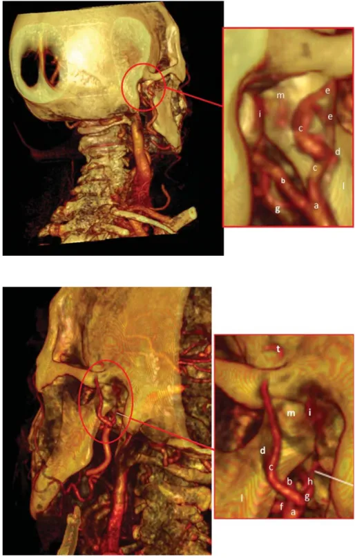
In this study, most of the patients demonstrated the fol- lowing features. Observing the temporomandibular region posteriorly (Fig. 1), it was possible to find the ECA (a) cur- ving somewhat anteriorly and then inclining dorsally to the space behind the neck of the mandible, where it divid- ed into the IMA (b) and STA (c); from the STA arose small diverging arteries (e). In this image, it was possible to iden- tify the transverse facial artery (TFA), a branch of the STA, which disappeared into the parotid gland (d) and the mid- dle meningeal artery (f), a branch of the mandibular por- tion of the MA.
Rotating the image and then observing the left TMJ (Fig.
2), it was possible to find the same vascular organization.
In addition, on this side, the inferior dental artery (IDA, f), a branch of the mandibular portion of the IMA, was visible until it disappeared behind the mandibular ramus and the anterior tympanic artery (ATA, h), branch of the IMA, which passed behind the TMJ to enter the tympanic cavity.
Modifying the parameters and magnifying the region of
the mandibular condyle, it was possible to better observe the relations of these vessels with bone structures (Fig. 3).
In particular, the ECA (a), the IMA (b), and the TFA (c), with their small diverging branches (d), a small portion of the IDA (e), the middle meningeal artery (MMA, f), and finally the ATA (g) were observed.
Removing the ramus of the mandible, all of the vessels of the region behind the ramus were visible. In particular,
Fig. 1.Posterior view of computerized tomography rendering of the temporo- mandibular joint of a 30-year-old heal- thy female. The external carotid artery (a), the internal maxillary artery (b), the superficial temporal artery (c), the trans- verse facial artery (d), with their small diverging arteries (e), the middle men- ingeal artery (g), the retrodiscal tissue (i), the ramus (l) and the condyle (m)
Fig. 2.Posterior and lateral view of computerized tomography rendering of the temporomandibular joint of a 30- year-old healthy female. The external carotid artery (a), the internal maxillary artery (b), the superficial temporal art- ery (c), the transverse facial artery (d), the inferior dental artery (f), the middle meningeal artery (g), the anterior tym- panic artery (h), the retrodiscal tissue (i), the ramus (l) and the condyle (m), the temporal posterior artery (t)
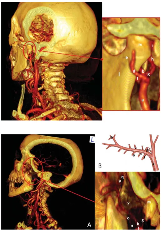
the entire extension of the IMA was observable with its branches (Fig. 4A). Here, the IMA (b), IDA (e), MMA (f), ATA (g), masseteric artery (MA), pterygoid artery (PA), sphenopalatine artery (SA), and deep temporal artery (DTA) were found. A schema of the IMA, with its branches, was also identified in order to demonstrate the fidelity of our study by 3D volume rendering (Figs. 4 and 5).
By observation of our image, it was clear that most of the vascular supply appeared to come from the lateral and medial aspects of the condylar head. The posterior disc attachment region was greatly vascularized, while the
intermediate zone and the anterior disc attachment region were relatively devoid of vessels.
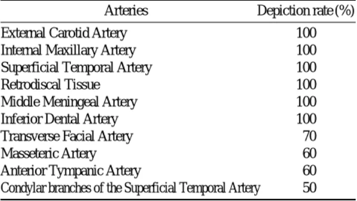
Besides the ECA, the arterial supply to the retrodiscal tissue (RT), the STA, the IMA, the IDA, and the MMA were seen in all cases (depiction rates all 100%). The TFA, the MA, the ATA, and the condylar branches of the STA were found in several but not all cases (depiction rates 70%, 100%, 60%, and 50%, respectively). The analysis of the depiction rates showed that the direct volume rendering technique consistently depicted larger arteries and the zone with a higher degree of vascular supply, but also arteries
Fig. 3.Posterior view of computerized tomography rendering of the temporo- mandibular joint of a 30-year-old heal- thy female. The external carotid artery (a), the internal maxillary artery (b), the transverse facial artery (d) with their small diverging arteries (e), the inferior dental artery (f), the middle meningeal artery (g), the anterior timpanic artery (h), the retrodiscal tissue (i), the ramus (l) and the condyle (m)
Fig. 4.Computerized tomography ren- dering of the left temporomandibular joint of a 28-year-old healthy male (A) and diagram (B) show the branches of maxillary artery. The external carotid artery (a), the internal maxillary artery (b), the inferior dental artery (f), the middle meningeal artery (g), the condyle (m), the masseteric artery (n), the ptery- goid artery (o), the sphenopalatine art- ery (p), the sphenopalatine foramen (q), the deep temporal artery (v)
B
A
with a thin diameter (Table 1).
Discussion
The knowledge of the blood supply around the TMJ is necessary in order to clarify how the blood supply works in the pathogenesis of TMD. Three-dimensional volume rendering has been used for evaluation of other arteries such as the cervicocranial arteries, intracranial aneurysms, and pulmonary artery.18-20However, these arteries have a larger size than those of the TMJ.
This study showed that three-dimensional volume render- ing of computed tomography angiography can successful- ly be used to delineate the vascular anatomy of the TMJ region in good detail.
By this technique, it could be observed that the lateral
and ventral region of the articulation was mainly vascular- ized by the STA and its branches. The anterior region was vascularized by the posterior DTA, the medial region by the ATA and medial meningeal arteries.21,22
The study by Wasicky and Pretterklieber revealed that while the left ATA originated as a singular vessel from either the MA or the STA with almost equal frequencies (44.7 and 45.9%, respectively), the right ATA predomi- nantly branched from the MA (77.8% of cases).23Mérida Velasco et al, in 18 adult cadavers, described the ATA as one of the main arteries of the RT and found that it is an ever-present artery.11Even though Takagi et al found that MR angiography depicted the ATA in only 25% of patients, probably because the artery was very thin in caliber, in our study, the three-dimensional volume rendering of comput- ed tomography angiography depicted the ATA in 60% of patients.6
Clinical relevance of the arterial supply of the TMJ
The circulatory systems of the TMJ can be compromis- ed by trauma, disease, changes of the head and neck posi- tion, and muscle spasm.
Trauma and post-surgical complications
Surgery of the TMJ (disc repair procedures, menisectomy with implant, condylotomy, condylectomy, arthroscopy, and other procedures) is an effective treatment for structu- ral disorders, and there are several general surgical indica-
Fig. 5.Lateral angiograms (A) and diagrams (B and C) of the internal maxillary artery and the superficial temporal artery arising from the external carotid artery. The external carotid artery (a), the internal maxillary artery (b), the superficial temporal artery (c), the transverse facial artery (d), the inferior dental artery (f), the masseteric artery (n), the pterygoid artery (o), the sphenopalatine artery (p), the occipital artery (r), the auricular posterior artery (s), the temporal posterior artery (t), the deep temporal anterior artery (u), the deep temporal artery (v)
A B C
Table 1.Depiction rate of arteries in the temporomandibular joint
Arteries Depiction rate (%)
External Carotid Artery 100
Internal Maxillary Artery 100
Superficial Temporal Artery 100
Retrodiscal Tissue 100
Middle Meningeal Artery 100
Inferior Dental Artery 100
Transverse Facial Artery 70
Masseteric Artery 60
Anterior Tympanic Artery 60
Condylar branches of the Superficial Temporal Artery 50
tions (documented refractory internal derangements, pain and dysfunction of such magnitude). Some of these pro- cedures require open surgery with full mandibular condyle exposure via large skin incisions (disc repair, menisectomy, bone reduction procedures), and some are performed per- cutaneously, with access to the mandibular condyle via multiple limited skin incisions (arthroscopy). The bleeding and vascular injuries associated with trauma to the carotid artery and its terminal branches, the STA, and the IMA are common complications.24
There have been a few reported cases of injury to the MA during intraoral vertical ramus osteotomies, subcondy- lar and condylar fractures, and TMJ surgery.25Those cases were associated with aggressive surgical techniques and necessitated ligation of the ECA to control hemorrhage.
Holmlund and Hellsing demonstrated the close proximi- ty of an arthroscopy puncture site to the STA and vein.26 The close proximity of the MMA to the medial capsule could increase the risk of hemorrhage during TMJ surgery (for example, in eminectomy, condylectomy, or during release of joint ankylosis).27Branches of the IMA may be damaged during extensive maxillary fractures (Le Fort I and II) or repair.
Temporomandibular disorders
The retroarticular region is very important to cushion from mechanical stress and to protect the tympanic wall.
A posterior condylar position, often related to the anterior displacement of the joint disk, reduces the posterior intra- articular space and represents a compression on the bila- minar zone.28,29
The symptomatology in this scenario can be expressed as pain of TMJ, limited movement or locking of the jaw, radiating pain in the face, neck or shoulders, painful click- ing, popping, or grating sounds in the jaw joint when open- ing or closing the mouth, tinnitus, vertigo, and otalgia.
Heffez and Jordan found a statistically significant associa- tion between superficial vascular changes (avascularity) in the RT and progressive anterior displacement of the TMJ disk.30
Vascular lesions
In the head and neck region, arteriovenous malforma- tions and arteriovenous fistulas are uncommon but pose serious therapeutic challenges. Most of the acquired arteri- ovenous fistula cases involve the internal carotid artery and are more often secondary to blunt, penetrating injury and
surgical procedures. The development of an arteriovenous fistula between the MA and the venous malformation has sometimes been reported after Le Fort I osteotomy.31Pseu- doaneurysm and arteriovenous malformation of the STA and vein have also been described after arthroscopy.32
Aneurysms
Aneurysms of the head vessels must be recognized as potential complications in the long-term follow-up of incidental trauma and iatrogenic injury. The branches of the ECA are protected from trauma in most regions of the head and neck by bone and soft tissue. However, in speci- fic areas, the vessels emerge from their protective buffer, consequently leaving them particularly vulnerable to trau- matic injury. Such areas are encountered as the vessels approach the surface and cross bony structures. For exam- ple, the STA traverses the zygomatic arch after it exits from the parotid gland, leaving the artery relatively unprotected.33 Conner et al revealed that, of the 386 reported cases of traumatic pseudoaneurysm of the face and temple, 85%
were found in the STA, 7% were in the IMA, and 8% were in the facial artery.34Cases of pseudoaneurysm of the IMA secondary to trauma have been reported.35,36
STA pseudoaneurysms due to iatrogenic injury have been reported to occur after cyst removal, TMJ excision, or arthroplast.37The post-traumatic development of an an- eurysm can lead to expansion under arterial pressure and potential rupture. Often, there is a delay between the initial injury and the development of the aneurysm, ranging from weeks to years.38
In conclusion, a number of imaging techniques have been used to assess the TMJs. The most prevalent alteration involving the TMJs are dysfunctional conditions (internal derangement) and non-dysfunctional diseases (arthritis, infections, coronoid process hyperplasia, secondary neo- plastic process, fractures, synovial chondromatosis, and avascular necrosis of the mandibular condyle).
Computed tomography and magnetic resonance imaging are important in the diagnosis of diseases of this region because they present a higher diagnostic accuracy as com- pared with conventional radiology, considering their high- er anatomical resolution.
The three-dimensional volume rendering of computed tomography angiography is a promising non-invasive dia- gnostic tool for evaluate the vascular anatomy of the TMJs, and widens the frontiers for the further understanding of TMJ disorders as related to vascular abnormality, in addi- tion to the planning of surgical procedures.
References
1. Molinari F, Manicone PF, Raffaelli L, Raffaelli R, Pirronti T, Bonomo L. Temporomandibular joint soft-tissue pathology, I:
Disc abnormalities. Semin Ultrasound CT MR 2007; 28 : 192- 204.
2. Patnaik VV, Bala S, Singla RK. Anatomy of temporomandibu- lar joint? A review. J Anat Soc India 2000; 49 : 191-7.
3. Ezure H, Mori R, Ito J, Otsuka N. Case of a completely absent facial artery. Int J Anat Var 2011; 4 : 72-4.
4. Putz R, Pabst R. Sobotta atlas of human anatomy. Vol. 1: head, neck, upper limb. 13 rev. ed. Munich: Urban & Fischer; 2001.
p. 86-7.
5. Hatcher DC, Blom RJ, Baker CG. Temporomandibular joint spatial relationships: osseous and soft tissues. J Prosthet Dent 1986; 56 : 344-53.
6. Takagi R, Shimoda T, Westesson PL, Takahashi A, Morris TW, Sano T, et al. Angiography of the temporomandibular joint. Description of an experimental technique with initial results. Oral Surg Oral Med Oral Pathol 1994; 78 : 539-43.
7. Piette E, Lametschwandtner A. The angioarchitecture of the rat mandibular joint synovium. Arch Oral Biol 1995; 40 : 487- 97.
8. Piette E, Lametschwandtner A. The angioarchitecture of the rat mandibular joint bilaminar zone. Arch Oral Biol 1995; 40 : 499-505.
9. Piette E, Lametschwandtner A. The fine vasculature of the rat mandibular joint. Acta Anat (Basel) 1995; 153 : 64-72.
10. Kvinnsland S, Kvinnsland I, Kristiansen AB. Effect of experi- mental traumatic occlusion on blood flow in the temporoman- dibular joint of the rat. Acta Odontol Scand 1993; 51 : 293-8.
11. Mérida Velasco JR, Rodríguez Vázquez JF, Jiménez Collado J. Anterior tympanic artery: course, ramification and relation- ship with the temporomandibular joint. Acta Anat (Basel) 1997;
158 : 222-6.
12. Pehling J, Schiffman E, Look J, Shaefer J, Lenton P, Fricton J.
Interexaminer reliability and clinical validity of the temporo- mandibular index: a new outcome measure for temporomandi- bular disorders. J Orofac Pain 2002; 16 : 296-304.
13. Huskisson EC. Measurement of pain. Lancet 1974; 2 : 1127- 31.
14. Takagi R, Westesson PL, Ohashi Y, Togashi H. MR angiogra- phy of the TMJ in asymptomatic volunteers. Oral Radiol 1998;
14 : 69-74.
15. Uysal II, Buyukmumcu M, Dogan NU, Seker M, Ziylan T. Cli- nical significance of maxillary artery and its branches: a cadav- er study and review of the literature. Int J Morphol 2011; 29 : 1274-81.
16. Boyer CC, Williams W, Stevens FH. Blood supply of the tem- poromandibular joint. J Dent Res 1964; 43 : 224-8.
17. Funakoshi K. Nutrient arteries of the temporomandibular joint:
an anatomical and a pathological study. Okajimas Folia Anat Jpn 2001; 78 : 7-16.
18. Sparacia G, Bencivinni F, Banco A, Sarno C, Bartolotta TV, Lagalla R. Imaging processing for CT angiography of the cer- vicocranial arteries: evaluation of reformatting technique.
Radiol Med 2007; 112 : 224-38.
19. Benvenuti L, Chibbaro S, Carnesecchi S, Pulera F, Gagliardi
R. Automated three-dimensional volume rendering of helical computed tomographic angiography for aneurysms: an advanc- ed application of neuronavigation technology. Neurosurgery 2005; 57(1 Suppl) : 69-77.
20. Ferretti GR, Arbib F, Bertrand B, Coulomb M. Haemoptysis associated with pulmonary varices: demonstration using com- puted tomographic angiography. Eur Respir J 1998; 12 : 989- 92.
21. Godlewski G, Bossy J, Giraudon M, Dussaud J, Pavart JC, Lopez JF. Arterial vascularization of the temporomandibular joint. Bull Assoc Anat (Nancy) 1978; 62 : 229-36.
22. Siéssere S, Vitti M, Semprini M, Regalo SC, Iyomasa MM, Dias FJ, et al. Macroscopic and microscopic aspects of the tem- poromandibular joint related to its clinical implication. Micron 2008; 39 : 852-8.
23. Wasicky R, Pretterklieber ML. The human anterior tympanic artery. A nutrient artery of the middle ear with highly variable origin. Cells Tissues Organs 2000; 166 : 388-94.
24. Cillo JE, Sinn D, Truelson JM. Management of middle menin- geal and superficial temporal artery hemorrhage from total temporomandibular joint replacement surgery with a gelatin- based hemostatic agent. J Craniofac Surg 2005; 16 : 309-12.
25. Rajab BM, Sarraf AA, Abubaker AO, Laskin DM. Masseteric artery: anatomic location and relationship to the temporomandi- bular joint area. J Oral Maxillofac Surg 2009; 67 : 369-71.
26. Holmlund A, Hellsing G. Arthroscopy of the TMJ. An autopsy study. Int J Oral Surg 1985; 14 : 169-75.
27. Talebzadeh N, Rosenstein TP, Pogrel MA. Anatomy of the structures medial to the temporomandibular joint. Oral Surg Oral Med Oral Pathol Oral Radiol Endod 1999; 88 : 674-8.
28. Weinberg LA. The etiology, diagnosis, and treatment of TMJ dysfunction-pain syndrome. Part II: Differential diagnosis. J Prosthet Dent 1980; 43 : 58-70.
29. Tallents RH, Macher DJ, Kyrkanides S, Katzberg RW, Moss ME. Prevalence of missing posterior teeth and intraarticular temporomandibular disorders. J Prosthet Dent 2002; 87 : 45- 50.
30. Heffez LB, Jordan SL. Superficial vascularity of temporoman- dibular joint retrodiskal tissue: an element of the internal der- angement process. Cranio 1992; 10 : 180-91.
31. Goffinet L, Laure B, Tayeb T, Amado D, Herbreteau D, Arb- eille P, et al. An arteriovenous fistula of the maxillary artery as a complication of Le Fort I osteotomy. J Craniomaxillofac Surg 2010; 38 : 251-4.
32. Manning MP, Marshall JH. Aneurysm after arthroscopy. J Bone Joint Surg Br 1987; 69 : 151.
33. Stewart CL, Cohen-Kerem R, Ngan BY, Forte V. Post-traumat- ic facial artery aneurysm in a child. Int J Pediatr Otorhinolar- yngol 2004; 68 : 1539-43.
34. Conner WC 3rd, Rohrich RJ, Pollock RA. Traumatic aneury- sms of the face and temple: a patient report and literature re- view, 1644 to 1998. Ann Plast Surg 1998; 41 : 321-6.
35. Bozkurt M, Kapi E, Karakol P, Yorgancilar E. Sudden rupture of the internal maxillary artery causing pseudoaneurysm (man- dibular part) secondary to subcondylar mandible fracture. J Craniofac Surg 2009; 20 : 1430-2.
36. Walker MT, Liu BP, Salehi SA, Badve S, Batjer HH. Super- ficial temporal artery pseudoaneurysm: diagnosis and preopera-
tive planning with CT angiography. AJNR Am J Neuroradiol 2003; 24 : 147-50.
37. Suzuki S, Kimura Y, Kanaji M, Sudo M, Igarashi M, Yama- moto H. Traumatic maxillary artery aneurysm with rupture into
the maxillary sinus. Pract Otol (Kyoto) 1999; 92 : 1107-10.
38. Mauldin FW, Cornay WJ 3rd, Mahaley MS Jr, Hicks JN. Sev- ere epistaxis from a false aneurysm of the external carotid art- ery. Otolaryngol Head Neck Surg 1989; 101 : 588-90.