Iatrogenic Iliac Vein Injury Following Extracorporeal Membrane Oxygenation
Cannulation in a Patient with May-Thurner Syndrome:
A Case Report and Literature Review
May-Thurner 증후군 환자에서 체외막산소공급 삽관 중 발생한 의인성 장골 정맥 손상: 증례 보고 및 문헌고찰
Seok Jin Hong, MD , Sang Min Lee, MD* , Jung Ho Won, MD
Department of Radiology, Gyeongsang National University School of Medicine, Gyeongsang National University Hospital, Jinju, Korea
A 53-year-old woman presented with dyspnea. She had undergone extended thymectomy for an invasive thymoma two months prior. CT revealed numerous small nodules in the lung. After that, she deteriorated owing to acute respiratory distress syndrome (ARDS), and the vascular surgeon planned veno-venous extracorporeal membrane oxygenation (ECMO). During percuta- neous cannulation through the left femoral vein, a vascular injury was suspected, and the pa- tient’s vital signs became unstable. Diagnostic angiography showed a ruptured left common ili- ac vein, and the bleeding was stopped by placement of a stent-graft. May-Thurner syndrome was diagnosed on abdominal CT. Here, we report a rare case of ECMO-related vascular injury in a patient with an unrecognized anatomical variant, May-Thurner syndrome.
Index terms May-Thurner Syndrome; Extracorporeal Membrane Oxygenation; Iliac Vein;
Wounds and Injuries
INTRODUCTION
Extracorporeal membrane oxygenation (ECMO) is a useful urgent treatment for pa- tients with severe respiratory failure and veno-venous ECMO is the most common ex-
Received April 28, 2020 Revised May 18, 2020 Accepted May 23, 2020
*Corresponding author Sang Min Lee, MD Department of Radiology, Gyeongsang National University School of Medicine,
Gyeonsang National University Hospital, 79 Gangnam-ro, Jinju 52727, Korea.
Tel 82-55-750-8211 Fax 82-55-758-1568 E-mail [email protected] This is an Open Access article distributed under the terms of the Creative Commons Attribu- tion Non-Commercial License (https://creativecommons.org/
licenses/by-nc/4.0) which permits unrestricted non-commercial use, distribution, and reproduc- tion in any medium, provided the original work is properly cited.
ORCID iDs Seok Jin Hong https://
orcid.org/0000-0002-5301-6140 Sang Min Lee
https://
orcid.org/0000-0001-8614-6753 Jung Ho Won
https://
orcid.org/0000-0002-4824-1933
tracorporeal life support technique (1). ECMO has improved with equipment and technology, which led to a decrease in treatment-related complications. Because percutaneous vascular cannulation has to be performed for the initiation of ECMO, vascular injuries remain as an important cause of morbidity and mortality related to ECMO. We present a case of cannula- tion-related vascular injury in a patient with an undetected anatomical variant.
CASE REPORT
Informed consent has been obtained from patient’s family for publication of the case re- port and accompanying images. A 53-year old female presented to our emergency room with chief complaints of dyspnea. She underwent extended thymectomy from invasive thymoma at the anterior mediastinum 2 months ago and the patient had been taking immunosuppres- sants such as cyclosporine and steroid because of the comorbidity like pure red cell aplasia.
At the time of admission, numerous small nodules were noted on the chest CT, confirmed as cytomegalovirus pneumonia by bronchial washing. Despite noninvasive mechanical ventila- tor support, the acute respiratory distress syndrome (ARDS) deteriorated and clinicians per- formed the ECMO. The vascular surgeon planned veno-venous ECMO which meant both femoral veins were used for drainage and perfusion. During the percutaneous venous can- nulation through the left femoral vein under ultrasound-guided, the vascular surgeon en- countered resistance. Afterwards, the patient’s vital signs became unstable and bleeding was suspected. Thus, after performing veno-arterial ECMO with right femoral artery and vein as the perfusion and drainage site because other venous access such as internal jugular vein was not possible due to the patient’s severe obesity, vascular surgeon asked the interventional radiologist for diagnostic angiography. The patient’s severe obesity prevented access to other veins such as internal jugular vein, so veno-arterial ECMO was performed through right fem- oral artery and vein as the perfusion and drainage site, and the vascular surgeon asked the interventional radiologist for diagnostic angiography.
The patient’s left common iliac vein (CIV) was compressed between right common iliac ar- tery (CIA) and lumbar vertebral body, which was confirmed by abdominal CT taken 8 days ago. Thus, we suspected May-Thurner syndrome. And there was an iliac vein variation that was a communicating vein from the right internal iliac vein (IIV) to the contralateral CIV (Fig. 1A, B) (2). An urgent angiography through the left common femoral venous access was performed using the 7-Fr vascular sheath, which revealed contrast extravasation arising from the left CIV (Fig. 1C). The vascular injury caused by an ECMO cannulation was strongly sus- pected. Thus, balloon tamponade for five minutes was initially attempted for the bleeding using a 10 mm × 60 mm Mustang balloon (Boston Scientific, Galway, Ireland). Because post- angiography showed persistent bleeding, a larger balloon (12 mm × 60 mm) (Boston Scientif- ic) was applied for 5 minutes. However, the bleeding was not stopped on the post-balloon an- giography. After that, we decided on placement of the stent-graft. The diameter and length of the proximal and distal landing zone were determined base on the abdomen CT. In general, an oversizing of 10–15% and a proper landing zone of > 20 mm are required. Because proxi- mal and distal diameter of left CIA were 12 mm and 10 mm, the largest, 12 mm × 60 mm, stent-graft (S&G Biotech, Seongnam, Korea) in our institution was selected. To avoid a back-
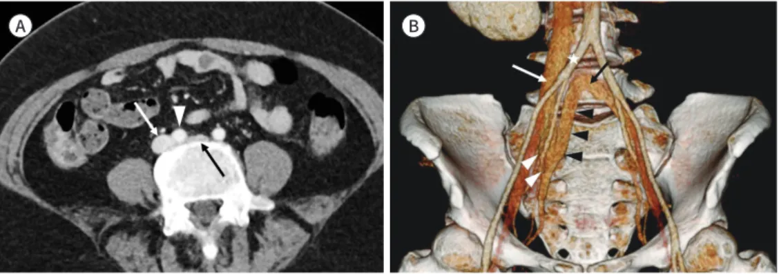
Fig. 1. Iatrogenic iliac vein injury with May-Thurner syndrome in a 53-year-old woman.
A. Axial CT shows compression of the left common iliac vein (black arrow) by the right common iliac artery (arrowhead). The white arrow indicates the right common iliac vein.
B. A 3D reconstructed CT scan shows communicating veins (black arrowheads) from the left common iliac vein (black arrow) to the right internal iliac vein (white arrowheads). The white asterisk and white arrow in- dicate the right common iliac artery and vein, respectively.
flow from the communicating vein, it was superselectively catheterized using 2.2-Fr micro- catheter (Progreat, Terumo, Tokyo, Japan) and was embolized using detachable microcoils such as three Interlock coil (Boston Scientific, Cork, Ireland) (size: 12 mm × 30 cm, 14 mm
× 30 cm, 18 mm × 50 cm) and one Concerto coil (Covidien, Plymouth, MN, USA) (size: 16 mm × 40 cm). Subsequently, the stent-graft were deployed over a 0.035-inch wire to cover the 15 mm distance between the proximal CIV and rupture site. After deployment, the stent- graft was dilated using a 12 mm × 40 mm Mustang balloon to achieve proper sealing. Post- procedure angiogram showed complete exclusion of the ruptured left CIV (Fig. 1D-F). Al- though the bleeding was successfully stopped with stabilized vital signs, the patient expired due to rapid aggravation of pulmonary infection three days after the procedure.
DISCUSSION
Veno-venous ECMO is a rescue therapy in patients with severe respiratory failure. Most of the patients are cannulated via both femoral veins due to the easy access in an emergency condition (3). Insertion of large cannula through the femoral vessels can potentially lead to vascular complications including arterial limb ischemia, retroperitoneal hemorrhage, thromboembolism, dissection, pseudoaneurysm, and the groin infection (4). Thus, the can- nulation should be performed under ultrasonography and/or fluoroscopic guidance. In case of anatomical problem, performing emergent ECMO on the bedside makes it difficult to avoid the vascular complications even with care. This case report described the significance of anatomical abnormality in cannulation process for ECMO. Upon reviewing CT, the patient showed compression of left CIV by overlying right iliac artery, widely recognized as May- Thurner syndrome (5). However, this was not realized at the time of ECMO cannulation be- cause the iliac vein was not only well visualized by ultrasound but also the patient did not present significant symptoms such as deep venous thrombosis or venous hypertension Re- cent imaging study describes that compression of the left iliac vein at arterial crossover point may be present in 66% of the general population without venous symptoms (6). As the inci-
A B
dence of the left iliac vein compression is not rare and ECMO is increasingly used in various clinical scenarios, the probability of encountering vascular complication will inevitably in- crease.
Although the venous flow is low, iliac vein injuries can result in mortality up to 51% (7). In Fig. 1. Iatrogenic iliac vein injury with May-Thurner syndrome in a 53-year-old woman.
C. The left iliac venogram shows contrast extravasation from the rupture site (arrow) of the left common ili- ac vein. A communicating vein (black arrowheads) is noted between the left common iliac vein and the right internal iliac vein (white arrowhead).
D. A balloon tamponade is initially attempted to stop the bleeding using a 10 mm × 60 mm balloon cathe- ter (arrow). Veno-arterial extracorporeal membrane oxygenation cannulas are inserted through the right femoral artery (white arrowhead) and vein (black arrowhead).
E. After the left internal iliac vein is embolized with detachable microcoils to avoid a backflow from the communicating vein, a 12 mm × 6 cm stent-graft (arrowheads) is deployed from the left common iliac vein to the external iliac vein.
F. After placement of the stent-graft, the venogram shows that there is no contrast extravasation and that patency of the left iliac venous system is restored.
C
E
D
F
the past, emergent surgical repair has been the traditional method for the hemodynamically unstable patients because of disruption of the iliac veins (8). However, the endovascular cov- ered stent placement for treatment of traumatic vascular injury has proven to be an effective alternative to open surgical repair (9, 10). In this case, continued hemorrhage was noted even after balloon tamponade. Thus, we chose the covered stent which made immediate cessation of extravasation and restoration of venous patency.
Although it is difficult to predict anatomical variants by bedside observation, the complica- tion could be avoided. This case raises the need to check the CT image for the anatomic vari- ant such as iliac vein compression before urgent cannulation. Also, we recommended using fluoroscopy and ultrasound during cannulation and stopping the procedure if the guidewire is not advanced due to resistance or not visible in the inferior vena cava.
Author Contributions
Conceptualization, L.S.M., W.J.H.; data curation, H.S.J., W.J.H.; formal analysis, H.S.J., L.S.M.; in- vestigation, all authors; resources, all authors; supervision, L.S.M.; visualization, H.S.J., L.S.M.; writ- ing—original draft, H.S.J., L.S.M.; and writing—review & editing, all authors.
Conflicts of Interest
The authors have no potential conflicts of interest to disclose.
REFERENCES
1. Shekar K, Mullany DV, Thomson B, Ziegenfuss M, Platts DG, Fraser JF. Extracorporeal life support devices and strategies for management of acute cardiorespiratory failure in adult patients: a comprehensive re- view. Crit Care 2014;18:219
2. Shin M, Lee JB, Park SB, Park HJ, Kim YS. Multidetector computed tomography of iliac vein variation: prev- alence and classification. Surg Radiol Anat 2015;37:303-309
3. Makdisi G, Wang IW. Extra corporeal membrane oxygenation (ECMO) review of a lifesaving technology. J Thorac Dis 2015;7:E166-176
4. Bisdas T, Beutel G, Warnecke G, Hoeper MM, Kuehn C, Haverich A, et al. Vascular complications in patients undergoing femoral cannulation for extracorporeal membrane oxygenation support. Ann Thorac Surg 2011;92:626-631
5. May R, Thurner J. The cause of the predominantly sinistral occurrence of thrombosis of the pelvic veins.
Angiology 1957;8:419-427
6. Kibbe MR, Ujiki M, Goodwin AL, Eskandari M, Yao J, Matsumura J. Iliac vein compression in an asymptom- atic patient population. J Vasc Surg 2004;39:937-943
7. Wilson RF, Wiencek RG, Balog M. Factors affecting mortality rate with iliac vein injuries. J Trauma 1990;
30:320-323
8. Tannous H, Nasrallah F, Marjani M. Spontaneous Iliac vein rupture: case report and comprehensive review of the literature. Ann Vasc Surg 2006;20:258-262
9. Avery LE, Stahlfeld KR, Corcos AC, Scifres AM, Ziembicki JA, Varcelotti J, et al. Evolving role of endovascular techniques for traumatic vascular injury: a changing landscape? J Trauma Acute Care Surg 2012;72:41-46;
discussion 46-47
10. Zieber SR, Mustert BR, Knox MF, Fedeson BC. Endovascular repair of spontaneous or traumatic iliac vein rupture. J Vasc Interv Radiol 2004;15:853-856
May-Thurner 증후군 환자에서 체외막산소공급 삽관 중 발생한 의인성 장골 정맥 손상: 증례 보고 및 문헌고찰
홍석진 · 이상민* · 원정호
52세 여자 환자가 호흡곤란을 주소로 내원하였다. 환자는 2개월 전에 침습성 흉선종으로 광 범위 흉선절제술을 받았다. 전산화단층촬영에서는 양측 폐에 수많은 소결절들이 발견되었 다. 급성 호흡곤란 증후군이 악화되어 혈관외과의는 정맥-정맥 체외막산소공급(extracorpo- real membrane oxygenation; 이하 ECMO)을 계획하였다. 왼쪽 대퇴동맥을 통해 경피적 삽 관술을 시행하는 도중에 혈관외과의는 혈관 손상을 의심하였고 환자의 활력 징후가 불안정 해졌다. 8일 전에 촬영한 복부 컴퓨터단층촬영에서 May-Thurner 증후군이 있었고, 이후 시 행한 혈관조영술에서 좌측 총장골정맥의 파열이 발견되어 stent-graft를 삽입하여 출혈을 멈 추었다. 8일 전 시행된 복부 전산화단층촬영을 확인해 보니 May-Thurner 증후군이 있었다.
이에 May-Thurner 증후군 환자에서 ECMO 삽관으로 인한 혈관 손상이 발생하여 스텐트 삽 입술을 시행하였던 드문 증례를 보고하고자 한다.
경상대학교 의과대학 경상대학교병원 영상의학과