130
Original Article
Korean Circulation J 2007;37:130-133
ISSN1738-5520
ⓒ2007, The Korean Society of Circulation CASE REPORT
Deep Vein Thrombosis and Pulmonary Embolism in the 8th Week of Pregnancy
Sung Hyung Ha, MD, Rak Kyeong Choi, MD, Ji Won Jang, MD, Dal Soo Lim, MD, Hweung Kon Hwang, MD and Young Moo Ro, MD Department of Internal Medicine, Sejong General Hospital, Bucheon, Korea ABSTRACT
A 29-year-old woman in her 8th week of pregnancy was referred to our hospital for swelling in the lower extremi- ties, rapid onset of dyspnea (1 hr) and pre-syncope. Severe right ventricular dysfunction and moderate pulmonary hypertension were detected using 2-dimentional Doppler echocardiography. In addition, left calf vein and proximal thromboses were detected by venous compression ultrasound imaging. After successful thrombolytic treatment, the patient quickly recovered and was discharged from hospital on subcutaneous low-molecular-weight heparin. She delivered a normal, healthy infant at full-term (40 weeks). (Korean Circulation J 2007;37:130-133)
KEY WORDS:Pulmonary embolism;Thrombolytic therapy;Pregnancy.
Introduction
Pulmonary embolism(PE) among pregnant and post- partum women remains a major cause of maternal morbi- dity and mortality. Deep vein thrombosis(DVT) usually starts in the calf veins, and may extend to the proximal veins and cause a pulmonary embolism.1)2) Available data show that the risk period for deep vein thrombosis and pulmonary embolism is greatest during the third trimes- ter and postpartum.3)4) However, herein is report a rare case of a patient who suffered from deep vein thrombo- sis and pulmonary embolism during the first trimester.
Case
A 29-year-old woman in her 8th week of pregnancy was referred to our hospital due to progressive swelling in the lower extremities over a 2-week period, acute onset of dyspnea(1 hr) and pre-syncope. She had a 5-year hi- story of hypothyroidism and daily Synthyroid(0.1 mg) administration. She had experienced a spontaneous abortion in the 12th week of pregnancy 1 year earlier.
Otherwise, she had no significant past medical history
and was taking no medication upon admission to the hospital. On examination, both her lower legs exhibited swelling and stasis, particularly the left lower leg. She weighed 87.6 kg and was 167 cm tall. Her blood pres- sure, heart rate and respiratory rate were 110/80 mmHg, 130 per minute and regular and 24 per minute, respec- tively. Arterial blood gas analysis on room air gave a pH of 7.43, PaCO2 30.9 mmHg, PaO2 56.5 mmHg, bicar- bonate 20.0 mmol/L, oxygen saturation 90.6% and P (A-a)O2 30.5 mmHg. The prothrombin time, partial thromboplastin time and protein C were all within nor- mal levels. Fibrin/fibrinogen degradation product and D-dimer values were 54.6 μg/mL and 6.7 mg/L, res- pectively, and the B-type natriuretic peptide(BNP) was 354.5 pg/mL. Serum titers of the anticardiolipin anti- body(IgG, IgM) were within normal levels. Antinuclear and antineutrophil cytoplasmic autoantibodies(ANCA) were both negative. No factor V Leiden or prothrom- bin G20210A mutations were found. Deficiencies were observed in both the antithrombin Ⅲ(18.0 mg/dl) and protein S activity(33%). ECG demonstrated sinus tachy- cardia and right axis deviation. A chest roentgenogram revealed mild hilar enlargement. A 2D echocardiogram revealed a normal left ventricular systolic function, di- mension with a dilated right heart, an impaired right ventricular systolic function and an estimated pulmo- nary artery pressure of 55 mmHg(Fig. 1A). Venous ultra- sonography demonstrated a left calf vein thrombosis, proximal vein thrombosis and incompressible posterior tibial veins(Fig. 2A-C). However, a ventilation perfusion
Received:October 25, 2006 Accepted:November 30, 2006
Correspondence:Rak Kyeong Choi, MD,Department of Internal Medicine, Sejong General Hospital, 91-121 Sosa bon 2-dong, Sosa-gu, Bucheon 422-232, Korea
Tel: 82-32-340-1102, Fax: 82-32-340-1236 E-mail: [email protected]
Sung Hyung Ha, et al:Deep Vein Thrombosis and Pulmonary Embolism in Pregnancy·131
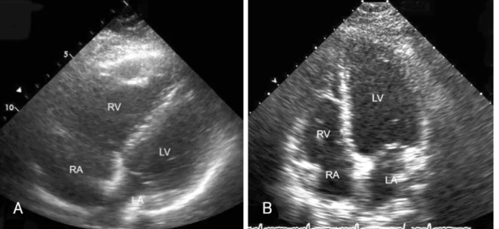
(V/Q) lung scan and CT pulmonary angiography were not performed due to a labor-management dispute at our hospital, and because of concerns about the effects of radiation on the fetal development and the risk of developing breast cancer.5) The patient received throm- bolytic therapy(tissue-type plasminogen activator: 50 mg for 1 hr, 50 mg for 2 hrs) followed by intravenous he- parinization, and was closely monitored in the intensive care unit. She improved dramatically following the thrombolytic therapy, with a marked reduction in the dyspnea and significant improvement in her exercise ca- pacity. Two weeks following the thrombolytic therapy, a 2D echocardiographic assessment revealed significant improvement in the right ventricular function, with re- duction in the right ventricular dimension and an esti- mated pulmonary artery pressure of 37 mmHg(Fig. 1B).
Her D-dimer level decreased to 0.4 mg/L. An obstetric assessment confirmed no evidence of fetal distress, with the baby continuing to progress. The patient responded very well over time and was continued on low molecular
weight heparin(Fraxiparine®: nadroparin, 86 U/kg, SC, 2 times/day). Three weeks later, she made an unevent- ful recovery and was discharged with antepartum pro- phylaxis treatment(Fraxiparine® 3800 U, SC, 2 times/
day). Finally, she delivered a normal, healthy infant at full-term(40 weeks).
Discussion
The overall incidence of deep vein thrombosis or pul- monary embolism during pregnancy has been reported as in 1 in 1000 to 1 in 2000 pregnancies. However, the relatively low incidence of venous thromboembolism (VTE) in Asians has been estimated, and may be rela- ted to a lower prevalence of genetic factors predisposing to VTE, such as factor V Leiden in Asians(0.5%) com- pared to Caucasians(5%).6) Several factors may be impor- tant in the development of VTE during pregnancy, such as hypercoagulability, venous stasis and vascular damage.
Hypercoagulability results from increased levels of coa-
A B
Fig. 1. Transthoracic echocardiograms in the apical 4-chamber view, both before and after the thrombolysis. A dilated, hypokinetic RV, and an increa- sed RV/LV ratio caused by interventricular septal bulging into the LV (A). Two weeks following thrombolytic therapy, note the significant improvement in the right ventricular function, with reduction in the right ventricular dimension (B). RV: right ventricle, LV: left ventricle, RA: right atrium, LA:
left atrium.
Fig. 2. Venous ultrasonography. Cross-section of the posterior tibial vein and artery demonstrating the thrombus (A) and incompressible vein (B). Color Doppler imaging of the femoral vein, showing the thrombus, with no phasic venous flow during respiration (C). A: artery, V: vein, SFA: superficial femoral artery, DFA: deep femoral artery.
C
A B
132·Korean Circulation J 2007;37:130-133
gulation factors(VIII and fibrinogen) during normal preg- nancy. Furthermore, around 40% of pregnancies acquire resistance to activated protein C. Reduction in protein S, the co-factor of protein C, is seen in normal pregnancy, and fibrinolysis is impaired by increased levels of plas- minogen activator inhibitors 1 and 2, which are derived from the placenta.7)8) In addition, one or more heritable or acquired thrombophilias are found in at least 50% of VTE cases during pregnancy. Deficiencies of antithrom- bin, protein C and protein S are uncommon. Factor V Leiden mutation, activated protein C resistance and pro- thrombin G20210A mutation are relatively common, and have been reported to carry an increased risk of VTE.7)9) Vascular damage of pelvic vessels caused by de- livery(vaginally or Caesarean section) probably contri- butes to postpartum venous thrombosis. Venous stasis begins by the end of first trimester, reaches a nadir at 36 weeks as a result of increased vein distensibility and compression of pelvic vessels by the gravid uterus, and takes about 6 weeks to return to the normal non-preg- nant flow rates.10)11) The majority(80-90%) of DVTs in pregnancy develop in the left leg. Compression of the left iliac vein due to the right iliac artery where they cross may explain the preponderance of left leg DVT. The pregnancy specific risk factors that have been demon- strated include prolonged bed rest, advanced maternal age(over 35), family history of thrombosis, parity, pre- vious thrombosis, thrombophilia, previous superficial phlebitis, pre-eclampsia, tobacco use and operative deli- very.12) In this case, obesity, prolonged bed rest and thrombophilia(decreased antithrombin Ⅲ and protein S activity) may have contributed to the pregnancy asso- ciated VTE.
The ventilation-perfusion scan and CT pulmonary angiography are the gold standards for the diagnosis of PE in both pregnant and non pregnant patients. Never- theless, physicians are often reluctant to perform radio- logic studies during pregnancy due to concerns over the effects of radiation on the fetal development. However, the estimated exposure of the fetus to radiation during these examinations is small. In most studies, exposure to radiation of less than 50,000 μGy(5 rad) has not been associated with significant risk of fetal injury.13) Therefore, many experts strongly recommend that venous throm- boembolism is aggressively investigated, with definitive studies, whenever suspected in a pregnant patient. Pre- sently, noninvasive echocardiographic and Doppler ana- lysis of the right heart dimension and RV function nei- ther definitively confirmation nor exclude suspected pulmonary embolism(PE). However, a typical echocardio- graphic picture of hemodynamically significant PE in- cludes dilated, hypokinetic RV, an increased RV/LV ratio caused by interventricular septal bulging into the LV, dilated proximal pulmonary arteries, increased velocity of the jet of tricuspid regurgitation(usually in the range
of 3-3.5 m/sec), a disturbed flow velocity pattern in the RV outflow tract and a dilated inferior vena cava(not collapsed on inspiration).14-16) The initial management of VTE confirmed during pregnancy necessitates im- mediate anticoagulation with unfractionated heparin or low molecular weight heparin(LMWH). According to available data, unless absolutely contraindicated, throm- bolysis should be given to all patients with a massive PE (shock and/or hypotension). In patients with normal blood pressure, normal tissue perfusion and clinical or echocardiographic evidence of RV dysfunction(submas- sive PE), thrombolytic therapy may be given in the ab- sence of contraindications. In our case, thrombolytic therapy, with immediate full dose anticoagulation, was decided for faster hemodynamic improvement compared to heparin alone, due to evidence of acute cor pulmo- nale, coinciding with high clinical suspicion of acute onset submassive PE, without previous cardiac or res- piratory disease. She received intravenous heparin for 5 days, and Subcutaneous LMWH was then continued, with a fulldose regimen, for 3 weeks. At the time of discharge, the LMWH was reduced to a half-dose regi- men, which was continued throughout the remainder of the pregnancy. Fortunately, she responded very well, without any serious side-effects.
Of the women with a history of VTE during preg- nancy, the incidence of recurrence during subsequent pregnancies has been estimated as 4 to 15 percent.17)18) The risk of recurrent VTE is the same for patients with proximal DVT or PE, but the risk of a fatal PE is 2- to 3-fold higher after an episode of PE than of DVT.19) Therefore, most experts recommend that pregnant wo- men at risk of VTE receive more aggressive prophylaxis than that traditionally recommended.
In conclusion, because of the gravity of maternal mor- bidity and mortality, the need for prolonged heparin therapy during pregnancy and prophylaxis during sub- sequent pregnancies, physicians should not be reluc- tant to perform definitive radiologic studies whenever VTE is suspected in a pregnant woman. Further studies will be required to make a confident recommendation in pregnancy associated VTE.
REFERENCES
1) Cogo A, Lensing AW, Prandoni P, Hirsh J. Distribution of throm- bosis in patients with symptomatic deep vein thrombosis: impli- cations for simplifying the diagnostic process with compression ultrasound. Arch Intern Med 1993;153:2777-80.
2) Moser KM, LeMoine JR. Is embolic risk conditioned by location of deep venous thrombosis? Ann Intern Med 1981;94:439-44.
3) Aaro LA, Juergens JL. Thrombophlebitis associated with preg- nancy. Am J Obstet Gynecol 1971;109:1128-36.
4) Heit JA, Kobbervig CE, James AH, Petterson TM, Bailey KR, Melton LJ 3rd. Trends in the incidence of venous thromboembo- lism during pregnancy or postpartum: a 30-year population-based study. Ann Intern Med 2005;143:697-706.
Sung Hyung Ha, et al:Deep Vein Thrombosis and Pulmonary Embolism in Pregnancy·133
5) Remy-Jardin M, Remy J. Spiral CT angiography of the pulmonary circulation. Radiology 1999;212:615-36.
6) Richard HW. The epidemiology of venous thromboembolism. Cir- culation 2003;107(23 Suppl 1):I4-8.
7) Clark P, Brennand J, Conkie JA, McCall F, Greer IA, Walker ID.
Activated protein C sensitivity, protein C, protein S and coagula- tion in normal pregnancy. Thromb Haemost 1998;79:1166-70.
8) Greer IA. Haemostasis and thrombosis in pregnancy. In: Bloom AL, Forbes CD, Thomas DP, Tuddenham EG, editors. Haemos- tasis and Thrombosis. Edinburgh: Churchill Livingstone; 1994.
p.987-1015.
9) Dahlback B, Hillarp A, Rosen S, Zoller B. Resistance to activa- ted protein C, the FV: Q506 allele, and venous thrombosis. Ann Hematol 1996;72:166-76.
10) Macklon NS, Greer IA, Bowman AW. An ultrasound study of gestational and postural changes in the deep venous system of the leg in pregnancy. Br J Obstet Gynaecol 1997;104:191-7.
11) Macklon NS, Greer IA. The deep venous system in the puerpe- rium: an ultrasound study. Br J Obstet Gynaecol 1997;104:198-200.
12) Rodger MA, Walker M, Wells PS. Diagnosis and treatment of venous thromboembolism in pregnancy. Best Pract Res Clin Hae- matol 2003;16:279-96.
13) Ginsberg JS, Hirsh J, Rainbow AJ, Coates G. Risks to the fetus of
radiologic procedures used in the diagnosis of maternal venous thromboembolic disease. Thromb Haemost 1989;61:189-96.
14) Nazeyrollas P, Metz D, Jolly D, et al. Use of transthoracic Dopp- ler echocardiography combined with clinical and electrocardio- graphic data to predict acute pulmonary embolism. Eur Heart J 1996;17:779-86.
15) McConnell MV, Solomon SD, Rayan ME, Come PC, Goldhaber SZ, Lee RT. Regional right ventricular dysfunction detected by echocardiography in acute pulmonary embolism. Am J Cardiol 1996;78:469-73.
16) Torbicki A, Kurzyna M, Ciurzynski M, et al. Proximal pulmonary emboli modify right ventricular ejection pattern. Eur Respir J 1999;13:616-21.
17) Tengborn L, Bergqvist D, Matzsch T, Bergqvist A, Hedner U.
Recurrent thromboembolism in pregnancy and puerperium: is there a need for thromboprophylaxis? Am J Obstet Gynecol 1989;
160:90-4.
18) Badaracco MA, Vessey MP. Recurrence of venous thromboem- bolic disease and use of oral contraceptives. Br Med J 1974;
1:215-7.
19) Murin S, Romano PS, White RH. Comparison of outcomes after hospitalization for deep venous thrombosis or pulmonary embo- lism. Thromb Haemost 2002;88:407-14.