Pulmonary vein stenosis is a rare congenital disease that occurs almost exclusively in young children with or without various forms of congenital heart disease (1).
Pulmonary vein stenosis in the adults is very rare, and the small number of reported cases has often been asso- ciated with mediastinal processes such as neoplasms or fibrosing mediastinitis (2, 3). Recently, stenosis has ap- peared as a complication of radiofrequency ablation pro- cedures around the pulmonary veins (4). It is a poor prognostic disease and survival to adulthood without treatment rarely occurs because the symptom emerges in infants with accompanying progressively worsening
pulmonary hypertension (5). Herein, the present au- thors report an asymptomatic patient who was diag- nosed as having congenital isolated pulmonary vein stenosis with mild pulmonary hypertension.
Case Report
A 31-year-old married Korean man with lower back pain that underwent surgery to repair a herniated inter- vertebral disc (HIVD) L5-S1 was transferred to our clinic because of a chest radiograph showing multiple small nodules with irregular pleural thickening in both upper lungs. The patient had no other previous diseases in- cluding pulmonary tuberculosis, chest trauma, or con- genital anomaly and was an office worker with no per- sonal or family history of exposure to drugs or smoking.
Upon physical examination, the man’s vital signs were stable and he appeared to be healthy. He also did not complain of any other respiratory symptoms. His chest had symmetric expansion without abnormal lung
Asymptomatic Primary Isolated Pulmonary Vein Stenosis in an Adult: A Case Report1
Ji Hyun Kim, M.D., Ho Sung Lee, M.D., Jae Sung Choi, M.D., Ju Ock Na, M.D., Yong Hoon Kim, M.D., Sung Shick Jou, M.D.2, Ki Hyun Seo, M.D.
1Department of Internal Medicine, College of Medicine, Soonchunhyang University, Cheonan, Korea
2Department of Diagnostic Radiology, College of Medicine, Soonchunhyang University, Cheonan, Korea
Received April 16, 2010 ; Accepted July 2, 2010
Address reprint requests to : Ki Hyun Seo, M.D., Department of Internal Medicine, Soonchunhyang University Cheonan Hospital, 23-20, Bongmyung-dong, Dongnam-gu, Cheonan 330-721, Korea.
Tel. 82-41-570-3665 Fax. 82-41-574-5762 E-mail: [email protected]
A 31-year-old man without respiratory symptoms was transferred to our clinic with incidentally detected small nodular densities in both the upper lung zones on chest ra- diography. Chest computed tomography and pulmonary angiography demonstrated that the entrance of the right inferior pulmonary vein to the left atrium was complete- ly blocked, and the venous return of the right lower lobe was achieved through the right superior pulmonary vein with a tortuous venous collateral complex in the venous phase. With echocardiography, mild pulmonary hypertension was detected. Here, we present an asymptomatic adult with isolated stenosis of the pulmonary vein with chronic compensation by venous collateral circulation in spite of mild pulmonary hy- pertension.
Index words :Pulmonary veins
Pulmonary Veno-Occlusive Disease Adult
sounds. His heart beat was normal and regular, and there was no cyanosis or finger clubbing; all other find- ings were unremarkable. His white blood count was 9,810 cells/mm3. Prothrombin time, cardiac markers, D- dimer and arterial blood gas analysis at room air were within the normal range and no abnormal electrocardio- gram readings were observed. Upon transthoracic echocardiography, mild pulmonary hypertension (sys- tolic pulmonary artery pressure 38.3 mmHg) (Fig. 1) was detected, but LV systolic function was normal and no structural abnormality was found except for mild tri- cuspid regurgitation.
A chest radiograph showed small nodular densities with irregular pleural thickening in both upper lung zones (Fig. 2). A spiral computed tomography pul- monary angiogram showed no evidence of a throm- boembolism, but demonstrated a markedly dilated right superior pulmonary vein without connection to the left atrium and multiple nodules, suggesting collateral vas- cular structures in the pleural area of both upper lung zones and in the superior segment of right lower lobe (Fig. 3). Thus, small nodular densities with irregular pleural thickening in chest radiography seemed to be collateral vascular structures on the chest CT scan. A volume rendering image showed that the right inferior pulmonary vein drained into the superior pulmonary vein through an intrapulmonary venous connection (Fig. 4). Upon undergoing a selective angiogram of right lower lobar pulmonary angiogram, the entrance of the right inferior pulmonary vein to the left atrium was found to be completely blocked, and the venous return of the right lower lobe was achieved through the right superior pulmonary vein with tortuous venous collater- al complex around the right hilar region in the venous
phase (Fig. 5). The patient was discharged without spe- cific treatment on the eighth post-operative day for HIVD because he had remained asymptomatic with an unlimited exercise tolerance.
Discussion
Pulmonary vein stenosis is a rare congenital disease seen in neonates and children comprises only 0.4% of all congenital heart disease cases (1). Affected patients most often become symptomatic in the first few months to years of life, and frequently have 1 or more additional cardiac anomalies. Approximately one half of patients
A B
Fig. 1. Echocardiogram shows only turbulent jet flows (arrow) originating from the pulmonary veins and entering the left atrium on apical four chamber view in figure 1A and mild pulmonary hypertension with 38.3 mmHg systolic pulmonary artery pressure (RA pressure 5 mmHg + maximal pressure gradient 33.3 mmHg) in figure 1B.
Fig. 2. Chest radiograph shows small nodular densities (ar- rows) with pleural thickening (arrow heads) in both upper lung zones and a dilated pulmonary vascular structure in the right hilar region (black arrow).
with primary pulmonary vein stenosis have some type of associated cardiac defect (6). Primary pulmonary vein stenosis is believed to result from the abnormal incorpo- ration of the common pulmonary vein into the left atri- um in the later stages of cardiac development (1). Recent studies have documented progression from normal pul- monary venous flow patterns in a significant number of patients who later developed progressive pulmonary vein stenosis (7). The evaluation of stenotic pulmonary veins is necessary in any young patient with severe pul- monary hypertension.
Pulmonary vein stenosis in adults is even rarer, and the small number of reported cases has often been asso- ciated with mediastinal processes such as neoplasms, sarcoidosis, or fibrosing mediastinitis (2, 3), and recently
has been identified as a complication of radiofrequency ablation procedures for atrial fibrillation (4). Lung parenchyma shows interstitial edema, alveolar edema, or fibrosis, resulting from long-standing insufficient ve- nous drainage in most pediatric patients (8, 9).
However, no definite pulmonary parenchymal abnor- malities in our patient were noted. Small nodular densi- ties with pleural thickening on chest radiograph were likely to be a collateral vascular structure on chest CT scans. A markedly dilated right superior pulmonary vein and collateral vascular structure in the superior segment of the right lower lobe without connection to left atrium on CT scan, suggests the complete compen- sation of anomalous venous drainage. The final diagno- sis could be made by angiography. Similar to the present
A B
C D
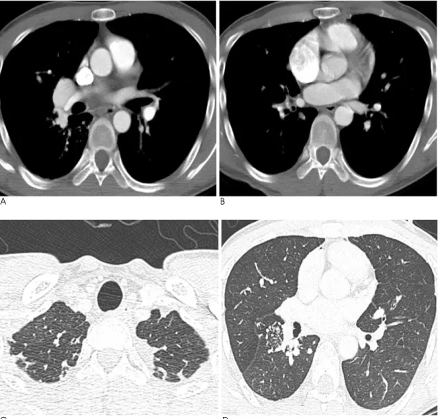
Fig. 3. Contrast-enhanced CT scan shows markedly a dilated right superior pulmonary vein (A) without connection to the left atri- um (B), and multiple nodules suggesting collateral vascular structures in the pleural area of both upper lung zones (C) and in the su- perior segment of right lower lobe (D).
case, a case described as isolated pulmonary vein steno- sis associated with full compensation of intrapulmonary venous drainage was reported by Saida (10). However, only a tortuous pulmonary vessel in the parahilar region of the right lung field on chest film in that study was noted, compared with collateral vascular structures in the peripheral region and dilated pulmonary vessels in the right hilar area region in our study. In addition, Saida didn’t mention whether the patient had pul- monary arterial hypertension or not. To knowledge of the presence of pulmonary arterial hypertension is very important because the severity of pulmonary hyperten- sion is part of the assessment of the condition’s progno- sis
Patients with the pediatric form of congenital pul- monary vein stenosis have a very poor prognosis.
Survival to adulthood without treatment rarely occurs because the symptom emerges in infants with accompa- nying progressively worsening pulmonary hypertension (5). The prognosis depends on the severity of the associ- ated cardiac disease and the number of the stenosed pul- monary veins (7). The cause of death is usually a pul- monary hypertensive crisis, recurrent pulmonary infec- tion, or massive hemoptysis. Although the precise natur- al history of milder forms of congenital pulmonary vein stenosis is not entirely clear, patients with only 1 or 2 pulmonary veins involved have a significantly more be- nign course. Breinholt (7) found a mortality rate of 83%
in pediatric patients with 1 or 2 stenosed pulmonary
veins. Our patient has no cardiac anomaly in spite of mild pulmonary hypertension and only a single pul- monary vein stenosis with collateral circulation. Thus, we consider that our patient would have a better prog- nosis. To our knowledge, asymptomatic patients with isolated pulmonary vein stenosis associated with chron- ic compensation through venous collateral drainage have been very extremely rare, and our patient is to date the oldest adult among those with a diagnosis of pul- monary vein stenosis. More asymptomatic patients of isolated pulmonary vein stenosis with chronic compen- sation could be diagnosed as a result of increased aware- ness and improvements in noninvasive imaging modali- ties.
References
1. Edward J. Congenital stenosis of the pulmonary veins: pathologic and developmental considerations. Lab Invest 1960;9:46-66 2. Omasa M, Hasegawa S, Bando T, Okano Y, Otani H, Nakshima Y,
et al. A case of congenital pulmonary vein stenosis in an adult.
Respiration 2004;71:92-94
3. Tan CW, Munfakh N, Helmcke F, Abourahma A, Caspi J, Glancy DL. Congenital bilateral pulmonary venous stenosis in an adult:
diagnosis by echo-Doppler. Catheter Cardiovasc Interv 2000;49:328- 330
4. Saad EB, Marrouche NF, Saad CP, Ha E, Bash D, White RD, et al.
Pulmonary vein stenosis after catheter ablation of atrial fibrilla- Fig. 4. Volume rendering image shows that the right inferior
pulmonary vein drains to the superior pulmonary vein an through intrapulmonary venous connection (arrows).
Fig. 5. Selective angiogram of right lower lobar pulmonary artery shows that the entrance of the right inferior pulmonary vein to the left atrium was completely blocked (arrow), and the venous return of right lower lobe was achieved through the right superior pulmonary vein with tortuous venous collat- eral complex around the right hilar region in venous phase.
tion: emergence of a new clinical syndrome. Ann Intern Med 2003;138:634-638
5. Van Son JA, Danielson GK, Puga FJ, Edwards WD, Driscoll DJ.
Repair of congenital and acquired pulmonary vein stenosis. Ann Thorac Surg 1995;60:144-150
6. Latson LA, Prieto LR. Congenital and acquired pulmonary vein stenosis. Circulation 2007;115:103-108
7. Breinholt JP, Hawkins JA, Minich L, Tani LY, Orsmond GS, Ritter S, et al. Pulmonary vein stenosis with normal connection: associat- ed cardiac abnormalities and variable outcome. Ann Thorac Surg
1999;68:164-168
8. Sun CC, Doyle T, Ringer RE. Pulmonary vein stenosis. Hum Pathol 1995;26:880-886
9. Belcourt CL, Roy DL, Nanton MA, Finley JP, Gillis DA, Krause VW, et al. Stenosis of individual pulmonary veins: radiologic find- ings. Radiology 1986;161:109-112
10. Saida Y, Eguchi N, Mori K, Tanaka YO, Ishikawa S, Itai Y. Isolated pulmonary vein stenosis associated with full intrapulmonary com- pensation. AJR Am J Roentgenol 1999;173:961-962
대한영상의학회지 2010;63:345-349
성인에서 무증상인 선천성 고립폐정맥협착증 1예1
1순천향대학교 부속병원 천안병원 내과
2순천향대학교 부속병원 천안병원 영상의학과
김지현∙이호성∙최재성∙나주옥∙김용훈∙조성식2∙서기현
호흡기 증세가 없는 31세 남자가 우연히 발견된 양쪽 폐상부에 작은 결절들이 관찰되어 내원하였다. 심초음파에서 경도의 폐고혈압이 확인되었다. 흉부 CT에서 현저한 우상부 폐정맥의 확장이 있었고 우하부 폐정맥은 좌심방과 연 결되지 않은 채 무수한 곁순환을 이루고 있었다. 폐혈관조영술에서는 우하부 폐정맥이 좌심방으로 가는 입구가 완전 히 막혀 있고 구불구불한 곁순환을 이루며 우상부 폐정맥으로 이어져 좌심방으로 연결되어 있었다. 결론적으로 저자 들은 경도의 폐고혈압이 있었지만, 만성적인 보상으로 무증상인 선천성 고립폐정맥협착증을 진단되어 보고하는 바 이다.