서 론
관상동맥 우회술 후 우회 이식편의 열림에 대한 가장 정 확한 판정 방법은 동맥을 통한 침습적인 관상동맥 조영술 이다. 그러나 수술 직전에 시행한 침습적인 관상동맥 조
영술을 이식편의 열림을 확인하기 위해 다시 시행하는 것 은 환자에게 부담이 되고 시술에 따른 위험도 발생할 수 있다[1]. 관상동맥 우회술 후 이식편의 조기열림을 판정하 기 위한 비침습적인 방법에는 여러 가지가 있다[2-5]. 그 중 CT 조영술은 최근 여러 개의 검출기(detector)를 가진
관상동맥우회술 후 CT 조영술을 이용한 이식편의 조기 열림의 판정
최종범*․이미경*․류대웅*․최순호*
Determination of Early Graft Patency Using CT Angiography after Coronary Artery Bypass Surgery
Jong Bum Choi, M.D.*, Mee Kyung Lee, M.D.*, Dae Woong Ryu, M.D.*, Soon Ho Choi, M.D.*
Background: CT angiography is now available to evaluate the early graft patency after coronary bypass surgery.
We investigated whether patency or occlusion of the bypass grafts can be visualized by CT angiography and what factors effect the visuality. Material and Method: Fifty patients underwent scanning with a 4-slice computed tomographic scanner (Somatom Volume ZoomTM; Siemens, Germany) before being discharged after coronary artery bypass grafting. To evaluate graft patency and relationship between the quality of graft image and the characteristics of the diseased coronary vessels, 50 internal thoracic artery grafts, 18 radial artery grafts, and 56 vein grafts were included in this study. Result: All vein grafts (24 grafts; 32 anastomoses) to left coronary artery system were well visualized, but 3 grafts (4.7%) of 30 vein grafts (35 anastomoses) to right coronary artery system were not visualized. The latter was also occluded in invasive coronary angiographic study. Thirty-nine (78%) internal thoracic artery grafts were well visualized, 8 (16%) faintly visualized, and 3 (6%) not visualized, but all the internal artery grafts were well patent in invasive coronary angiographic study. Conclusion: Unvisualized vein grafts in CT angiography means occlusion of the grafts, but unvisualized arterial grafts in CT angiography may not mean occlusion of the graft but result from competitive flow between the graft and coronary artery. To confirm patency of the unvisualized arterial grafts, invasive coronary angiography is needed.
(Korean J Thorac Cardiovasc Surg 2004;37:570-577) ꠏꠏꠏꠏꠏꠏꠏꠏꠏꠏꠏꠏꠏꠏꠏꠏꠏꠏꠏꠏꠏꠏꠏꠏꠏꠏꠏꠏꠏꠏꠏꠏꠏꠏꠏꠏꠏꠏꠏꠏꠏꠏꠏꠏꠏꠏꠏꠏꠏꠏꠏꠏꠏꠏꠏꠏꠏꠏꠏꠏꠏꠏꠏꠏꠏꠏꠏꠏꠏꠏꠏꠏꠏꠏꠏꠏꠏꠏꠏꠏꠏꠏꠏꠏꠏꠏꠏꠏꠏꠏꠏꠏ Key words: 1. Coronary artery bypass
2. Computed tomography 3. Coronary angiography 4. Vascular patency
*원광대학교 의과대학 흉부외과학교실
Department of Thoracic and Cardiovascular Surgery, Wonkwang University School of Medicine, Iksan, Jeonbuk
†이 논문 내용은 2003년 대한흉부외과학회 제 35차 추계학술대회 포름에서 구연되었음.
‡이 논문은 2004년 원광대학교의 교비 지원에 의해서 수행됨.
논문접수일:2004년 3월 27일, 심사통과일:2004년 5월 17일
책임저자 : 최종범 (570-711) 전북 익산시 신용동 344-2번지, 원광의대부속병원 흉부외과 (Tel) 063-850-1275, (Fax) 063-857-0252, E-mail: [email protected] 본 논문의 저작권 및 전자매체의 지적소유권은 대한흉부외과학회에 있다.
CT기가 나오면서 검사시간이 빨라지고 안전하여 환자에 게 주는 정신적 신체적 부담도 거의 없어 수술 후 이식편 의 열림을 조사하기 위해서 많이 이용되고 있다. 본 저자 들은 관상동맥 우회술 후 조기에 이러한 CT 조영술을 이 용하여 이식편의 열림을 판정할 수 있는지를 조사하고 조 영에 미치는 인자들을 찾아보았다.
대상 및 방법
2003년 1월 1일부터 2003년 7월 31일까지 관상동맥 우 회술을 받은 50예를 대상으로 하였다. 남자가 31예(62%) 이고 여자가 19예(38%)였으며 평균연령은 62.6±7.0세(범
위, 46∼75세)였다. 29예(58%)는 과거나 최근에 심근경색 증을 앓았고, 관상동맥 병변의 정도는 30예(60%)에서 3-혈 관질환, 7예(14%)에서 2-혈관질환, 13예(26%)에서 좌주관 동맥 병변과 부가병변을 가지고 있었다. 30예(60%)에서는 체외순환 및 심정지 상태(on-pump)에서 수술했고 20예 (40%)에서는 심박동 상태(off-pump)에서 수술하였다. 환자 당 평균 원위부 문합수는 2.86±0.83개소였고, on-pump 수 술 환자에서는 평균 2.75±0.71개소, off-pump 수술 환자에 서는 평균 2.93±0.91개소였다(p=0.57). 관상동맥 우회술 외에 동반수술로서 5예(10%)에서 승모판막 성형술, 2예 (4%)에서 승모판막 치환술을 시행하였다(Table 1).
우회이식편으로는 내흉동맥편(좌 49편, 우 1편), 요골동 맥편(18편) 및 대퇴부위의 복재정맥편을 사용하였다. 좌전 하행지에는 좌 내흉동맥편만을 문합했고, 80% 이상의 회 선지 병변에는 내흉동맥에 y-복합된 요골동맥편을 문합했 으며, 80% 미만의 회선지 병변에는 정맥편을 이용하였다.
우관상동맥 병변에는 모두 정맥편을 사용하였다.
요골동맥편 18편 중 16편은 내흉동맥편에 붙여 Y-복합 문합편을 만들어 사용하였고 1편은 정맥편을 두건으로 하 여 상행대동맥에 연결하였으며 다른 1편은 우 내흉동맥에 연결하여 I-형 이식편으로 사용하였다. 요골동맥 18편으로 21부위의 원위부 문합을 하였고 이 중 19문합은 회선지 분지에, 2문합은 우 후하행지(right posterior descending artery)에 시행되었다(Table 2).
정맥편은 전체 환자의 84%(42예)에서 사용되었고, 환자 당 평균 1.33±0.57편(평균 1.62±0.88문합)이 사용되었다.
좌 관상동맥 병변에는 24편의 정맥편을 사용했으며 이 중 7편에서 연속 문합하여 총 32문합이 이루어졌으며, 우 관
2-vessel disease 7 (14%)
Left main lesion only 1 (2%) Left main+other lesion 12 (24%) Operation
On-pump/off-pump 30/20 (60/40%)
Average grating no 2.86±0.83
Off-pump 2.75±0.71
On-pump 2.93±0.91 (p=0.57)
Combined procedures 7 (14%)
Mitral valve repair 5 Mitral valve replacement 2
ꠏꠏꠏꠏꠏꠏꠏꠏꠏꠏꠏꠏꠏꠏꠏꠏꠏꠏꠏꠏꠏꠏꠏꠏꠏꠏꠏꠏꠏꠏꠏꠏꠏꠏꠏꠏꠏꠏꠏꠏꠏꠏꠏꠏꠏꠏꠏꠏꠏꠏꠏꠏꠏꠏꠏ MI=Myocardial infarction.
Table 2. Shapes and graft sites of radial artery grafts
ꠚꠚꠚꠚꠚꠚꠚꠚꠚꠚꠚꠚꠚꠚꠚꠚꠚꠚꠚꠚꠚꠚꠚꠚꠚꠚꠚꠚꠚꠚꠚꠚꠚꠚꠚꠚꠚꠚꠚꠚꠚꠚꠚꠚꠚꠚꠚꠚꠚꠚꠚꠚꠚꠚꠚ Graft shape of total 18 graftsY-composite graft to LITA 16 grafts
Vein hood 1 graft
I-graft to RITA 1 graft
Graft sites of total 21
to RPDA 2 sites
to OM (±RPDA or PL) 19 sites
ꠏꠏꠏꠏꠏꠏꠏꠏꠏꠏꠏꠏꠏꠏꠏꠏꠏꠏꠏꠏꠏꠏꠏꠏꠏꠏꠏꠏꠏꠏꠏꠏꠏꠏꠏꠏꠏꠏꠏꠏꠏꠏꠏꠏꠏꠏꠏꠏꠏꠏꠏꠏꠏꠏꠏ
*LITA=Left internal thoracic artery; RITA=Right internal thoracic artery; RPDA=Right posterior descending artery; OM=
Obtuse marginal artery; PL=Posterolateral branch.
(±RPDA or PL) 24 grafts (32 anastomoses) Right coronary system 32 grafts (36 anastomoses) to right coronary artery 9 grafts
to right posterior descending 23 grafts artery
ꠏꠏꠏꠏꠏꠏꠏꠏꠏꠏꠏꠏꠏꠏꠏꠏꠏꠏꠏꠏꠏꠏꠏꠏꠏꠏꠏꠏꠏꠏꠏꠏꠏꠏꠏꠏꠏꠏꠏꠏꠏꠏꠏꠏꠏꠏꠏꠏꠏꠏꠏꠏꠏꠏꠏ
*RPDA=Right posterior descending artery; PL=Posterolateral branch.
상동맥에는 32정맥편으로 36문합을 시행하고 주로 우 후 하행지에 문합되었다(Table 3).
1) 수술 후 CT 조영술 및 우회이식편의 영상 판정 4개의 검출기를 가진 컴퓨터 단면촬영기(4 detector-row computed tomography, MDCT; multi-slice computed tomo- graphy, MSCT; Siemens, Forchheim, Germany)를 이용하여 심전도 동조하에 관상동맥 이식편의 영상을 얻었다. 하나 의 심장 주기에서 얻어지는 영상은 갠트리 2/3에 해당되 는 데이터로 만들어지고 실제 영상에 기여하는 데이터는 180도 회전(회전시간 250 ms) 시 얻어진다. 갠트리 회전속 도는 500 ms이고 1.25 mm절편 두께로 4개의 영상을 동시 에 얻어 전체 심장 영상을 얻는데 약 70초가 걸렸다. 그리 고 좀 더 정확한 영상을 얻기 위하여 이 시간 동안 환자가 숨을 참을 수 있는 정도까지 가능한 한 길게 참도록 하였 다. 이때 조영제 170 mL (농도 400 mg/iodine mL) 이상을 4 mL/sec의 속도로 빠르게 주입하였다. 이식편의 영상은 R-R 간격의 40∼70%에서 후향적으로 재구성하였으며 재 구성된 data를 컴퓨터에 옮겨 3D-VertuosuTM (Siemens, For- chheim, Germany) 프로그램으로 3차원 영상을 얻었다.
CT 조영술은 수술 후 평균 10.4±3.2일(범위, 6∼13일) 에 시행되었으며, 50개의 내흉동맥편, 18개의 요골동맥편, 56개의 대퇴정맥편의 조영 및 개존 상태를 조사하였다.
우회이식편의 조영상태를 ‘조영 안 됨’, ‘희미함’, ‘양호’,
‘우수’의 4개로 분류했고 ‘희미함’은 조영혈류가 끊기고 좁아진 것처럼 조영된 상태를 정의했고 ‘양호함’은 조영 혈관의 두께는 만족하나 혈류의 끊김이 있는 것을 정의하
였다(Fig. 1). 이식편들의 조영상태와 열림상태의 관계를 조사하고, 관상동맥(문합된 혈관)의 직경, 부행혈류(‘많음’,
‘적음’, ‘없음’) 및 협착병변의 정도(<70%, 70∼90%, >90%) 등이 이식편의 조영상태에 영향을 미치는지 조사하였다.
문합될 관상동맥의 직경은 수술 시 사용된 션트 카뉼라의 크기로 측정했다. 부행혈류의 ‘많음’은 관상동맥 조영술에 서 후하행지와 후하행지의 중격분지가 조영되는 경우를 정의했고 부행혈류의 ‘적음’은 후하행지의 일부만 조영되 는 경우를 정의했다.
CT 조영술에서 정상적인 조영을 보이지 않는 동맥편이 나 정맥편은 대퇴동맥을 통한 침습적인 관상동맥 조영술 을 퇴원 전(CT 조영술 후 수일 내)에 시행하여 열림을 다 시 판정하였다.
2) 결과에 대한 통계처리
연속변수는 평균 ± 표준편차로 나타냈고 ‘SPSS 7.5 한 글 프로그램’을 이용하여 통계처리를 했다. 연속변수의 비 교를 위해서 비모수적 방법으로 Mann-Whitney U-검정을 이용하였으며 비연속변수에 대해서는 3×3 이상의 교차 분석(카이제곱 검정)으로 선형대선형 결합(linear by linear association)으로 경향분석을 하였다.
결 과
좌 내흉동맥편의 조영상태에서 50편 중 32편(64%)은 우 수, 7편(14%)은 양호, 8편(16%)은 희미한 조영을 보였고, 3편(8%)은 전혀 조영이 되지 않았다. 전혀 조영되지 않은
Fig. 1. CT angiographic findings of internal thoracic artery grafts. The internal thoracic artery grafts (black arrows) were well (A),
fairly (B) and faintly (C) visualized.A B C
3편은 침습적인 관상동맥 조영술에서 잘 조영되어 열려있 음을 보여주었다(Fig. 2). 내흉동맥의 조영상태는 내흉동맥 의 협착병변의 정도와 관계가 있었고(Crosstable: Linear by Linear Association p<0.001), 부행혈류의 정도와는 무관하 였다(Crosstable: Linear by Linear Association p=0.572) (Table 4, 5).
요골동맥편 18편(21문합) 중 1편(y-형)이 CT 조영술에서 조영되지 않았으나 침습적인 관상동맥 조영술에서는 열
려 있었고 상경혈류를 보였다(Fig. 3).
좌관상동맥에 문합한 24 정맥편(32문합)은 CT 조영술에 서 모두(100%) 조영되었고 우 관상동맥계에 문합한 32 정 맥편(36문합) 중 29편(91%)은 잘 조영되었으나 3편은 조 영되지 않았다. CT 조영술에 조영되지 않은 이 3편의 정 맥편은 침습적인 관상동맥 조영술에서 이식편이 완전히 폐쇄된 소견을 보였으며 모두 우 하행지에 문합했던 정맥 편으로 문합된 우후하행지의 직경은 1.5 mm 이하였다
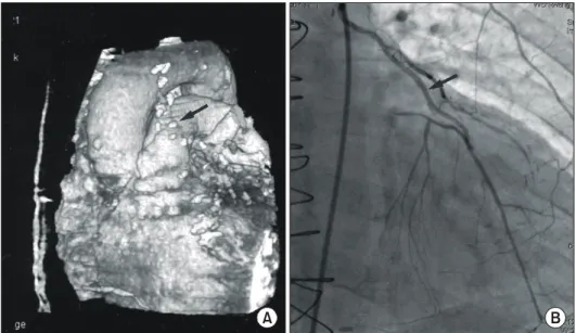
Fig. 2. In CT angiography, the
internal thoracic artery graft (black arrow) was not visualized and only the metal clips on the arterial graft were shown (A). In invasive coronary angiography, the internal thoracic artery graft showed good patency (B).A B
Fig. 3. In CT angiography, the internal thoracic artery graft (long black arrow) was not visible and the radial artery graft (black
arrow head) that was y-grafted to the internal thoracic artery was not also seen (A). In invasive coronary angiography, both arterial grafts were well shown and patent, but the flow in the radial arterial graft was relatively slow (B).A B
(Fig. 4). 정맥편의 이러한 폐쇄는 병변을 가진 관상동맥의 직경과 부행혈류 정도와 밀접한 관계를 보였다(Linear by linear association에서 각각 p=0.003, p<0.001)(Table 6).
고 찰
관상동맥우회술 후 이식도관의 열림을 판정하는 데에 는 관상동맥 조영술이 가장 정확한 방법이다. 그러나 수 술 전에 진단 목적으로 침습적인 관상동맥 조영술을 시행 한 환자에서 수술 직후 이식편의 열림을 확인하기 위해서 다시 침습적인 검사를 시행한다는 것은 아직 혈역학 상태 나 정신적으로 안정이 덜 된 환자에게 부담이 될 수도 있 고 위험도 따를 수 있다[1]. 따라서 수술 후 퇴원 전에 이
식편의 열림을 판정할 수 있는 비침습적인 방법이 간단하 고 더 유용할 수 있다. 이 중에서도 4개의 검출기를 가진 CT (multi-detector CT, MDCT; multi-slice CT, MSCT)는 다 른 비침습적인 검사법인 MRI 등에 비해 검사시간이 짧고 간단하며 안전하다는 장점을 가지고 있어[4,6,7] 관상동맥 우회로술 후 이식편의 주행, 열림 및 문합상태를 쉽게 파 악할 수 있다. CT 조영술로 이식편의 조영상태를 침습적 인 관상동맥 조영술에 근거하여 그 정확도를 측정한 결과 그 민감도(sensitivity)는 86% 이상이고 특이도(specificity) 도 100%를 보이고 있어서[8,9] CT 조영술로 열림이 확인 된 이식편에 대해서는 더 이상의 침습적인 관상동맥 조영 술이 필요하지 않고 조영되지 않는 이식편에 대해서만 열 림 상태를 확인하기 위해서 침습적인 조영술이 필요하다 고 생각한다. 본 연구에서도 CT 조영술의 100% 특이도를
Fig. 4. In CT angiography, the
left internal thoracic artery graft and the vein graft to the cir- cumflex artery were well visu- alized (A), but the vein graft to the right coronary artery showed only the proximal stump (black arrow) (B). The latter showed complete occlusion in invasive coronary angiography.A B
Table 4. Effect of the stenotic degree of left anterior
descending artery on the image of left internal thoracic artery graft.ꠚꠚꠚꠚꠚꠚꠚꠚꠚꠚꠚꠚꠚꠚꠚꠚꠚꠚꠚꠚꠚꠚꠚꠚꠚꠚꠚꠚꠚꠚꠚꠚꠚꠚꠚꠚꠚꠚꠚꠚꠚꠚꠚꠚꠚꠚꠚꠚꠚꠚꠚꠚꠚꠚꠚ LAD stenosis
CT angio
ꠏꠏꠏꠏꠏꠏꠏꠏꠏꠏꠏꠏꠏꠏꠏꠏꠏꠏꠏꠏꠏꠏꠏꠏꠏꠏꠏꠏꠏꠏ Total findings
<70% 70∼90% >90%
ꠚꠏꠏꠏꠏꠏꠏꠏꠏꠏꠏꠏꠏꠏꠏꠏꠏꠏꠏꠏꠏꠏꠏꠏꠏꠏꠏꠏꠏꠏꠏꠏꠏꠏꠏꠏꠏꠏꠏꠏꠏꠏꠏꠏꠏꠏꠏꠏꠏꠏꠏꠏꠏꠏꠏ
Invisible 2 1 0 3 (6%)
Thin 5 1 2 8 (16%)
Fair 1 4 2 7 (14%)
Good 1 6 25 32 (64%)
ꠏꠏꠏꠏꠏꠏꠏꠏꠏꠏꠏꠏꠏꠏꠏꠏꠏꠏꠏꠏꠏꠏꠏꠏꠏꠏꠏꠏꠏꠏꠏꠏꠏꠏꠏꠏꠏꠏꠏꠏꠏꠏꠏꠏꠏꠏꠏꠏꠏꠏꠏꠏꠏꠏꠏ Crosstabs: linear by linear association p<0.001; CT=Computed tomographic; LAD=Left anterior descending artery.
Table 5. Effect of the degree of collaterals to the left
anterior descending artery on the image of left internal thoracic artery graftꠚꠚꠚꠚꠚꠚꠚꠚꠚꠚꠚꠚꠚꠚꠚꠚꠚꠚꠚꠚꠚꠚꠚꠚꠚꠚꠚꠚꠚꠚꠚꠚꠚꠚꠚꠚꠚꠚꠚꠚꠚꠚꠚꠚꠚꠚꠚꠚꠚꠚꠚꠚꠚꠚꠚ LAD collaterals
CT angio
ꠏꠏꠏꠏꠏꠏꠏꠏꠏꠏꠏꠏꠏꠏꠏꠏꠏꠏꠏꠏꠏꠏꠏꠏꠏꠏꠏꠏꠏꠏ Total findings
Much Less No
ꠏꠏꠏꠏꠏꠏꠏꠏꠏꠏꠏꠏꠏꠏꠏꠏꠏꠏꠏꠏꠏꠏꠏꠏꠏꠏꠏꠏꠏꠏꠏꠏꠏꠏꠏꠏꠏꠏꠏꠏꠏꠏꠏꠏꠏꠏꠏꠏꠏꠏꠏꠏꠏꠏꠏ
Invisible 3 3 (6%)
Thin 8 8 (16%)
Fair 2 1 4 7 (14%)
Good 1 4 27 32 (64%)
ꠏꠏꠏꠏꠏꠏꠏꠏꠏꠏꠏꠏꠏꠏꠏꠏꠏꠏꠏꠏꠏꠏꠏꠏꠏꠏꠏꠏꠏꠏꠏꠏꠏꠏꠏꠏꠏꠏꠏꠏꠏꠏꠏꠏꠏꠏꠏꠏꠏꠏꠏꠏꠏꠏꠏ Crosstabs: linear by linear association p=0.572.
근거로 이식편의 열림이 의심되는 경우, 즉 협착이나 폐 쇄된 소견을 보이는 경우에만 침습적인 관상동맥 조영술 을 시행하였다. 그러나 4개의 검출기를 가진 CT의 조영술 에도 검사상 몇 가지 문제점이 있다. 정확한 영상을 얻기 위해서는 환자의 심박동이 65∼70회 이하이고 규칙적이어 야 하며[8,10] 한 번의 호흡을 참는 동안에 가슴의 움직임 이 없는 상태에서 영상을 얻어야 정확한 영상을 얻을 수 있다[6,11,12]. 따라서 심박동이 빠르거나 매우 불규칙한 환자에서는 심박동을 조절하기 위해서 CT 조영술 전에 처치가 필요할 수 있다. 그러나 본 연구에서는 수술 후 짧 은 기간 내에 아직 심장 기능이 안정되지 않은 상태에서 검사의 목적으로 약물을 사용한다는 것은 심기능에 영향 을 줄 것으로 생각되어 검사 전에 특별한 처치를 하지 않 았다. 또 더 좋은 영상을 얻기 위해 1.25 mm 두께의 절편 을 얻으면서 일반적으로 호흡을 참을 수 있는 30∼40초 (심장 기저부로부터 첨부까지 모든 영상을 얻기 위한 CT 테이블의 속도)에 검사를 끝낼 수 있으나[8,12], 본 연구의 대상 환자들에서는 그 보다 더 긴 70초 정도에 걸쳐 검사 를 시행하였다. 본 연구의 대상 환자들은 수술 후 얼마 안 되어 약 30초까지 호흡을 참아낼 수가 없어 신체의 정지 상태에서 촬영을 끝낸다는 것이 불가능하므로 차라리 70 초 정도로 촬영시간을 연장하여 더 좋은 선명도를 기대했 다. 이 방법으로 심장 기저부에 있는 이식편의 선명도는 우수하나 횡격막 쪽의 후하행지나 회선지 부위의 이식편 의 선명도는 떨어지는 단점이 있었다[6,12]. 그러나 최근 에는 한 번의 회전으로 16절편을 얻을 수 있는 CT가 나와 절편의 두께가 얇아지고 선명도가 더 우수하여 조영의 질 이 더 나아졌을 뿐 아니라 심장 움직임에 의한 영향도 매
지 않고 8편이 희미하게 보였으나 침습적인 관상동맥 조 영술에서 폐쇄나 협착 소견이 없는 정상적인 열림을 보여 주었다. 이러한 거짓 양성(false positive)은 내흉동맥편이 문합된 좌전하행지의 병변의 정도가 경할수록 더욱 심했 으며, 이런 현상의 원인을 상경혈류로도 설명한다[9]. 특 히 18개의 요골동맥편 중 거의 조영이 안 된 1편에서는 문합된 관상동맥 주위의 많은 부행혈류에 의한 상경혈류 때문에 이식편이 조영되지 않았으며, 침습적인 관상동맥 조영술에서 열려 있었으나 혈류는 상당히 느린 상태였다.
정맥편이 좌 관상동맥계에 문합된 경우 모두 잘 조영되어 열려 있는 소견을 보였다. 그러나 후하행지에 문합된 23 정맥편 중 3편은 CT 조영술에서 조영되지 않았고 침습적 인 관상동맥 조영술에서도 모두 폐쇄된 소견을 보여주었 으며, 이러한 폐쇄는 모두 직경이 1.5 mm 이하의 후하행 지에 문합된 경우에 발생했다. 내경이 작은 내흉동맥편과 는 달리 정맥편들은 그 열림에 대한 CT 조영술에서 100%
의 민감도와 특이도를 보여주었다. 이와 같이 동맥편에 비해 정맥편의 조영이 더 우수하다는 것은 이미 보고된 바[8] 있으며 5∼7 mm 이상의 정맥 편은 양질의 영상을 보이지만 5 mm 이하의 동맥편은 조영제의 증강(enhance- ment)이 충분하지 않아 양질의 영상을 얻는 데 한계가 있 다고 하였다. 더욱이 금속 클립 때문에 동맥편절(arterial segment)의 정확한 검사가 어려울 수 있다고 하였다. 최근 들어 한 회전으로 16 절편을 얻을 수 있는 더 발달된 CT 기가 나오면서 이식혈관의 내면뿐 아니라 그 벽의 소견까 지 얻을 수 있을 수 있고 요골동맥편에서 연축과 내면증 식(intimal hyperplasia)을 구별할 수 있다고 한다[14]. 향후 더 많은 수의 검출기를 가진 CT기의 출현으로 검사시간 은 더욱 짧아 심박동과 움직임에 영향을 받지 않고 이식 편뿐 아니라 관상동맥 병변에서도 정확한 영상을 얻을 수 있으리라 생각된다.
결 론
관상동맥 우회로술 후 이식편(우회로관)의 조기 열림을 확인하기 위해 4개의 검출기를 가진 CT를 이용한 조영술 에서 조영되지 않는 정맥편에 대해서는 침습적인 관상동 ꠏꠏꠏꠏꠏꠏꠏꠏꠏꠏꠏꠏꠏꠏꠏꠏꠏꠏꠏꠏꠏꠏꠏꠏꠏꠏ ꠏꠏꠏꠏꠏꠏꠏꠏꠏꠏꠏꠏꠏꠏꠏꠏꠏꠏꠏꠏꠏꠏꠏꠏꠏꠏ
>2.5 mm 3 No 22
2.0∼2.5 mm 13 Little 5
1.5∼2.0 mm 11 Much 2 3
<1.5 mm 2 3
ꠏꠏꠏꠏꠏꠏꠏꠏꠏꠏꠏꠏꠏꠏꠏꠏꠏꠏꠏꠏꠏꠏꠏꠏꠏꠏ ꠏꠏꠏꠏꠏꠏꠏꠏꠏꠏꠏꠏꠏꠏꠏꠏꠏꠏꠏꠏꠏꠏꠏꠏꠏꠏ Linear by linear association Linear by linear association p=0.003; RCA=Right coronary p<0.001.
artery.
맥 조영술 없이도 정맥편의 폐쇄를 단정할 수 있으나, 조 영되지 않는 동맥 이식편에 대해서는 폐쇄로 단정하지 말 고 열림의 여부를 확인하기 위하여 동맥을 통한 침습적인 관상동맥 조영술이 필요할 것으로 생각된다.
참 고 문 헌
1. Kennedy JW. Complications associated with cardiac cathe-
terization and angiography. Cathet Cardiovasc Diagn 1982;8:
5-11.
2. Brundage BH, Lipton MJ, Herfkens RJ, et al. Detection of
patent coronary bypass grafts by computed tomography. A preliminary report. Circulation 1980;61:826-31.
3. Vrachliotis TG, Bis KG, Aliabadi D, Shetty AN, Safian R, Simonetti O. Contrast-enhanced breath-hold MR angio-
graphy for evaluating patency of coronary artery bypass grafts. Am J Roentgenol 1997;168:1073-80.
4. Nieman K, Oudkerk M, Rensing BJ, et al. Coronary
angiography with multi-slice computed tomography. Lancet
2001;357:599-603.5. Achenbach S, Moshage W, Ropers D, Nossen J, Bachmann K. Noninvasive, three-dimensional visualization of coronary
artery bypass grafts by electron beam tomography. Am J
Cardiol 1997;79:856-61.6. Ko YG, Choi DH, Jang YS, et al. Assessment of coronary
artery bypass graft patency by multislice computed tomo- graphy. Yonsei Med J 2003;44:438-44.
7. de Feyter PJ, Nieman K, van Ooijen P, Oudkerk M.
Non-invasive coronary artery imaging with electron beam
computed tomography and magnetic resonance imaging.
Heart 2000;84:442-8.
8. Burgstahler C, Kuettner A, Kopp AF, et al. Non-invasive
evaluation of coronary artery bypass grafts using multi-slice computed tomography: initial clinical experience. Int J
Cardiol 2003;90:275-80.9. Yoo KJ, Choi D, Choi BW, Lim SH, Chang BC. The
comparison of the graft patency after coronary artery bypass grafting using coronary angiography and multi-slice com- puted tomography. Eur J Cardiothorac Surg 2003;24:86-91.
10. Schroeder S, Kopp AF, Kuettner A, et al. Influence of heart
rate on vessel visibility in noninvasive coronary angiography using new multislice computed tomography: experience in 94 patients. Clin Imaging 2002;26:106-11.
11. Giesler T, Baum U, Ropers D, et al. Noninvasive visuali-
zation of coronary arteries using contrast-enhanced multi detector CT: influence of heart rate on image quality and stenosis detection. Am J Roentgenol 2002;179:911-6.
12. Achenbach S, Giesler T, Ropers D, et al. Detection of coro-
nary artery stenoses by contrast-enhanced, retrospectively electrocardiographically-gated, multislice spiral computed tomography. Circulation 2001;103:2535-8.
13. Ropers D, Baum U, Pohle K, et al. Detection of coronary
artery stenoses with thin-slice multi-detector row spiral com- puted tomography and multiplanar reconstruction. Circula-
tion 2003;107:664-6.14. Gurevitch J, Gaspar T, Orlov B, et al. Noninvasive evalu-
ation of arterial grafts with newly released multi-detector
computed tomography. Ann Thorac Surg 2003;76:1523-7.
방법: 관상동맥 우회술을 받은 50예에서 수술 후 퇴원 전에 4개의 검출기를 가진 CT기로 조영술을 시행하고 내흉동맥 이식편(50편), 요골동맥편(18편), 대퇴정맥편(56편)의 조영상태로 이식편의 열림을 조사하고 이식편의 조영상태와 문합혈관의 직경, 부행혈류 및 협착정도와의 관계를 분석하였다. 결과 : 좌관상동맥계에 문합되는 정맥편(24편, 32문합부)은 모두 잘 조영되었으나, 우관상동맥계에 문합된 정맥편(30편, 35문합부) 중 3편(4.7%)은 CT 조영술에서 조영되지 않았고 대퇴동맥을 통한 침습적인 관상동맥 조영술에서도 폐쇄된 소견을 보였다. 내흉동맥편 중 39편(78%)은 CT 조영술에서 잘 조영되 었고 8편(16%)은 일부 좁아지거나 끊기는 불완전한 조영을 보였으며 3편(6%)은 조영되지 않았으나 동맥을 통한 침습적인 관상동맥 조영술에서는 모두 정상적인 열림 소견을 보였다. 결론: 관상동맥조 영술 후 CT 조영술에서 조영되지 않는 정맥편은 폐쇄를 의미하나, CT조영술에서 조영되지 않는 동 맥편은 폐쇄가 아닌 관상동맥과 이식편 사이의 상경혈류 때문에 일어날 수 있으므로 조영이 안 되는 동맥편의 열림을 확인하기 위해서는 침습적인 관상동맥조영술이 필요할 것으로 생각된다.
중심 단어:1. 관상동맥 우회로술 2. 전산화단층촬영 3. 관상동맥 조영술 4. 혈관 개방률