Copyrightⓒ 2008, The Korean Academy of Oral Biology
131
Journal of Oral Biology
Effects of substance P on mineralization markers and heme oxygenase-1 Expression in human immortalized periodontal ligament cells
You-Min Cho, Chung-Hwan Suh, Sang Woo Chun1, Eun-Cheol Kim2, Kyung-Hwa Kang*
Dept. of Orthodontics, College of Dentistry, Wonkwang University
1Dept. of Oral Physiology, College of Dentistry, Wonkwang University, 2Department of Oral & Maxillofacial Pathology.
(Received November 21, 2008 ; Revised November 28, 2008 ; Accepted December 5, 2008)
Substance P (SP) is known to be expressed in the nerve fibers of dental pulp and periodontal tissues. It was recently reported that SP expression increased in response to orthodontic force. In the present study, we investigated the effect of SP on expression of mineralization markers and heme oxygenase-1 (HO-1) in human immortalized periodontal ligament (IPDL) cells. Cell viability was measured using a 3,4,5-dimethylthiazol-2-yl-2,5-diphenyl tetrazolium bromide (MTT) assay. The expression of mineralization markers, including alkaline phosphatase (ALP), osteonectin (ON) and bone sialoprotein (BSP), and heme oxygenase-1 (HO-1) was assessed by reverse transcription-polymerase chain reaction (RT-PCR) and Western blot analysis. SP did not significantly change human IPDL cell viability, with the exception of the 24 hour treatment group. Treatment of human IPDL cells with 10-10 to 10-4 M SP upregulated mineralization marker and HO-1 expression in a time- and concentration-dependent manner.
Our results suggest that SP may modulate osteoblastic cell differentiation of human IPDL cells through a mechanism involving HO-1 expression.
Key words : Periodontal ligament cell, Substance P, Mi- neralization marker, Heme oxygenase-1
INTRODUCTION
Periodontal ligament (PDL) is a unique structure which is a typical soft connective tissue and lies between the tooth cementum and the alveolar bone. With bone remodeling phenomenon, PDL cells are involved in tooth movement by orthodontic force (Irie et al., 2002). PDL cells that were applied orthodontic force release neuropeptides, cytokines and growth factors, and differentiate osteoblasts and osteoclasts (Nicolay et al., 1990; Jeon, 1997; Long et al., 2002). Several studies have revealed osteoblastic pheno- types of PDL cells, such as high alkaline phosphatase (ALP) activity, osteonectin (ON), bone sialoprotein (BSP), para- thyroid hormone responsiveness, production of bone-like matrix proteins, and formation of mineralized nodules (Nojima et al., 1990; Cho et al., 1992; Nohutcu et al., 1997).
But the mechanisms that mediate osteoblastic or ost- eoclastic differentiation are not completely understood, especially in human PDL cells.
Heme oxygenase (HO) is a rate-limiting enzyme in the oxidative degradation of free heme (Li et al., 2006). Two isoforms of HO have been identified: the oxidative stress- inducible HO-1 and the constitutively expressed HO-2 (Choi and Alam, 1996). Accumulating evidence suggests that up-regulation of HO-1 expression and subsequent increase of HO activity may confer adaptive survival response to oxidative insults both in vitro (Vile et al. 1994) and in vivo (Otterbein et al., 1995).
The neuropeptides including substance P (SP) are known to be present in the nerve fibers of dental pulps and periodontal tissues (Lutherman et al., 1988; Kvinnsland and Heyeraas, 1992). Recently, it was found that the expression of SP increased in dental pulps and PDLs in response to orthodontic force in experimental studies (Nicolay et al.,
*Corresponding author: Kyung-Hwa Kang, Department of Ortho- dontics, College of Dentistry, Wonkwang University, 344-2, Shinyong-Dong, Iksan 570-749, Korea.
Tel.: +82-63-850-6635, E-mail: [email protected]
132 You-Min Cho, Chung-Hwan Seo, Sang Woo Chun, Kyung-Hwa Kang 1990; Kvinnsland and Heyeraas, 1992; Jeon, 1997). It was
revealed that SP stimulated the receptor activator of nuclear factor kappa B ligand (RANKL) in human pulp cells (Kojima et al., 2006), osteoclast precursors (Sohn, 2005) and synovial fibroblastic cells (Matayoshi et al., 2005). But, there is little information on possible correlation with HO-1 and osteoblastic differentiation of SP in human PDL cells.
The aim of this study was to investigate the effects of SP on the expression of mineralization markers and HO-1 in human immortalized PDL (IPDL) cells in vitro. We focused on mineralization markers such as ON, BSP and ALP, because these are known to affect osteoblastic diffe- rentiation.
MATERIALS AND METHODS
Cell culture / Samples
HPV16-immortalized periodontal ligament cells (IPDL 17) were derived by transfecting normal human PDL cells with PLXSN vector containing the E6/E7 open reading frames of HPV type 16 (Pi et al., 2007). Human IPDL 17 cells were previously characterized for expression of genes normally expressed by primary PDL cells, including ALP, ON, osteopontin (OPN), BSP, BMP-2, periostin, S-100A4, and PDLs 17. All experiments were performed using human IPDL 17 cells between 60 and 70 passages.
Cell viability analysis
To examine the viability with various concentration, serial dilutions of SP in 1 ml volume were added and cells were incubated for 2 hours. And to examine the viability with various incubation time, 10-5 M SP were added and cells were incubated for serial times. Viable cells were detected using 3,4,5-dimethylthiazol-2-yl-2,5-diphenyl tetrazolium bromide (MTT) dye, which forms blue formazan crystals that are reduced by the mitochondrial dehydrogenase present in living cells.
Statistical evaluation of the result of MTT assay was performed with analysis of variance (ANOVA) followed by the Tukey's multiple comparison test. Significance was inferred when the p value was < 0.05.
Reverse transcription polymerase chain reaction (RT- PCR)
The ALP expression was identified by RT-PCR analysis.
Cells were subjected to SP at each concentration for 30 minutes, or to 10-7 M SP for various incubation time. Total RNA was isolated from cells using easy-Blue (iNtRON Biotechnology, Daejeon, Korea). Reverse transcription of the RNA was performed using AccuPower RT PreMix (Bioneer, Daejeon, Korea). One microgram of RNA and 20 pmol primers were preincubated at 70oC for 5 minutes and transferred to a mixture tube. The reaction volume was 20µl ß°. cDNA synthesis was performed at 42oC for 60 minutes,
followed by RT inactivation at 94oC for 5 minutes. Then, the RT-generated DNA (2-5µl) was amplified using AccuPower PCR PreMix (Bioneer). The primer used for cDNA amplification and PCR condition was as follow : ALP, 475 bp, (F) 5'-ACGTGGCTAAGAATGTCATC-3', (R) 5'-CTGGTAGGCGA TGTCCTTA-3'. The PCR con- dition was performed with annealing temperature of 55oC After 30 cycles, aliquots of the PCR products were resolved into 0.5 % TAE-agarose gels, were stained with ethidium bromide, and were photographed under ultraviolet light.
Western blot analysis
ON, BSP and HO-1 were identified by immunoblotting with each polyclonal antibody.Cells were incubated according to the experimental design and were lysed in a lysis buffer. Protein concentrations were determined with Bradford reagent (Bio-rad Laboratories, USA) using bovine serum albumin as standards. Equivalent amounts of total proteins per each sample of cell extracts were denatured and were run on 10 % SDS-polyacrylamide gel electrophoresis.
Proteins in the gel were stained with silver or electro- phoretically transferred to a PVDF membrane (Millipore Corp., Bedford, MA, USA). After the membranes were blocked with blocking solution (Zymed, San Francisco, CA, USA), they were reacted with anti-ON, anti-BSP and anti- HO-1 polyclonal antibodies (1:2500 dilution) at 37oC for 1 hour. And the membranes were incubated with each anti- rabbit IgG in blocking solution for 40 minutes at 37oC.
Reactive bands were visualized using enhanced chemilu- minescence kit (Amersham Corp., Arlington Heights, IL, USA).
RESULTS
Effects of SP on cell viability of human IPDL cells After treating SP with concentration of 10-10 M to 10-4 M for 2 hours, cell viabilities were decreased in proportion to concentration, especially in 10-5 and 10-4 M SP treated groups. But there were no significant differences in all experimental groups (Fig. 1A).
We treated 10-5 M SP for 10 minutes to 24 hours to examine time-dependent cell viability. The growth of human IPDL cells was progressively decreased until 2 hours. After 2 hours, the cell growth was progressively recovered. But there were no significant differences except the 24 hours group (Fig. 1B).
Effects of SP on mineralization markers and HO-1 in human IPDL cells
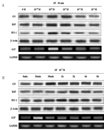
When cells were treated with 10-10 to 10-4 M SP for 30 minutes, mineralization markers and HO-1 were up- regulated. ALP and HO-1 were particularly up-regulated in 10-8 M group, and ON and BSP in 10-8 M and 10-6 M group (Fig. 2A).
In time-dependent manner, all mineralization markers and HO-1 expressions were up-regulated by 10-7 M SP treatment. But the time which expressions were the highest were different. The maximal induction of ALP was seen after 10 minutes, ON after 30 minutes, BSP after 2 hours, respectively. And HO-1 was most abundant in 1 and 2 hours group (Fig. 2B).
DISCUSSION
Orthodontic tooth movement is created by the response of periodontal tissues to orthodontic force, which leads to modeling and remodeling of the surrounding alveolar bone (Hayashi et al., 2002). PDL is a source of pluripotential cells and molecular factors controlling cellular events in surr- ounding tissues (Norton et al., 1995). The bony response is primarily mediated by PDL, so we focused to PDL cells in the present study.
SP is a multifunctional neuropeptide that transmits pain signals, regulates the immune system, and may modulate bone metabolism (Basdra and Domposch, 1997). SP stimulates bone resorption activity of osteoclasts, and the
expression of SP increases in dental pulp and PDL in response to orthodontic force (Nicolay et al., 1990; Jeon, 1997; Norevall et al., 1998). Jeon (1997) or Nicolay et al.
(1990) suggested that SP was increased in PDL cells by application of orthodontic force. It was increased on tension site and especially on compression site. Therefore, SP may be involved in the initial pulpal and periodontal inflammation that occurs after the application of orthodontic force.
In the present study, we found that human IPDL cells stimulated by SP were decreased in their viability in both time and dose dependent manner up to 2 hours. But it was not statistically significant, and was readily recovered (Fig.
1). These results suggest that SP does not significantly change the growth of human IPDL cells and that the cellular protective mechanism works immediately.
Human IPDL cells exhibit phenotypic characteristics in vitro consistent with osteoblast-like cells (Basdra and Domposch, 1997). The extracellular matrix (ECM) com- Fig. 1. Effects of SP on cell viability of human IPDL cells by MTT
assay. A, Dose-dependent cell viability after each concentration SP treatment for 2 hours. B, Time-dependent effect after 10-5 M SP treatment. *Significant difference compared with control (p < 0.05).
Fig. 2. Expression of mineralization markers and HO-1 in human IPDL cells treated with SP. A, Dose-dependent expression of ON, BSP, and HO-1 by western blot analysis and ALP by RT-PCR anal- ysis after SP treatment for 30 minutes. Maximal induction of ON and BSP were seen in 10-8 M and 10-6 M. ALP and HO-1 were most expressed in 10-6 M. B, Time-dependent expression of ON, BSP, and HO-1 by western blot analysis and ALP by RT-PCR anal- ysis after 10-7 M SP treatment. The maximal expression of ALP, ON, BSP could be seen in 10 minutes, 30 minutes, 2 hours, respec- tively. HO-1 was most expressed in 1 and 2 hours.
134 You-Min Cho, Chung-Hwan Seo, Sang Woo Chun, Kyung-Hwa Kang ponents in bone includes collagen and non-collagenous
protein, as in other connective tissues. We examined ON, BSP and ALP as markers of osteoblast-like differentiation by western blot and RT-PCR analysis. It is known that ALP activity in mineralizing chondrocytes and osteoblasts prior to the onset of mineralization is increased in the process of differentiation of osteoblasts (Oh and Choi, 1995). It seems to have a positive role for catalytic activity in initiating mineralization. ON is a major noncollagenous matrix protein and is related with bone cell development (Termine et al., 1981). It has been shown to play various roles in the initiation of mineralization in various experimental models.
BSP is considered to be a marker of osteoblastic cell differentiation in the late stage of differentiation (Mizuno et al., 2000). It is a major noncollagenous protein in miner- alized connective tissues. Expression of BSP is highly specific in mineralizing tissues, including bone, mineralized cartilage, dentin and cementum.
We found that the expressions of ALP, ON and BSP in human IPDL cells were increased by SP treatment. 10-7 M SP was treated, ALP expression level was most abundant in 10 minutes. The highest expression level of ON was seen in 30 minutes, and BSP in 2 hours (Fig. 2). These differences are concerned with the fact that ALP was participated in the initiation of mineralization, and then ON concerned, and BSP had a part in the late stage of differentiation (Termine et al., 1981; Mizuno et al., 2000). Our results are consistent with rat osteoblast, in which SP stimulated bone colony formation or cAMP production (Bjurholm et al., 1992). But there are many reports that SP stimulates formation of the resorption pit by cultured osteoclasts and up-regulates osteoprotegerin (OPG) and RANKL (Mori et al., 1999;
Imasi and Matsusue, 2002). Goto et al. (2002) suggested that SP stimulates the bone formation activity of osteoblasts and the bone resorption activity of osteoclasts. So it might be probably that SP came into baphsic effects in human IPDL cells: the osteoblastic effect and the osteoclastic effect.
HO-1 is a rate-limiting enzyme in heme catabolism (Terry et al., 1999). The role of heme oxygenase in different tissues has not been fully characterized, but it is becoming evident that it is involved with a variety of cellular regulatory and protective mechanisms. As shown in Fig. 2, HO-1 expre- ssion was up-regulated by SP treatment, and was most abundant in 1 hour group and 10-6 M group. The HO-1 expression patterns to SP treatment were similar to osteoblastic markers. So, it could be thought that HO-1 protein is related with differentiation of human IPDL cells.
Moreover, Zwerina et al. (2005) suggested that HO-1 induction is related with osteoclast formation in vitro and in vivo. But there are no reports about correlation with osteoblastic differentiation and HO-1. So, another research is needed for certain relationship in human IPDL cells.
Collectively, SP has biological effect on osteoblastic differentiation and HO-1 expression in human IPDL cells.
Namely, SP can promote cell defferentiation and have cytoprotective effect. It might be affected by the amount of SP produced by activated cells.
Acknowledgment
This paper was supported by Wonkwang University in 2007.
REFERENCES
Basdra EK and Domposch G. Osteoblast-like properties of human periodontal ligament cells : an in vitro analysis. Eur J Orthod. 1997;19:615-21.
Bjurholm A, Kreicberg A, Schulzberg M. Neuroendocrine regulation of cyclic AMP formation in osteoblastic cell lines (UMR-106-01, ROS 17/2.8, MC3T3-E1, ans Saos2) and primary bone cells. J Bone Miner Res. 1992;7:1011-9.
Cho MI, Matsuda N, Lin WL, Moshier A, Ramakrishnan PR.
In vitro formation of mineralized nodules by periodontal ligament cells from the rat. Calcif Tissue Int. 1992;50:459-67.
Choi AM and Alam J. Heme oxygenase-1: function, regulation, and implication of a novel stress-inducible protein in oxidant-induced lung injury. Am J Respir Cell Mol Biol. 1996;15:9-19.
Goto T and Tanaka T. Tachykinins and tachykinin receptors in bone. Microsc Res Tech. 2002;58:91-7.
Hayashi K, Igarashi K, Miyoshi K, Shinoda H, Mitani H.
Involvement of nitric oxide in orthodontic tooth movement in rats. Am J Orthod Dentofacial Orthop. 2002;122:306-9.
Imai S and Matsusue Y. Neuronal regulation of bone meta- bolism and anabolism: calcitonin gene-related peptide-, substance P-, and tyrosine hydroxylase-containing nerves and the bone. Microsc Res Tech. 2002;58:61-9.
Irie K, Hara-Irie F, Ozawa H, Yajima T. Calcitonin gene- related peptide (CGRP)-containing nerve fibers in bone tissue and their involvement in bone remodeling. Microsc Res and Tech. 2002;58:85-90.
Jeon IS. An immunohistochemical study on substance P and calcitonin gene-related peptide in the experimental tooth movement of rat. J Wonkwang Dent Res. 1997;7:63-84.
Kojima T, Yamaguchi M, Kasai K. Substance P stimulates release of RANKL via COX-2 expression in human dental pulp cells. Inflamm Res. 2006;55:78-84.
Kvinnsland I and Heyeraas KJ. Effect of traumatic occlusion on CGRP and SP immunoreactive nerve fiber morphology in rat molar pulp and periodontium. Histochemistry.
1992;97:111-20.
Li MH, Cha YN, Surh YJ. Peroxynitrite induces HO-1 expression via PI3K/Akt-dependent activation of NF-E2- related factor 2 in PC12 cells. Free Radic Biol Med.
2006;41:1079-91.
Long P, Liu F, Piesco NP, Kapur R, Agarwal S. Signaling by mechanical strain involves transcriptional regulation of proinflammatory genes in human periodontal ligament cells
in vitro. Bone. 2002;30:547-52.
Lutherman J, Dahlof G, Modeer T, Johansson O. Immuno- histochemical study of neuronal markers in human gingiva with phenytoin-induced overgrowth. Scand J Dent Res.
1988;96:339-46.
Matayoshi T, Goto T, Fukuhara E, Takano H, Kobayashi S, Takahashi T. Neuropeptide substance P stimulates the formation of osteoclasts via synovial fibroblastic cells.
Biochem Biophys Res Commun. 2005;327:756-64.
Mizuno M, Imai T, Fujisawa R, Tani H, Kuboki Y. Bone sialoprotein (BSP) is a cucial factor for the expression of osteoblastic phenotypes of bone marrow cells cultured on type I collagen matrix. Calcif Tissue Int. 2000;66:388-96.
Mori T, Ogata T, Okumura H, Shibata T, Nakamura Y, Kataoka K. Substance P regulates the function of rabbit cultured osteoclast; increase of intracellular free calcium concentration and enhanced bone resorption. Biochem Biophys Res Commun. 1999;262:418-22.
Nicolay OF, Davidovitch Z, Shanfeld JL, Alley K. Substance P immunoreactivity in periodontal tissues during orthodontic tooth movement. Bone Miner. 1990;11:19-29.
Nohutcu RM, Mc Cauley LK, Koh AJ, Somerman MJ.
Expression of extracellular matrix proteins in human periodontal ligament cells during mineralization in vitro. J Periodontol. 1997;68:320-7.
Nojima N, Kobayashi M, Shionome M, Takahashi N, Suda T, Hasegawa K. Fibroblastic cells derived from bovine periodontal ligaments have the phenotypes of osteoblasts. J Periodont Res. 1990;25:179-85.
Norevall LI, Matsson L, Forsgren S. Main sensory neuropeptides, but not VIP and NPY, are involved in bone remodeling during orthodontic tooth movement in the rat.
Ann NY Acad Sci. 1998;865:353-9.
Norton LA, Andersen KL, Arenholt-Bindslev Dorthe, Andersen L, Melsen B. A methodical study of shape changes in human
oral cells perturbed by a stimulated orthodontic strain in vitro. Arch Oral Biol. 1995;40:863-72.
Oh JH and Choi BJ. Effects of substance P and VIP on the alkaline phosphatase activity and the gene expression of bone proteins in osteoblast-like cells. J Oral Biol.
1995;19:33-9.
Otterbein L, Sylvester SL, Choi AM. Hemoglobin provides protection against lethal endotoxemia in rats: the role of heme oxygenase-1. Am J Respir Cell Mol Biol. 1995;13:595- 601.
Pi SH, Lee SK, Hwang YS, Choi MG, Lee SK, Kim EC.
Differential expression of periodontal ligament-specific markers and osteogenic differentiation in HPV16-immor- talized human gingival fibroblasts and periodontal ligament cells. J Perio Res. 2007;42:104-13.
Sohn SJ. Substance P upregulates osteoclastogenesis by activating nuclear factor kappa B in osteoclast precursors.
Acta Otolaryngol. 2005;125:130-3.
Termine JD, Belcourt AB, Conn KM, Kleinman HK. Mineral and collagen binding proteins of fetal calf bone. J Biol Chem. 1981;256:10403-8.
Terry CM, Clickeman JA, Hoidal JR, Callahan KS. TNF-α and IL-1α induce heme oxygenase-1 via protein kinase C, Ca2+ and phospholipase A2 in endothelial cells. Am J Physiol. 1999;276:1493-501.
Vile GF, Basu-Modak S, Waltner C, Tyrrell RM. Heme oxygenase-1 mediates an adaptive response to oxidative stress in human skin fibroblasts. Proc Natl Acad Sci.
1994;91:2607-10.
Zwerina J, Tzima S, Hayer S, Redlich K, Hoffmann O, Hanslik-Schnabel B, Smolen JS, Kollias G, Schett G. Heme oxygenase 1 (HO-1) regulates osteoclastogenesis and bone resorption. FASEB J. 2005;19:2011-3.