Effect of Diallyl Disulfide on Heme Oxygenase-1 Expression in Human Hepatoma Cell Line HepG2
Kang Mi Kim
1, Sang Kwon Lee
2and Young Chul Park
1*
1
Department of Microbiology & Immunology, Pusan National University School of Medicine, Yangsan, Gyeongnam 626-870, Korea
2
Cardiovascular Center, Pusan National University Yangsan Hospital, Yangsan, Gyeongnam 626-770, Korea Received June 10, 2011 /Revised July 12, 2011 /Accepted July 15, 2011
Diallyl disulfide (DADS), the most prevalent oil-soluble organosulfur compound in garlic, is known to have diverse biological activities, including anticarcinogenic, antiatherosclerotic, antiinflammatory, and antioxidant actions. In this study, we investigated the effect of DADS on the expression of heme oxygenase-1 (HO-1) in human liver hepatoma cell line HepG2. Treatment of HepG2 cells by DADS evoked a dose-dependent growth inhibition without significant toxicity to the cells, and also induced the expression of transcription factor Nrf2. However, DADS did not have any enhancing effect on transcription and translation of HO-1 expression in HepG2 cells. In addition, DADS efficiently blocked protein synthesis of HO-1 in HepG2 cells stimulated by CoPP or hemin. But, DADS did not decrease the content of transcripts of HO-1 gene stimulated by CoPP, with accumulation of Nrf2 and small Maf in the nucleus. Based on these results, we conclude that DADS inhibits HO-1 expression by modu- lation of translational level of CoPP or hemin-induced HO-1 expression in HepG2 cells.
Key words : Diallyl disulfide (DADS), heme oxygenase-1 (HO-1), Nrf2, small Maf, HepG2 cells
*Corresponding author
*Tel:+82-51-510-8093, Fax:+82-55-382-8090
*E-mail : [email protected]
서 론
마늘은 많은 문화권에서 오랜 역사 동안 식용과 약용으로 이용되고 있으며, 최근에는 건강보조식품 또는 의약품으로 인 식이 확대되고 있는 실정이다. 실제로 여러 가지 질병을 예방 하고 치료하는데 효과가 있어 많은 연구의 대상이 되고 있는 데, 이는 마늘이 함유하고 있는 다량의 유기 황화합물 (organosulfur compounds) 때문인 것으로 알려지고 있다 [5,10]. 특히, diallyl disulfide (DADS)는 마늘의 주요한 유기 황화합물 성분으로서 화학적 간독성에 대한 보호와[17,28] 항 박테리아[3] 및 항동맥경화[19] 등에 효과를 보이는 것으로 보 고되고 있다. 또한 DADS는 다양한 암세포의 G2/M 세포주기 를 억제시키고[12,20,22], 미토콘드리아의 Bcl-2 family 및 re- active oxygen species (ROS)의 증가를 유도함으로써 세포사 멸(apoptosis)을 일으켜 결과적으로 암세포의 증식을 억제하 는 것으로도 알려져 있다[9,15,23,29]. 최근에는 DADS는 세포 내 항산화(antioxidant) 인자인 glutathione의 양을 크게 증가 시킴으로써 세포 내 항산화 기능에도 기여한다는 결과가 발표 되었다[2,27]. 이들 상반된 결과는 아마도 사용한 DADS 농도 의 세포 유형에 따른 민감성도 한가지 원인일 수 있지만, 세포 내 조절 인자들간의 상호관련성을 통한 정확한 분자기전을 규명함으로써 해결될 심도있는 연구가 필요한 부분이라고 여
겨진다.
Heme oxygenase-1 (HO-1)은 일반적으로 heme을 대사시 키는 효소로 잘 알려져 있으며, biliverdin, carbon monoxide 및 ferrous iron을 산물로 방출한다. Biliverdin은 biliverdin re- ductase에 의해 강력한 항산화 인자로 알려져 있는 bilirubin 으로 뒤이어 전환된다[25]. HO-1은 저산소증[16], metal ions[24] 및 염증성 cytokines 등과 같은 산화적 스트레스 (oxidative stress) 환경을 유도하는 자극[6,14]에 의해 합성이 유도된다. HO-1은 산화적 스트레스에 대항하여 세포 내 re- dox (reduction-oxidation) 균형을 유지하는 항산화 인자로서 크게 기여한다. HO-1의 발현은 일차적으로 전사단계에서 조 절되며 전사인자 Nrf2에 의해 이루어지고, JNK, ERK 및 p38 이 포함되어 있는 mitogen-activated protein kinases (MAPKs) 에 의해서도 HO-1의 유전자 발현이 조절되는 것으로 알려져 있다[21,30].
최근의 보고에 따르면 DADS를 포함한 마늘의 주요 유기
황화합물이 Nrf2의 활성화를 통해 HO-1의 발현을 증가시킨
다는 연구결과가 있다[4,11]. 본 연구에서는 유기 황화합물 중
에서 가장 강력한 약리작용을 보이는 DADS가 HO-1과 그 전
사인자인 Nrf2의 발현에 미치는 영향을 인간 간암세포주
HepG2에서 조사하였다. 또한 HO-1의 강력한 유도제로 알려
져 있는 CoPP와 hemin에 의해 유도되는 HO-1의 발현과정에
서 DADS의 효과에 대해서도 조사하였다.
재료 및 방법
시약
DADS는 Fluka Chemical Co. (Bucha, Switzerland) 제품을 사용하였다. Protease inhibitor cocktail, trypan blue, CoPP, hemin은 Sigma Chemical Co. (St. Louis, MO, USA) 제품을 구입하여 사용하였다. Nrf2, HO-1, Bach1, β-actin에 대한 항 체는 Santa Cruz Biotechnology (Santa Cruz, CA, USA) 제품 을 사용하였다. Poly (ADP-ribose) polymerase (PARP), Bach1, Keap1, small Maf에 대한 항체는 Cell Signaling Technology (Beverly, MA, USA) 제품을 구입하여 사용하였 다. Enhanced chemiluminescence (ECL) Western blotting kit 는 Amersham Pharmacia Biotech (Piscataway, NJ, USA) 제품 을 사용하였고, RT-PCR에 필요한 reverse transcriptase, dNTP mixture, DNA polymerase 등의 시약은 Invitrogen (Carlsbad, CA, USA) 제품을 사용하였다.
세포 배양
실험에서 사용된 인간 간암세포주 HepG2는 한국세포주은 행에서 구입하였고, 세포배양에 필요한 minimum essential medium (MEM), fetal bovine serum (FBS), 항생제, tryp- sin-EDTA는 HyClone Laboratories Inc. (Logan, UT, USA) 제 품을 구입하여 사용하였다. HepG2 세포는 10% FBS, 100 μg/
ml streptomycin/100 U/ml penicillin이 함유된 MEM 배양액 에서 5% CO
2조건에서 37℃ incubator에서 배양하였다.
세포 생존율 분석
HepG2 세포를 60-mm dish에 1×10
5cells/ml의 농도로 5 ml씩 분주하여 12시간 배양한 후 DADS를 농도별로 처리하였 다. 24시간 배양 후 상층액과 0.01% trypsin-EDTA을 이용하여 부착한 세포를 수확하여 FBS를 포함하는 PBS로 2번 세척한 후 300× g로 원심분리를 시행하였다. 세포를 배양액에서 se- suspension 시킨 후 trypan blue dye exclusion 방법을 이용하 여 hemocytometer 상에서 살아있는 세포와 죽은 세포의 수를 측정하여 생존율을 분석하였다. 실험은 3번 이상 반복하였으 며, 결과의 수치는 3개의 독립적인 dish에서 수행한 값을 means±SD 값으로 나타내었다.
Western blot analysis
약물을 처리한 세포를 PBS로 세척한 후, 단백질 분해효소 저해제(protease inhibitor cocktail)를 포함한 lysis buffer (50 mM Tris-HCl pH 7.4, 150 mM NaCl, 1% Nonidet P-40)를 첨가하여 세포의 전체 단백질을 추출하였다. Bicinchoninic acid를 이용하여 정량하고, 동일한 양의 단백질을 10%
SDS-polyacrylamide gel에 전기영동한 후 electroblotting ap- paratus (Bio-rad, Richmond, CA, USA)를 이용하여 단백질을
nitrocellulose membrane에 이동시켰다. Membrane은 항체의 비특이적 부착을 막기 위하여 5% skim milk에 1시간 동안 두었고, 그 후 필요한 일차 항체 및 horse radish peroxide (HRP)가 부착되어 있는 2차 goat 항체를 반응시켰다. 부착된 면역복합체는 ECL kit를 이용하여 X-ray 필름에 표현하여 단 백질 발현을 분석하였다.
세포질 및 핵 단백질의 분리
세포를 PBS로 세척한 후, Fermetas (Waltham, MA, USA) 제품인 ProteoJET™ cytoplasmic/ nuclear protein extraction kit를 이용하여 제조사가 추천하는 protocol에 의거하여 세포 질과 핵 단백질을 분리하였다. 분리된 각각의 단백질은 정량 한 후 Western blot analysis를 이용하여 분석하였다. 세포질 및 핵 단백질의 오염을 측정을 위하여 histone과 β-actin에 대 한 항체를 각각 사용하였다.
RNA 분리 및 RT-PCR
약물을 처리한 세포를 PBS로 세척한 후, TRIzol reagent (Invitrogen, Carlsbad, CA, USA)를 이용하여 RNA를 분리하 고 spectrophotometer를 이용하여 RNA를 정량하였다. 각 2 μ g의 RNA를 사용하여 제조사의 지침에 따라 superscript re- verse transcriptase를 이용하여 cDNA를 제작하였고, 합성된 cDNA는 -20℃에 보관하였다. PCR은 pre-heating time을 95℃
에서 5분간 반응한 다음, denaturation 95℃에서 10초, anneal- ing은 57℃에서 10초, extension은 72℃에서 10초간 실시하여 30 cycles을 시행하였다. 마지막으로 extension 72℃에서 5분 간 반응하였다. DNA 증폭양상을 확인하기 위하여 1.5% agar- ose gel에서 전기영동한 후, ethidium bromide에 염색하여 UV 램프 상에서 확인하였다. DNA polymerase의 primer는 다음과 같다: HO-1, 5’-CGGGCCAGCAACAAAGTG-3’
(sense) and 5’-AGTGTAAGGACCCATCGGAGAA-3’
(antisense), Nrf2, 5’-GAGAGCCCAGTCTTCATTGC-3’
(sense) and 5’-TGCTCAATGTCCTGTTGCAT-3’ (antisense), and glyceraldehyde-3-phosphate dehydrogenase (GAPDH), 5’-TGAAGGTCGGAGTCAACGGATTTGGT-3’ (sense) and 5’-CATGTGGGCCATGAGGTCCACCAC-3’ (antisense).
결과 및 고찰
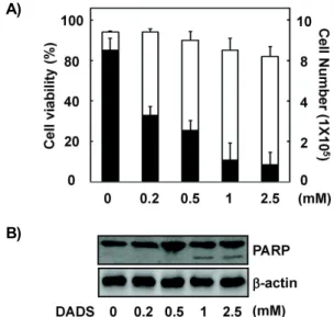
간암세포주 HepG2의 증식 및 세포사멸에 대한 DADS의 효과
마늘의 주요 약리 성분인 유기 황화합물 DADS에 의한 인
간 간암세포주 HepG2의 생존율에 미치는 영향을 분석하기
위하여, 배양중인 세포에 다양한 농도(0.2, 0.5, 1, 2.5 mM)의
DADS를 처리하였다. 그 후 24시간 동안 배양한 다음 trypan
blue dye exclusion 방법으로 살아있는 세포와 죽은 세포의
수를 측정하였다. 세포의 생존율에서는 1 mM DADS 까지는 큰 영향을 보이지 않았다. 그러나, 세포의 증식면에서는 DADS 처리에 의한 세포수의 감소가 농도의존적으로 현저히 관찰되었다(Fig. 1A). 그러나 1 mM 이상에서 세포사멸 관련 단백질인 활성형 PARP의 분절화(fragmentation)가 명확하게 확인되었는데(Fig. 1B), 이는 DADS가 1 mM 이상의 고농도에 서는 HepG2 세포에 독성을 주어 세포사멸을 일으킬 수 있다 는 것을 시사한다. 따라서 본 연구에서는 DADS가 HepG2 세 포의 세포사멸을 유도하지 않는 범위의 농도를 사용하였다.
간암세포주 HepG2의 HO-1 발현에 대한 DADS의 효과 HO-1과 그 대사산물은 항염증 및 항세포사멸에 관련된 유 전자 발현과 효소의 활성에 영향을 주는 중요한 항산화 효소 로 알려져 있으며[26], 다양한 세포에서 HO-1의 과발현은 염 증 및 세포사멸의 완화에 기여한다고 보고되고 있다[7,8]. 실제 로 마늘의 황화합물 성분이 HepG2 세포의 DNA 손상을 보호 한다는 최근의 연구결과와[1] 전사인자 Nrf2의 활성화를 통하 여 HO-1 등의 전사단계에서의 증가를 보고한 논문이 발표되 었다[4]. 또한, diallyl sulfide (DAS)가 ROS 경유 Nrf2 및 MAPK의 활성을 통하여 HO-1의 발현을 유도한다는 연구결 과도 보고되었다[11]. 그러나 본 연구에서는 상반된 결과가 나
A)
B)
Fig. 1. Effects of DADS on viability, proliferation, and apoptosis in HepG2 cells. Cells were plated at 1×10
5cells per 60-mm dish, incubated for 12 hr, and treated with differ- ent concentrations of DADS for 24 hr. (A) Cell viability (□, viable cells/total cells) and cell proliferation (■, via- ble cells) were assessed by trypan blue dye exclusion assay. The results are reported as mean±SD of three in- dependent experiments. (B) Whole cell extracts were an- alyzed by Western blot analysis using an anti-PARP antibody. β-Actin was used as an internal control for equal protein loading.
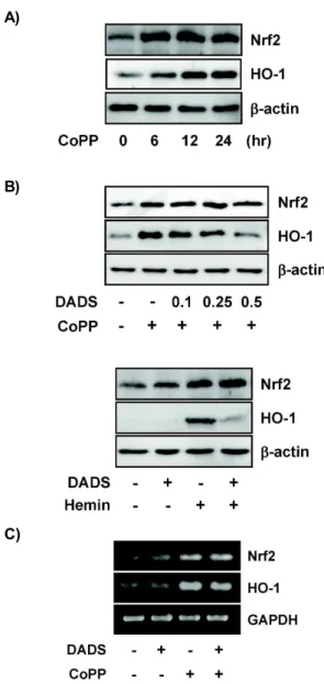
타났는데, DADS를 농도별로 HepG2 세포에 단독 처리하였을 때, HO-1의 발현은 RNA 뿐만 아니라 단백질 수준에서도 유도 되지 않는다는 것을 알 수 있었다. 이는 세포사멸을 일으키는 높은 농도의 경우에서도 같은 결과를 볼 수 있었다(data not shown). 그러나, HO-1의 전사인자인 Nrf2는 1 mM 이상 고농 도의 DADS 처리 때 RNA 및 단백질 수준에서의 증가를 RT-PCR과 Western blot analysis로 확인할 수 있었다(Fig. 2).
CoPP에 의해 유도되는 HO-1 발현에 대한 DADS의 효과 HO-1 유도제의 자극으로 HO-1의 발현을 증가시키는 경우 DADS가 HepG2 세포에 미치는 영향을 조사하였다. 본 연구 를 위하여 강력한 HO-1 유도제로 널리 사용되는 cobalt proto- porphyrin (CoPP)과 hemin을 사용하였다. 배양중인 HepG2 세포에 50 μM CoPP를 처리하면 HO-1과 전사인자 Nrf2의 발 현이 6시간부터 시간이 지남에 따라 증가됨을 관찰할 수 있었 다(Fig. 3A). 그러나, DADS가 1시간 전처리 된 HepG2 세포에 CoPP를 처리한 경우에는 HO-1 단백질의 양이 현저하게 감소 하는 것을 확인하였다(Fig. 3B). 이때 전사인자 Nrf2 단백질의 양은 DADS 처리에 의해 억제되지 않고 발현이 유지되었다.
다른 HO-1 유도제로 사용되는 porphyrin 계열의 hemin을 처 리하였을 때도 CoPP의 경우와 같은 결과를 관찰할 수 있었다 (Fig. 3B). 다음으로 HO-1 유도제에 의한 HO-1의 단백질 합성 의 DADS에 의한 감소가 유전자 수준에서도 단백질과 같은 발현 양상을 나타내는지 조사하기 위하여 RNA를 분리하여
A)
B)
Fig. 2. Effect of DADS on the induction of Nrf2 and HO-1 ex-
pression in HepG2 cells. Cells were stimulated with vari-
ous concentrations of DADS for 24 hr. (A) Whole cell
extracts were analyzed by Western blot analysis using
an appropriate antibodies. β-Actin was used as an in-
ternal control for equal protein loading. (B) Total RNA
was prepared. Nrf2 and HO-1 mRNA level was ana-
lyzed by RT-PCR. GAPDH was used as a control to
monitor equal loading.
A)
B)
C)
Fig. 3. Effect of DADS on the CoPP-induced HO-1 expression in HepG2 cells. (A) Cells were stimulated with CoPP at 50 μM for different time periods. (B) Cells were pre- treated for 1 hr in the absence or presence of DADS (0.1, 0.25, and 0.5 mM DADS), and then incubated for an ad- ditional 24 hr with CoPP or hemin at 50 μM. Whole cell extracts were analyzed by Western blot analysis using an appropriate antibodies. β-Actin was used as a control.
(C) Total RNA was prepared. Nrf2 and HO-1 mRNA level was analyzed by RT-PCR. GAPDH was used as a control to monitor equal loading.
RT-PCR을 이용해 분석하였다. 흥미롭게도, CoPP에 의해 유 도되는 HO-1과 전사인자 Nrf2의 RNA 수준에서는 DADS에 의해 억제되지 않고 발현이 유지됨을 확인할 수 있었다(Fig.
3C). 이러한 결과를 볼 때, CoPP 같은 HO-1 유도제로 자극한 HepG2 세포의 HO-1 발현 과정에서, DADS는 HO-1 유도제에 의해 자극된 HepG2 세포에서 HO-1 유전자의 전사 후 단백질 합성과정을 억제하는데 역할을 하는 것으로 볼 수 있다. 그러
므로, HO-1 유전자의 전사 후 단백질로의 번역 과정에 관여하 는 세포내 인자들과 DADS의 상호관련성을 살펴보는 것이 필 요하다고 여겨진다.
HO-1 발현에 관련된 전사인자들에 대한 DADS의 영향 COPP의 자극에 의한 HO-1 유전자 발현 과정에 관여하는 Nrf2 외 다른 조절인자들의 활성에 DADS가 미치는 영향을 조사하였다. HO-1 유전자 발현을 조절하는 여러 전사인자 중 하나인 Nrf2는 세포질에서 Keap1과 결합하여 불활성 상태로 있다가 적합한 자극에 의해 Keap1이 thiol기를 산화시켜 Nrf2 을 떨어지게 하여 핵내로 이동하게 해준다. Keap1은 평소에 세포질에서 Nrf2의 붕괴를 막는 역할을 담당하고 있다[18,31].
핵내로 이동한 Nrf2는 small Maf와 이량체를 이루어 HO-1 유전자 promoter의 antioxidant response element (ARE)라는 특정염기서열 부위에 결합함으로써 HO-1의 발현을 개시하는 것으로 알려져 있다[21,30]. 또한 HO-1 발현은 전사 억제인자 로 알려져 있는 Bach1의 불활성화가 요구되는데, Bach1은 자 극이 없는 경우에는 HO-1 유전자 promoter의 ARE 부위에 결합하고 있다가 자극이 오면 떨어져 세포질로 나가면서 Nrf2/small Maf 이량체가 ARE에 결합이 가능하도록 하여 HO-1 유전자 발현이 개시된다[13]. 본 연구에서 DADS 단독 처리는 핵내 전사조절 억제 인자인 Bach1의 감소와 small Maf 의 증가를 보였으며, Nrf2와 결합하고 있는 Keap1의 변화는 없는 것으로 나타났다(Fig. 4). 그리고, DADS 전처리는 CoPP 에 의해 유도되는 Bach1의 세포질로의 이동과 small Maf의 핵내의 축적을 더욱 증가시켰다. 이러한 DADS가 HO-1 유전 자 발현에 관련된 전사인자들의 활성에는 영향을 주지 못하는 것을 확인하였는데, 이는 DADS가 CoPP 처리에 의한 HO-1 유전자 발현을 억제하지 못한다는 결과와 부합한다.
Fig. 4. Effect of DADS on Bach1, Keap1, and small Maf ex-
pression in CoPP-treated HepG2 cells. HepG2 cells were
pretreated with 1 hr for 0.5 mM DADS. And, cells were
stimulated with 50 μM CoPP for 24 hr. Nuclear and cyto-
plasmic proteins were isolated and used for Western blot
analysis. Antibodies against β-actin and histone were
used for contamination of the nuclear and cytoplasmic
proteins, respectively.
감사의 글
이 논문은 부산대학교 자유과제 학술연구비(2년)에 의하여 연구되었음.
References