Introduction
A fundamental goal of periodontal treatment is the functional reha- bilitation of damaged periodontal tissue and the regeneration of tis- sue from the periodontal ligament (PDL). To accomplish these goals several procedures, such as bone grafting and guided tissue regen- eration have been used.
However, the results of these therapies are not always pre- dictable. Recently, tissue engineering technology using an extracellular
matrix, such as enamel matrix derivatives (EMDs) and growth fac- tors have been used for periodontal regeneration.
1The composition of an enamel matrix derivative is 90% amelo- genin and 10% hydrophobic protein. Protein matrix released from ameloblast during the generation of tooth enamel forms enamel by organizing a crystal layer, amelogenin is the protein that com- poses the intercellular layer. Thus, amelogenin evolute and play an important role in cementogenesis.
2Acellular cementogenesis begins at the inner epithelium of Hertwig's epithelial root sheath,
1 http://dx.doi.org/10.4047/jkap.2016.54.3.203 ORIGINAL ARTICLE법랑기질유도체가 인간 치주인대세포의 증식 및 성장인자 발현에 미치는 영향
정겨운1∙방은경2*
1고려대학교 안암병원 치주과, 2이화여자대학교 의학전문대학원 치주과학교실
Effects of enamel matrix derivatives on the proliferation and the release of growth factors of human periodontal ligament cells
Gyu-Un Jung1, Eun-Kyoung Pang2*
1Department of Periodontology, Korea University Anam Hospital, Seoul, Republic of Korea
2Department of Periodontology, School of Medicine, Ewha Womans University, Seoul, Republic of Korea
Purpose: Stimulating the proliferation and migration of periodontal ligament cells (PDLCs) has become the main goal of periodontal regeneration. To accomplish this goal, regeneration procedures have been developed, but results have not been predictable. Recently, tissue engineering using enamel matrix derivatives (EMDs) and growth factors has been applied to periodontal regeneration; however, the mechanism of EMDs is largely unknown. The aim of this study was to investigate the effects of EMDs on the pro- liferation and release of growth factors from PDLCs. Materials and methods: Human PDLCs were removed from individually extracted 3rd molars of healthy young adults, and cultured in the media containing EMDs (Emdogain, Biora, Malmo, Sweden) at concentration of 0, 12.5, 25, 50, 100, and 200 μg/mL each. Cell proliferation and ALP (alka- line phosphatase) activity were measured. The evaluation of growth factors released by PDLCs was also performed by one-way analysis of variance (ANOVA) and Bonferroni's multiple comparison test. Results: Significantly increased proliferation and ALP activity were observed in PDLCs treated with over 25 μg/mL and 50 μg/mL EMDs, respectively. Additionally, treatment of PDLCs with 50 μg/mL resulted in significantly increased release of vascular endothelial growth factor (VEGF) and transforming growth factor (TGF)-βafter 24 h and 48 h, respectively. Conclusion: EMDs enhance the proliferation and ALP activity of PDLCs, and promote the release of growth factors, includ- ing VEGF and TGF-β, from PDLCs. Therefore EMDs could be one of the effective methods for periodontal regeneration. (J Korean Acad Prosthodont 2016;54:203-9) Keywords: Enamel matrix derivatives; Periodontal ligament; Periodontal regeneration; Tissue engineering; Growth factor
c cc
2016 The Korean Academy of Prosthodontics
This is an Open Access article distributed under the terms of the Creative Commons Attribution Non-Commercial License (http://creativecommons.org/licens- es/by-nc/3.0) which permits unrestricted non-commercial use, distribution, and reproduction in any medium, provided the original work is properly cited.
*Corresponding Author: Eun-Kyoung Pang
Department of Periodontology, School of Medicine, Ewha Womans University 1071 Anyancheon-ro, Yangcheon-gu, Seoul 07985, Republic of Korea +82 (0)2 2650 2725: e-mail, [email protected]
Article history: Received January 25, 2016 / Last Revision April 2, 2016 / Accepted April 14, 2016
and the protein found in the acellular cementum is similar to enamel matrix derivative.
3Amelogenin is also known not only as cell adhesion protein which has high compatibility with both hydrox- yapatite and collagen, but also promoting cell spreading property.
4When EMDs are applied on exposed root surfaces, tissue regen- eration is achieved by osteogenesis, and acellular cementogenesis, histologically.
5According to a clinical experiment which EMD was applied, Heiji et al.
6reported a bone gain of 2.6 mm in one- or two- wall bone defect. Froum et al.
7reported that in case of horizontal bone loss, the periodontal ligament attachment level was retained by open flap debridement with EMDs application. Sculean et al.
8reported a clinical result of periodontal surgery using EMDs showing a favor- able long term outcome over more than 4 years. And Van der Pauw et al.
9reported that EMDs increase the stimulation of periodontal attachment by increasing alkaline phosphate (ALP) level. However, it is not yet clear how the EMDs imitate root and supporting tissue formation.
Periodontal regeneration is associated with a serial procedure, which is proliferation of periodontal ligament cells (PDLCs), and composition and calcification of an organic substance, such as enamel matrix deriv- ative.
10PDLCs are thought to play an important role in maintenance, regeneration and recovery of the tooth attachment apparatus.
11For the successful regeneration, PDLCs and probiont should inter- act with each other, and there may be some factors that influence pro- ductivity and growth powerfully.
Therefore, we intended to identify the effects of enamel matrix derivatives on the proliferation and the release of growth factors of human PDLCs and ALP activity, by culturing human PDLCs at dif- ferent EMD concentrations.
Materials and Methods Cell isolation and culture
PDLCs were prepared from extracted third molars that were removed for orthodontic reason in young healthy volunteers. The study protocol was approved by the Institutional Review Board of Ewha Womans University Mokdong Hospital (No. 2015-02-022). Using sharp blades or curettes, PDLCs were obtained from the middle one- third of individual extracted teeth and placed in Dulbecco's modi- fied minimum essential medium F-12 (DMEM/F12; GIBCO BRL, Grand Island, NY, USA) supplemented with 10% fetal bovine serum (FBS; GIBCO BRL, Grand Island, NY, USA), 100 U/mL peni- cillin, 100 μg/mL streptomycin and were overlaid with cover slips. Cultures were performed in an incubator with an environment of 37℃, 95% air, and 5% CO
2for 24 hours. While waiting for the release of PDLCs from the tissue for about 5 days, culture medium
was replaced once in two days. Outgrowing cells were sub-cultured in fresh culture medium. Cells of the fourth to sixth passage were used for the experiments described below.
Preparation of EMDs
Emdogain (Biora, Malmo, Sweden), a mixture of freeze-dried enam- el matrix proteins harvested from the developing crowns of 6- month-old swine, is a unique material that came into the market as a device promoting regeneration of PDLCs4, used as EMDs in this study. Gel-type Emdogain was completely dissolved in 10 mM glacial acetic acid, then was added to culture media.
Effect of EMDs on the proliferation of PDLCs
The PDLCs were seeded in 96-well plate at a density of 1×10
4cells/well and incubated in 100 μL DMEM containing 10% FBS.
After 24 hours, the medium was replaced with serum-free DMEM.
After 24 hours, they were subcultured into two groups. The medi- um was replaced with serum-free DMEM containing EMD in the test group, and serum-free DMEM in the control group. EMDs were present in each test media at concentration of 0, 12.5, 25, 50, 100, and 200 μg/mL.
2After 3 days of incubation, analysis was per- formed using cell proliferation assay kit (Cell counting kit-8, Donjindo Lab., Kumamoto, Japan). All samples were prepared every 3 units.
Effect of EMDs on the ALP activity of PDLCs
The PDLCs were seeded in 96-well plate at a density of 1×10
4cells/well and incubated in 100 μL DMEM containing 10% FBS.
After 24 hours, the medium was replaced with serum-free DMEM.
After 24 hours, they were subcultured into two groups and the medi- um was replaced with DMEM containing EMDs in test group, and serum-free DMEM in the control group. Five subgroups were set in the EMD group by concentrations of 0%, 2.5%, 10%, 20%, and 40% EMD.
ALP activity in the cell layer was determined by ALP analyzing reagent (BioAssay Sys., Hayward, CA, USA). The cells from test and control groups were washed twice with Dulbecco's phosphate buffered saline (DPBS) and 50 μL of 0.2% Triton X-100 was added and shaked on a plate-shaker for 20 minutes. 150 μL of ALP analyzing reagent (1 M p-nitrophenyl phosphate and 0.2 M Mg acetate) was added and incubated at 37℃ for 30 minutes.
The ALP disintegrated p-nitrophenyl phosphateto p-nitrophenol
and phosphate forming yellow color. After stopping the reaction by
adding 100 μL of 1 M NaOH, p-nitrophenol formed was measured
at 405 nm using a microplate reader (Synergie HT, Bio-Tek, Winooski, VT, USA). The more it appears brown, the higher activity it shows.
Effect of EMDs on the release of growth factors from PDLCs
3×10
5cells of PDLCs were added to each 35 mm culture dish- es and incubated in DMEM medium supplemented with 10%
FBS. After 24 hours, culture medium was replaced with serum-free DMEM. Two groups were set after 24 hours. The test group was incu- bated in DMEM medium supplemented with EMD of minimum con- centration that increase cell proliferation and ALP activity at former experiment and the control group was incubated in serum-free DMEM medium. The conditioned media was harvested after 0, 8, 24, 48, and 72 hour and kept in -70℃. Then, platelet derived growth factor (PDGF)-AB, PDGF-BB, transforming growth factor (TGF)-β1, vascular endothelial growth factor (VEGF) level of each conditioned media according to time were measured by ELISA method. Average data of 3 unit samples were used.
Statistical analysis
Data were analyzed using GraphPad Prism version 5.0 (GraphPad Software, San Diego, CA, USA).
In cell proliferation assay according to EMD concentration and ALP activity analysis, statistical analysis was performed by one-way analysis of variance (ANOVA) and Bonferroni's multiple comparison test (P < .05).
Results
A. Effect of EMDs on the proliferation of PDLCs
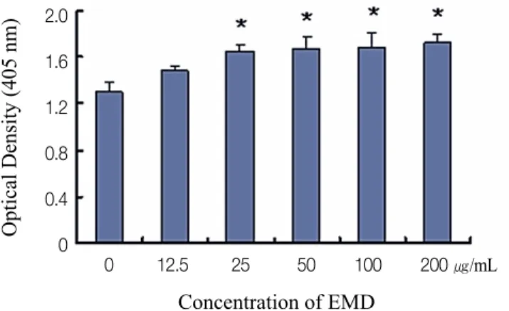
The effect of different EMD concentrations on the proliferation of PDLCs was examined. The results represent that cell prolifera- tion of PDLCs cultured in more than 25 μg/mL EMD was signif- icantly higher than control group (P < .05) (Fig. 1).
B. Effect of EMD on the ALP activity of PDLCs
Effect of different EMD concentration on the ALP activity of PDLCs was examined. The results represent that ALP activity of PDLCs cul- tured in 50 μg/mL EMDs was significantly higher than control group (P < .05) (Fig. 2).
C. Effect of EMDs on the release of growth factors of PDLCs
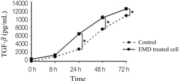
According to the result about an effect of different EMD con- centrations on the ALP activity and proliferation of PDLCs, the con- centration of 50 μg/mL EMD was selected to evaluate the release of VEGF and TGF-β. The growth factors of PDLCs were measured and evaluated over time for 72 hours (Fig. 3, Fig. 4). VEGF and TGF- βreleased from PDLCs cultured in 50 μg/mL EMD were significantly higher than control group (P < .05).
Fig. 1. The effect of different enamel matrix derivatives (EMDs) concentra- tions on the proliferation of PDLCs. The results represent the means ± SD of three replicates. * Significantly different from the control group (P < .05).
Fig. 2. The effects of different EMDs concentrations on the ALP activity of PDLCs.
The results represent the means ± SD of three replicates. * Significantly different from the control group (P < .05).
2.0 1.6 1.2 0.8 0.4 0
0 12.5 25 50 100 200 ㎍/mL
2.0 1.6 1.2 0.8 0.4 0
0 12.5 25 50 100 200 ㎍/mL
Concentration of EMD Concentration of EMD
Optical Density (405 nm) Optical Density (405 nm)
1. VEGF
The release of VEGF relevant to angiogenesis, increased over time.
The release of VEGF was significantly higher than control group after 48 hours (P < .05) (Fig. 3).
2. TGF-β
The release of TGF-βincreased over time. TGF-βreleased from PDLCs cultured in 50 μg/mL EMD were significantly higher than the control group after 24 hours (P < .05) (Fig. 4).
Discussion
PDLCs are essential in supporting and healing the tooth attach- ment apparatus. Thus, the ultimate goal of periodontal regeneration is to promote PDLC productivity and calcified tissue by stimulat- ing growth of PDLC. As PDLC can proliferate and migrate, they pro- mote periodontal tissue regeneration by differentiating to osteoblast- like and cementoblast-like cells.
1During this procedure, EMDs are thought to play a considerable role in promoting PDLC attachment and growth.
4There have been reports that EMD control intracellular cAMP signals which are associated with PDLC proliferation and metabolism activity and secretion of numerous self releasing growth factor.
12EMD is composed of 2 protein families, 90% of amelogenins which are thought to form the periodontal attachment during tooth devel- opment, and the non-amelogenins such as ameloblastin, enamelin, and tuftelin.
13The theory that EMDs play a considerable role in tis- sue regeneration to promote PDLC proliferation and repair has been applied clinically. There have been reports that EMDs promote the proliferation of PDLC and stimulate osteogenesis by production of collagen and reinforcement of calcification,
14but there has been no definite answer about the mechanism how EMDs influence PDLC.
It was identifed that EMD didn't contain the growth factor includ-
ing PDGF, TGF-β, and insulin like growth factor.
4The previous study demonstrated that EMDs enhanced proliferation, increased protein production, and promoted mineral nodule formation.
4Thus, we observed ALP activity which reflects the change of cell prolifera- tion rate and osteogenetic ability when EMD was applied to PDLC, and examined optimum concentration of EMD.
As a result, cell proliferation of PDLCs cultured in >25 μg/mL EMDs were significantly higher than the control group, and ALP activity of PDLCs cultured in 50 μg/mL EMDs was significantly high- er than the control group (P < .05). Accordingly, 50 μg/mL EMD was used in the next experiment which evaluated the effect of EMDs on release of growth factor from PDLCs over time.
In this experiment, >25 μg/mL EMDs promoted cell proliferation of PDLCs, 50 μg/mL EMDs increased ALP activity of PDLCs. As ALP activity is the marker of early osteogenesis, this result shows validity of the theory that EMDs play a primary role in periodon- tal regeneration by PDLCs. Van der Pauw et al. announced that EMDs increase ALP which is not proliferating PDLCs, but stimulating attach- ment of PDLCs.
9According to in vitro experiment of Chong et al.
about which area among damaged site is most effective when EMDs was applied, EMDs did not reveal significant wound closure at center of wounds, and showed obvious proliferation of PDLCs and increased wound closure only when PDGF-BB was added.
2One of the problems of regeneration of PDLCs is to promote proliferation of PDLCs and to suppress grouping epithelial cells at the same time, in this procedure PDGF is effective to heal wound.
In this experiment we examined growth factors released from cul- tured PDLCs. Results showed that VEGF and TGF-βwere detect- ed significantly, but PDGF-AB and PDGF-BB were not detected sig- nificantly. According to the experiment of Hoang et al. which is about wounds, PDLCs showed significant proliferation when both EMDs and PDGF-BB were applied.
1In the experiment of Boyan et al. about rats, EMDs took part in the proliferation and differentiation of
Fig. 3. Time-dependent effect of EMDs (50 μg/mL) on the release of VEGF fromPDLCs. The results represent the means ± SD of three replicates. * Significantly different from the control group (P < .05).
Fig. 4. Time-dependent effect of EMDs (50 μg/mL) on the release of TGF-βfrom PDLCs. The results represent the means ± SD of three replicates. * Significantly different from the control group (P < .05).
VEGF (pg/mL)
Time
240200 160 120 80 40 0
0 h 8 h 24 h 48 h 72 h
Control EMD treated cell
TGF-β(pg/mL)
Time
1400012000 1000 8000 6000 4000 2000 0
0 h 8 h 24 h 48 h 72 h
Control EMD treated cell
osteoblasts, and when EMDs and demineralized freeze-dried bone allografts (DFDBA) were used together, EMDs strengthened the release of TGF-βand IL-6.
11Mirastschiljski reported that EMDs promote the production of VEGF, which is an angiogenic growth factor that forms blood vessels by stimulating fibroblasts in adult skin.
15That is, EMDs directly pro- mote angiogenesis by stimulating endothelial cells, and indirectly promotes release of growth factor which participate in angiogenesis.
It has been known that TGF-βwhich is powerful growth factor associated with platelet and osteogenesis, stimulates synthesis and accumulation of extracellular matrix. Especially, TGF-β1 influences proliferation, chemotaxis, and differentiation of osteoblast.
16TGF-βis an additional factor used in the regenerative treatment of periodontal tissue for recovering and regenerating calcified tissue, but the expression effect of TGF-β, which is a self-releasing growth factor, is not obvious.
17,18In the experiment of Lyngstadaas et al., production and release of growth factor, such as TGF-βbegan in medium on which PDLCs were cultured for several days, TGF-βwas released faster and showed more abundant concentration in medium which EMDs were added.
12In this experiment the release of VEGF and TGF-βwas detect- ed significantly, the amount of each growth factor detected was dif- ferent according to time. VEGF relevant to angiogenesis, was released significantly after 48 hours culturing, and detected amount of TGF-βwas significantly higher than that in the control after 24 hours culturing.
Therefore, the positive effect of EMDs on periodontal regener- ation is achieved by promoting the release of growth factors like VEGF and TGF-βof PDLCs.
Our results suggest that EMDs are important for the success of peri- odontal tissue regeneration, but we can not apply these in vitro results to in vivo experiments. Thus, more exact cytological experiments and clinical studies will be needed in the future.
Conclusion
Conclusively, EMDs enhanced the proliferation of PDLCs, the ALP activity of PDLCs, and the release of growth factors such as VEGF and TGF-βfrom PDLCs. Therefore, EMDs could be one of the effective methods for periodontal regeneration.
ORCID
Gyu-Un Jung http://orcid.org/0000-0001-5622-1126 Eun-Kyoung Pang http://orcid.org/0000-0002-2633-109X
References
1. Hoang AM, Oates TW, Cochran DL. In vitro wound healing re- sponses to enamel matrix derivative. J Periodontol 2000;71:1270- 7.
2. Chong CH, Carnes DL, Moritz AJ, Oates T, Ryu OH, Simmer J, Cochran DL. Human periodontal fibroblast response to enam- el matrix derivative, amelogenin, and platelet-derived growth fac- tor-BB. J Periodontol 2006;77:1242-52.
3. Slavkin HC. Towards a cellular and molecular understanding of periodontics. Cementogenesis revisited. J Periodontol 1976;47:249- 55.
4. Gestrelius S, Andersson C, Lidström D, Hammarström L, Somerman M. In vitro studies on periodontal ligament cells and enamel matrix derivative. J Clin Periodontol 1997;24:685-92.
5. Hammarström L. Enamel matrix, cementum development and regeneration. J Clin Periodontol 1997;24:658-68.
6. Heijl L, Heden G, Svärdström G, Ostgren A. Enamel matrix de- rivative (EMDOGAIN) in the treatment of intrabony peri- odontal defects. J Clin Periodontol 1997;24:705-14.
7. Froum SJ, Weinberg MA, Rosenberg E, Tarnow D. A comparative study utilizing open flap debridement with and without enamel matrix derivative in the treatment of periodontal intrabony defects:
a 12-month re-entry study. J Periodontol 2001;72:25-34.
8. Sculean A, Donos N, Miliauskaite A, Arweiler N, Brecx M.
Treatment of intrabony defects with enamel matrix proteins or bioabsorbable membranes. A 4-year follow-up split-mouth study. J Periodontol 2001;72:1695-701.
9. Van der Pauw MT, Van den Bos T, Everts V, Beertsen W.
Enamel matrix-derived protein stimulates attachment of periodontal ligament fibroblasts and enhances alkaline phosphatase activi- ty and transforming growth factor beta1 release of periodontal ligament and gingival fibroblasts. J Periodontol 2000;71:31-43.
10. Melcher AH. On the repair potential of periodontal tissues. J Periodontol 1976;47:256-60.
11. Boyan LA, Bhargava G, Nishimura F, Orman R, Price R, Terranova VP. Mitogenic and chemotactic responses of hu- man periodontal ligament cells to the different isoforms of platelet-derived growth factor. J Dent Res 1994;73:1593-600.
12. Lyngstadaas SP, Lundberg E, Ekdahl H, Andersson C, Gestrelius S. Autocrine growth factors in human periodontal ligament cells cultured on enamel matrix derivative. J Clin Periodontol 2001;28:181-8.
13. Grandin HM, Gemperli AC, Dard M. Enamel matrix derivative:
a review of cellular effects in vitro and a model of molecular arrange- ment and functioning. Tissue Eng Part B Rev 2012;18:181-202.
14. Schlueter SR, Carnes DL, Cochran DL. In vitro effects of enamel matrix derivative on microvascular cells. J Periodontol 2007;78:141-51.
15. Mirastschijski U, Konrad D, Lundberg E, Lyngstadaas SP, Jorgensen LN, Agren MS. Effects of a topical enamel matrix de- rivative on skin wound healing. Wound Repair Regen 2004;12:100- 8.
16. Pfeilschifter J, Oechsner M, Naumann A, Gronwald RG, Minne
HW, Ziegler R. Stimulation of bone matrix apposition in vitro by
local growth factors: a comparison between insulin-like growth
factor I, platelet-derived growth factor, and transforming growth factor beta. Endocrinology 1990;127:69-75.
17. Gao J, Symons AL, Bartold PM. Expression of transforming growth factor-beta receptors types II and III within various cells in the rat periodontium. J Periodontal Res 1999;34:113-22.
18. Hobbs HC, Rowe DJ, Johnson PW. Periodontal ligament cells
from insulin-dependent diabetics exhibit altered alkaline phos-
phatase activity in response to growth factors. J Periodontol
1999;70:736-42.
ORIGINAL ARTICLE
법랑기질유도체가 인간 치주인대세포의 증식 및 성장인자 발현에 미치는 영향
정겨운1∙방은경2*
1고려대학교 안암병원 치주과, 2이화여자대학교 의학전문대학원 치주과학교실
목적: 치주조직재생을 위해서는 치주인대세포의 증식 및 이주를 촉진시키는 것이 필수적이나 현재까지 이것을 만족시키는 재생술식은 없다. 최근 법랑기질유도체(enamel matrix derivatives)가 치주조직 재생 술식에 적용되고 있으나 그 기전에 대해서는 완전히 알려지지 않았다. 따라서 본 연구의 목적은 법랑기질유도체가 치주인대세포의 증식과 성장인자 발현에 미치는 영향에 대하여 알아보고자 함이다.
재료 및 방법: 건강한 성인의 제 3 대구치로부터 치주인대세포를 추출한 후, 법랑기질유도체(Emdogain (Biora, Malmo, Sweden))의 농도가 각각 0, 12.5, 25, 50, 100, and 200 μg/mL인 배지에서 배양시켰다. 치주인대 세포증식과 알칼리성 인산분해효소(alkaline phosphatase) 활성도를 측정하고, 발현되는 성 장인자를 평가하였다.
결과: 치주인대세포의 세포증식은 25 μg/mL이상의 농도의 법랑기질유도체를 첨가한 군에서, 알칼리성 인산분해효소 활성도는 50 μg/mL의 법랑기 질유도체를 첨가한 군에서 각각 유의하게 증가하였고, 50 μg/mL의 법랑기질유도체를 첨가한 군에서 치주인대세포의 VEGF (vascular endothelial growth factor)와 TGF (transforming growth factor)-β의 발현이 유의하게 증가하였다.
결론: 법랑기질 유도체는 인간 치주인대세포의 세포증식과 알칼리성 인산분해효소 활성을 증진시키고, VEGF와 TGF-β등 성장인자의 발현을 촉 진함으로써 치주조직 재생에 기여할 수 있을 것이다. (대한치과보철학회지 2016;54:203-9)
주요단어: 법랑기질유도체; 치주인대; 치주조직재생; 조직공학; 성장인자
*교신저자: 방은경
07985 서울 양천구 안양천로 1071 이화여자대학교 의학전문대학원 치주과학교실 02 2650 2725: e-mail, [email protected]
원고접수일: 2016년 1월 25일 / 원고최종수정일: 2016년 4월 2일 / 원고채택일: 2016년 4월 14일
2016 대한치과보철학회
이 글은 크리에이티브 커먼즈 코리아 저작자표시-비영리 3.0 대한민국 라이선스에 따라 이용하실 수 있습니다.
c cc