343
책임저자: 최수진나, 광주시 동구 학1동 5
501-746, 전남대학교 의과대학 외과학교실 Tel: 062-220-6473, Fax: 062-227-1635
E-mail: [email protected]
접수일:2009년 12월 9일, 게재승인일:2010년 2월 17일 이 연구는 2007년도 전남대학교 의과학 연구소 연구비의 지원으 로 이루어졌음.
Mycophenolic Acid에 의해 유도된 Jurkat 세포주 세포자멸사에서 Heme Oxygenase-1의 발현이 미치는 영향
1전남대학교 의과대학 외과학교실, 2전남대학교 의과학연구소
이호균
1ㆍ최수진나
1,2Effects of Heme Oxygenase-1 Expression in Mycophenolic Acid Induced Apoptosis of Jurkat Cell Lines
Ho Kyun Lee1, Soo Jin Na Choi1,2
1Department of Surgery, Chonnam National University Medical School, 2Research Institute of Medical Science, Chonnam National University, Gwangju, Korea
Purpose: This study demonstrates that pharmacologic induction of heme oygenase-1 (HO-1) along with catalytic activation significantly modulated apoptosis of Jurkat cells induced by mycophenolic acid (MPA).
Methods: Cells were cultured with the presence or absence of MPA. Flow cytometric analysis was performed after propidium iodide staining. Western blotting of HO-1, Bcl, and Bax was also performed. Cells were stained 4'-6-Diamidino-2-phenylindole (DAPI) and measured by flow cytometry in the absence or presence of CoPPIX.
Results: Treatment of MPA decreased cell viability in a dose- and time-dependent manner. MPA-induced cell death was confirmed as apoptosis characterized by sub G0/G1 phase arrest. Expression of HO-1 assumes a pattern of decline after rising at the initial phase. CoPPIX, HO-1 inducer, significantly inhibited the cisplatin-induced apoptosis. Treatment of MPA resulted in reactive oxygen species (ROS) generation in Jurkat cells. CoPPIX attenuated ROS production in MPA-treated cells.
Conclusion: This result suggests that the protective mechanism of HO-1 on MPA-induced cytotoxicity is associated with direct inhibition of ROS generation and mitochondrial permeability transition. (J Korean Surg Soc 2010;
78:343-349)
Key Words: Mycophenolic acid, Jurkat cell, Heme oxygenase-1 중심 단어: 마이코페놀릭산, Jurkat 세포, 헴 옥시게나아제
서 론
진균류인 Penicillium stoloniferum에서 추출된 mycophe- nolic acid (MPA)는 면역억제제로 이식된 장기에 급성 거부
반응의 발생을 억제하기 위해 사용된다.(1) MPA는 B 및 T 림프구 증식에 사용되는 퓨린의 합성에 중요한 guanine monophosphate의 생성을 조절하는 효소인 inosine monopho- sphate dehydrogenase (IMPDH)를 억제하며, guanylate synthe- tase의 활성을 저해하여 세포증식을 방해한다.(2,3) 특히, 림 프구는 핵산합성을 퓨린 신합성에 의존하므로 MPA에 의해 선택적으로 증식이 억제된다고 한다. IMPDH는 type I, II 두 가지 isoform로 나뉘며, 이 중 활성화된 림프구는 주로 type II에 대해 더욱 반응하는 것으로 알려져 있다.(4-6) MPA에 의한 GTP의 고갈은 림프구 증식 억제 외에 림프구와 단핵
방어기전이 존재하며, 대표적인 예로 헤모글로빈을 비롯한 heme 단백질의 보결분자족인 heme을 담즙색소(biliverdin, bilirubin)와 일산화탄소와 환원철(Fe2+)로 분해하는 heme oxygenase (HO)를 들 수 있다. HO는 최소 두 가지의 isozyme (HO-1과 HO-2)이 보고되고 있다. HO-2는 구성형 효소이고 HO-1은 각종 스트레스원에 반응하여 세포 내에서 유도 발 현되는 효소로 이 효소의 반응을 조사하여 스트레스 검출 에 이용하기도 한다.(8,9)
또한, 이러한 세포 내 활성산소는 미토콘드리아 기능이 상에 의해 초래되거나, 반대로 미토콘드리아 기능이상을 초래한다고 알려져 있다.(10)
저자들은 이 활성산소에 반응하여 항산화효소인 HO-1의 역할이 어떠한 작용 기전을 가질 것인지 연구해보기로 하 였다. 따라서 본 연구에서는 MPA에 의한 Jurkat 세포의 죽 음에서 활성산소 생성, HO-1의 발현 변화, 미토콘드리아 기 능 등을 조사하였고 HO-1 발현 증가를 통한 세포자멸사 조 절 기전을 규명하고자 하였다.
방 법
1) 재료
(1) 세포주: 사람 T 림프구 세포주인 Jurkat 세포는 한국세포 주은행(KNCC, 서울대학교)으로부터 분양 받아 실험하였다.
(2) 시약 및 기기: RPMI 1640, 항생제 및 우태아 혈청(fetal bovine serum)은 GIBCO BRL사(Grand Island, NY, USA)에 서, 배양용기(24 well plate와 10 cm dish)는 Falcon사(San Jose, CA, USA)에서 구입하였다. 3-[4,5-dimethylthiazol-2-yl]- 2,5-diphe-nyltetra zolium bromide (MTT), 4'-6-Diamidino-2- phenylindole (DAPI), bicinchoninic acid, dimethyl sulfoxide, CoPPIX는 Sigma사(St. Louis, MO, USA)에서 구입하였다.
MPA는 10 mM 농도로 phosphate buffered saline (PBS)에 녹 여 −70oC에 보관하였고, RPMI 1640 배지에 희석하였다.
2) 방법
(1) Jurkat 세포주 배양 및 시약처리: Jurkat 세포를 CO2 세
용하였다. 세포 배양판(24-well plate)에 세포(1×10⁵ 세포수/
ml)를 1 ml씩 분주하여 3시간 이상 CO2 세포배양기 안에서 안정시킨 후 시료를 각각의 조건에 따라 처리하였다. MTT 용액(5 mg/ml, PBS, pH 7.4)은 각각의 배양세포에 배양액 의 1/10을 첨가하여 4시간 반응시켰다. 살아있는 세포에 의해 생성된 보라색 formazan은 10% SDS가 포함된 0.01N HCl 용액을 1 ml/well에 첨가하여 용해시킨 후, 분광광도계 (THERMO max, USA)를 이용하여 540 nm 파장에서 흡광도 를 측정하였다. 세포 생존율은 정상 대조군과 비교하여 백 분율(%)로 표시하였다.
(3) Western blot analysis: 배양된 Jurkat 세포에 MPA를 처리한 후 세포를 채취하여, 냉 PBS로 2회 세척하였다. 채 취된 세포는 파쇄용액(50 mM HEPES, pH 7.4, 150 mM NaCl, 1% deoxycholate, 1 mM EDTA, 1 mM PMSF, 1μg/ml aprotinin)과 4oC에서 30분 반응시켰다. 동량의 세포파쇄액 (200μg)과 2배의 sample buffer를 혼합하여 100oC에서 3분 동안 가열하여, 12.5% 및 15% SDS-polyacrylamide gel elec- trophoresis (SDS-PAGE)에 전개 분리시켰다. 이후 electro- transfer system (Ellard Inc, Seattle, WA, USA)을 이용(0.8 mA/cm)하여 nitrocellulose membrane으로 이동시키고, block- ing buffer (5% skim milk)와 상온에서 2시간 반응시켰다.
HO-1, Bcl-2, Bax 및 β-actin에 대한 항체는 0.05% (v/v)의 tween-20이 함유된 Tris-buffered saline (TBS-T)에 1:1,000으 로 희석하여 nitrocellulose membrane과 상온에서 2시간 반응 시켰으며, 각 항체에 대한 이차항체 anti-rabbit IgG conju- gated horse radish peroxidase (HRP)와 anti-mouse IgG con- jugated HRP는 TBS-T로 희석(1:3,000)하여 상온에서 1시간 반응시킨 후, enhanced chemilluminescence (ECL) kit (Amers- ham, England)를 이용하여 현상하였다.
결 과
1) MPA 처리시 Jurkat 세포의 세포 생존율 측정 및 세포 죽음의 성격
MPA의 농도에 따라 Jurkat 생존율을 조사한 결과 MPA
Fig. 1. MPA decreased the viability of Jurkat cells in a dose- and time-dependent manner. (A) Cells were treated with various concentrations of MPA for 24 hr and cell viability measured by MTT assay. (B) Cell viability was measured by MTT assay after 5μM MPA treatment for various times. *P<0.05 by student t-test, compared to control group.
Fig. 2. MPA increased the sub-G0/G1 fraction in Jurkat cells. (A) Cells were treated with 5μM MPA for various periods.
After PI staining, sub-G0/G1 fraction (%) was measured by flow cytometry. (B) Apoptosis (%) in (A) was measured and plotted against time. Data represent the mean±S.D. of quadruplicates. *P<0.05 by Student's t-test, compared to control group.
0.1μM에서부터 세포의 생존율은 22% 감소하기 시작하여 5μM에서는 대조군에 비교하여 70% 감소하였다(Fig. 1A).
또한, MPA에 의한 Jurkat 세포 죽음의 시간의존적인 변화를 확인하기 위하여 5μM 농도의 MPA를 처리한 후 시간별로 세포 생존율을 조사한 결과 MPA 처리 6시간 이후부터 유 의한 감소가 시작되어 18시간 이후에는 약 50%로 유의한
세포 생존율의 감소를 보였다(Fig. 1B). Jurkat 세포에서 MPA에 의한 세포 죽음이 세포자멸사 기전에 의해서 매개 됨을 확인하기 위하여, MPA (5μM)를 처리한 세포의 DNA 를 PI 염색 후 유식 세포분석기를 이용하여 세포자멸사 정 도를 조사하였다. MPA (5μM)를 18시간 처리한 Jurkat 세포 의 sub-G0/G1 분획은 11.3% 정도로 증가하였으며, 24시간 후
Fig. 3. Production of H2O2 in MPA treated Jurkat cells. Cells were treated with 5μM MPA for various periods. Then, cells were incubated with the dye 2', 7'-dichlorofluorescin diacetate (5μM) and the fluorescence intensity of more than 10,000 cells was analyzed using flow cytometry.
Fig. 4. Effects of HO-1 expression on MPA treated-Jurkat cells in a time-dependent manner. Cells were treated with 5μM MPA for various periods. Cell lysates were separated on 15% SDS-PAGE and immunoblotted for anti-HO-1 and β- actin. The immunoreactive signals were visualized by ECL detection kit.
Fig. 5. Effect of CoPPIX on expression of HO-1 proteins in MPA treated Jurkat cells. Cells were treated with 5μM MPA in the absence and presence of 20μM MPA for 36 hr. Equal amounts of protein from cell lysate were subjected on 15%
SDS-PAGE, transferred onto nitrocellulose membrane and immunoblotted with anti-HO-1 and anti-β-actin antibo- dies. The immunoreactive signals were visualized by ECL detection kit.
에는 40%, 그리고 38시간 후에는 45%로 점차 시간의존적 으로 증가하였다(Fig. 2).
이상의 결과는 MPA가 Jurkat 세포의 생존율을 농도 및 시간의존적으로 저하시키며, 이러한 MPA에 의한 Jurkat
세포 독성은 세포자멸사 기전에 의하여 매개됨을 시사하 였다.
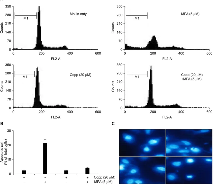
Fig. 6. Effect of CoPPIX, HO-1 inducer, in MPA induced apoptosis on Jurkat cells. (A) Cells were treated with 5μM MPA in the absence and presence of 20μM MPA for 48 hr. After PI staining, the fluorescence intensity of more than 10,000 cells was analyzed using a flow cytometry. (B) Apoptosis (%) in A was measured and plotted against dose. Data represent the mean±standard deviation (S.D.) of quadruplicates. (C) Cells were stained with 10μg/ml DAPI and visualized under fluorescence microscope.
2) MPA에 의한 Jurkat 세포자멸사에서 세포 내 활성산소 생성(reactive oxygen species)
MPA에 의한 Jurkat 세포의 사멸이 H2O2에 의한 세포사멸 임을 밝히기 위하여 2', 7'-dichlorofluorescin diacetate 염색으 로 세포 내 H2O2의 생성을 flow cytometry를 통하여 조사, 분 석하고 M1 값으로 표시하였다. 그 결과, 5μM 농도의 MPA 처리시 대조군인 48.8%에 비하여, 12시간에 56.2%, 24시간에 68.1%, 48시간에 94.0%로 시간의존적인 증가를 보였다(Fig.
3). 이상의 결과는 MPA에 의한 세포 내 활성산소인 H2O2 생 성이 세포자멸사에 기여하였을 가능성을 시사하였다.
3) MPA에 의한 Jurkat 세포자멸사에서 HO-1 발현 변화 에 미치는 영향
산화적 손상에 의한 방어기작으로 세포 내 발현이 증가 되는 HO-1 단백질에 대한 MPA의 효과를 Western blot 방법 으로 조사하였다. Jurkat 세포의 HO-1 단백질 발현은 5μM MPA 처리시 24시간에 약간 증가하였다가 36시간 이후부터
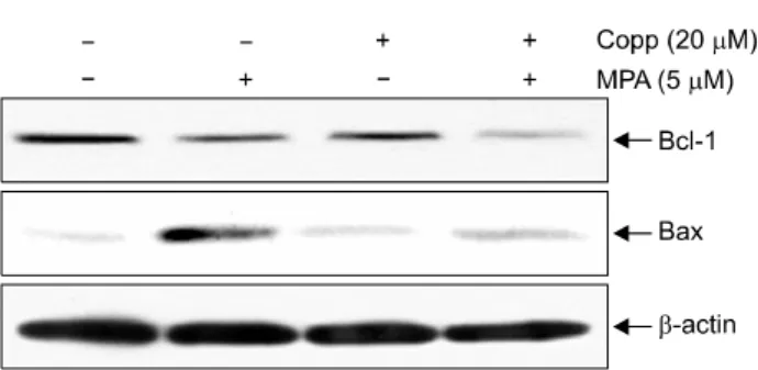
Fig. 7. Effects of CoPPIX on expression of Bcl-2 and Bax proteins in MPA treated Jurkat cells. Cells were treated with MPA for 36 hr in the absence or presence of CoPPIX. Equal amounts of protein from cell lysate were subjected on 15%
SDS-PAGE, transferred onto nitrocellulose membrane and immunoblotted with anti-Bcl-2, anti-Bax and anti-β-actin antibodies. The immunoreactive signals were visualized by ECL detection kit.
감소하였다(Fig. 4).
4) HO-1 inducer가 MPA에 의한 Jurkat 세포자멸사에 미 치는 영향
앞에서 MPA 처리시 HO-1의 발현이 감소하였으므로 MPA 에 의한 Jurkat 세포의 세포 죽음에서 HO-1의 역할을 조사 하고자 하였다. 먼저 HO-1 inducer인 CoPPIX (20μM)를 6시 간 전 처리한 후 MPA를 36시간 처리하여 세포를 포집한 후 HO-1의 발현 정도를 Western blot 분석법으로 조사하였 다. 그 결과 CoPPIX는 HO-1의 발현을 현저하게 증가시켰으 며 MPA 처리에 의해 감소된 HO-1의 발현도 현저하게 증가 시켰다(Fig. 5). 또한 MPA에 의한 세포자멸사 정도를 유식 세포분석기로 조사한 결과, 5μM의 MPA 48시간 처리시 대 조군 3.53%에 비하여 23%의 세포자멸사를 보였으나 CoPPIX 처리시 5.8%로 감소하였다(Fig. 6A). 이때 CoPPIX 자체는 세포독성을 보이지 않았으며 이를 histogram으로 표시하였 다(Fig. 6B).
MPA에 의한 Jurkat 세포의 DNA 분절에 미치는 영향을 DAPI 염색을 통해 핵의 형태를 형광현미경으로 관찰하였 다. 그 결과 대조군과 CoPPIX 단독처리군에서는 타원형의 균일한 형광세기를 나타냈지만 MPA 처리군에서는 핵산이 분절되었다. 그러나 CoPPIX 전처리에 의하여 핵의 분절이 억제되었다(Fig. 6C). 이상의 결과는 CoPPIX는 Jurkat 세포 에서 HO-1의 발현을 증가시켰으며, HO-1의 발현 증가는 MPA에 의한 세포사멸을 억제하였으므로, MPA에 의한 Jur- kat 세포사멸은 HO-1의 발현 감소에 의해 매개되었을 가능 성을 시사하였다.
을 실시하였다. 그 결과 MPA에 의해 감소되었던 Bcl-2 단 백질의 발현양에는 차이가 없었으며, MPA 처리에 의해 발 현이 현저히 증가되었던 Bax 단백질은 발현양이 감소하여 대조군 수준의 발현 양상을 보였다(Fig. 7).
고 찰
MPA는 T 림프구와 B 림프구의 증식을 선택적으로 억제 한다.(11) 이는 MPA가 GTP뿐만 아니라 ATP 생성까지 영향 을 주는 IMPDH의 활성을 억제하고 GTP의 고갈을 유도하 기 때문이다.(12)
활성산소종은 활성화된 호중구, xanthine oxidase, 미토콘 드리아 호흡 및 아라키돈산 대사 과정을 통해서 생성된다.
(7) 비록 superoxide dismutase, catalase, glutathione peroxidase 효소 등이 세포 내에서 생성된 활성산소종을 제거하기 위 한 생리적 방어기전으로 작용하지만, 과도한 활성산소종은 이들 방어체계를 초과하여 세포손상을 초래한다.(10) 신체의 각 장기는 환경에서 오는 다양한 스트레스로부터 항상성을 유지하기 위하여 즉각적으로 반응한다. 카드뮴, 납, 수은과 같은 중금속이나 endotoxin이 체내에 들어오거 나, heat shock, 자외선, 활성산소, 저산소 상태에 노출되면, 장기에서는 스트레스에 대한 반응으로 다양한 단백질과 효소가 유도되면서 생체 방어 작용이 일어난다. 대표적인 예로 헤모글로빈을 비롯한 heme 단백질의 보결분자족인 heme을 담즙색소(biliverdin, bilirubin)와 일산화탄소와 환원 철(Fe2+)로 분해하는 HO를 들 수 있다. HO는 최소 두 가지 의 isozyme (HO-1과 HO-2)이 보고되고 있는데, HO-2는 구 성형 효소이고, HO-1은 각종 스트레스원에 반응하여 세포 내에서 유도 발현되는 효소로 이 효소의 반응을 조사하여 스트레스 검출에 이용할 수 있다.(13) 본 연구에서 MPA에 의한 Jurkat 세포자멸사는 세포 내 활성산소종인 H2O2의 생 성 증가와 HO-1 단백질의 발현 감소를 초래하였다. 특히, 외부 스트레스에 반응하여 항산화 작용을 하는 HO-1이 MPA 처리 24시간에 일시적으로 증가하였으나 MPA 처리 48시간에 현저히 감소하였으며, 세포 내 H2O2는 48시간에
최대의 생성량 증가를 보였다.
저자들의 연구 결과에서는 MPA는 Bcl-2의 발현 감소와 Bax의 발현 증가 혹은 그에 따른 비율의 감소를 초래하여 Jurkat 세포의 세포자멸사를 유도하였을 것으로 판단된다.
특히 이러한 세포자멸사 현상은 HO-1 inducer인 CoPPIX로 HO-1 단백질의 발현을 증가시켰을 경우 MPA에 의한 Jurkat 세포 내 H2O2의 생성을 억제하였고, Bax 단백질의 발현 증 가를 억제하여 세포자멸사를 억제하였다. 따라서, MPA는 Jurkat 세포에서 활성산소의 생성을 유도하고 이를 위한 방 어기작인 HO-1의 발현 감소를 유도하여 세포자멸사를 초 래하였다고 판단된다. 특히 HO-1의 발현은 Nrf2라는 전사 활성인자에 의해 조절되는 것이 잘 알려져 있으며,(14) MPA 에 의한 Jurkat 세포자멸사에서 이에 대한 정확한 조절 기전 연구가 요구된다.
결 론
MPA는 Jurkat 세포에서 활성산소의 생성을 유도하고 이 를 위한 방어기작인 HO-1의 발현 감소를 유도하여 세포자 멸사를 초래하는 한 원인으로 판단된다.
REFERENCES
1) Florey HW, Jennings MA, Gilliver K, Sanders AG. Myco- phenolic acid: an antibiotic from penicillium brevicompactum Dierckx. Lancet 1946:247;46-9.
2) Allison AC, Kowalski WJ, Muller CD, Eugui EM. Mechan- isms of action of mycophenolic acid. Ann N Y Acad Sci 1993;696:63-87.
3) Allison AC, Eugui EM. Mycophenolate mofetil and its mecha- nisms of action. Immunopharmacology 2000;47:85-118.
4) Natsumeda Y, Carr SF. Human type I and II IMP dehydrogen- ases as drug targets. Ann N Y Acad Sci 1993;696:88-93.
5) Konno Y, Natsumeda Y, Nagai M, Yamaji Y, Ohno S, Suzuki
K, et al. Expression of human IMP dehydrogenase types I and II in Escherichia coli and distribution in human normal lym- phocytes and leukemic cell lines. J Biol Chem 1991;266:
506-9.
6) Nagai M, Natsumeda Y, Konno Y, Hoffman R, Irino S, Weber G. Selective up-regulation of type II inosine 5'-monophosphate dehydrogenase messenger RNA expression in human leuke- mias. Cancer Res 1991;51:3886-90.
7) Allison AC, Eugui EM. The design and development of an immunosuppressive drug, mycophenolate mofetil. Springer Se- min Immunopathol 1993;14:353-80.
8) Goda N, Suzuki K, Naito M, Takeoka S, Tsuchida E, Ishimura Y, et al. Distribution of heme oxygenase isoforms in rat liver.
Topographic basis for carbon monoxide-mediated microvascu- lar relaxation. J Clin Invest 1998;101:604-12.
9) Hayashi S, Takamiya R, Yamaguchi T, Matsumoto K, Tojo SJ, Tamatani T, et al. Induction of heme oxygenase-1 sup- presses venular leukocyte adhesion elicited by oxidative stress:
role of bilirubin generated by the enzyme. Circ Res 1999;
85:663-71.
10) Gao Z, Huang K, Xu H. Protective effects of flavonoids in the roots of Scutellaria baicalensis Georgi against hydrogen peroxide-induced oxidative stress in HS-SY5Y cells. Pharma- col Res 2001;43:173-8.
11) Lee WA, Gu L, Miksztal AR, Chu N, Leung K, Nelson PH.
Bioavailability improvement of mycophenolic acid through amino ester derivatization. Pharm Res 1990;7:161-6.
12) Sollinger HW. Mycophenolate mofetil for the prevention of acute rejection in primary cadaveric renal allograft recipients.
U.S. Renal Transplant Mycophenolate Mofetil Study Group.
Transplantation 1995;60:225-32.
13) Lemos FB, Ijzermans JN, Zondervan PE, Peeters AM, van den Engel S, Mol WM, et al. Differential expression of heme oxy- genase-1 and vascular endothelial growth factor in cadaveric and living donor kidneys after ischemia-reperfusion. J Am Soc Nephrol 2003;14:3278-87.
14) Park SH, Jang JH, Li MH, Na HK, Cha YN, Surh YJ.
Nrf2-mediated heme oxygenase-1 induction confers adaptive survival response to tetrahydropapaveroline-induced oxidative PC12 cell death. Antioxid Redox Signal 2007;9:2075-86.