The effect of tumor necrosis factor (TNF)-α to induce matrix metalloproteinase (MMPs) from the human dental pulp, gingival, and periodontal ligament cells
Eun-Mi Rhim1, Sang-Hyuk Park2,3, Duck-Su Kim2, Sun-Young Kim2,3, Kyoung-Kyu Choi2,3, Gi-Woon Choi2,3*
1Department of Conservative Dentistry, St. Paul’s Hospital, The Catholic University School of Medicine,
2Oral Biology Institutue, Kyung Hee University, 3Dept. of Conservative Dentistry, Kyung Hee University School of Dentistry, Seoul, Korea
Objectives: In the present study, three kinds of tissues cells (pulp, gingiva, and periodontal ligament) were investigated if those cells express MMP and TIMP when they were stimulated with neuropeptides (sub- stance P, CGRP) or proinflammatory cytokine, TNF-α.
Materials and Methods: The cells cultured from human dental pulp (PF), gingiva (GF) and periodontal liga- ment were (PDLF) stimulated with Mock, SP, TNF-α, and CGRP for 24 hrs and 48 hrs. for an RNase pro- tection assay and Enzyme Linked Immunosorbent Assay.
Cells (PF, GF and PDLF) seeded in 100 mm culture dish were stimulated with SP (10-5, 10-8M) or only with medium (Mock stimulation) for 4hrs and for 24 hrs for RNase Protection Assay, and they were stim- ulated with CGRP (10-5M) and TNF-α(2 ng/mL) for 24 hrs and with various concentraion of TNF-α(2, 10, and 100 ng/mL) for Rnase Protection Assay with a human MMP-1 probe set including MMP 1, 2, 8, 7, 8, 9, 12, and TIMP 2, 3.
In addition, cells (PF, GF and PDLF) were stimulated with Mock and various concentraion of TNF-α(2, 10, and 100 ng/mL) for 24 hrs and with TNF-α(10 ng/mL) for 48 hrs, and the supernatents from the cells were collected for Enzyme Linked Immunosorbent Assay (ELISA) for MMP-1 and MMP-13.
Results: The expression of MMPs in PF, GF, PDLF after stimulation with SP and CGRP were not changed compared with Mock stimulation for 4 hrs and 24 hrs. The expression of MMP-1, -12, -13 24 hrs after stimulation with TNF-αwere upregulated, however the expression of TIMP-3 in PF, GF, PDLF after stim- ulation with TNF-αwere downregulated. TNF-α(2 ng/mL, 10 ng/mL, 100 ng/mL) increased MMP-1 and MMP-12 expression in PF dose dependently for 24 hrs.
Conclusions: TNF-αin the area of inflammation may play an important role in regulating the remodeling of dentin, cementum, and alveolar bone. [J Kor Acad Cons Dent 2011;36(1):26-36.]
Key words:Gingival fibroblasts; Matrix metalloproteinases (MMPs); Periodontal ligament fibroblasts; Pulp fibroblasts; Substance P; Tumor necrosis factor-alpha
-Received 6 October 2005; revised 17 October 2005; accepted 11 June 2010- ABSTRACT
1Rhim EM, DMD, MSD, Ph D, Clinical Professor, Dept. of Conservative Dentistry, St. Paul's Hospital, The Catholic University of Korea
2Park SH, DMD, MSD, PhD, Associate Professor; Kim DS, DMD, MSD, PhD, Clinical Instructor; Kim SY, DDS, MSD, PhD, Assistant Professor;
Choi KK, DMD, MSD, PhD, Professor; Choi GW, DMD, MSD, PhD, Professor, Oral Biology Institutue, Kyung Hee University
3Park SH, DMD, MSD, PhD, Associate Professor; Kim SY, DDS, MSD, PhD, Assistant Professor; Choi KK, DMD, MSD, PhD, Professor;
Choi GW, DMD, MSD, PhD, Professor, Dept. of Conservative Dentistry, Kyung Hee University School of Dentistry, Seoul, Korea
*Correspondence to Gi-Woon Choi, DMD, MSD, PhD
Professor of Division of Dentistry, Graduate school of KyungHee University, 1, Hoegi-dong, Dongdaemun-gu, Seoul, Korea, 130-702 TEL, +82-2-956-9330; FAX, +82-2-956-5108; E-mail: [email protected]
Introduction
Dental pulp is a one of the most important tissues among all of the organs in human body, and inflam- matory process in it is complex anatomically and his- tologically, which is thought to be closely related together in neurogenic, inflammatory, osteoblastic/
osteoclastic processes in dental pulp. Recently, SP has been introduced to induce inflammatory cytokines in human dental pulp cells, and several cytokines have been detected in dental pulp during inflammation including interleukin and tumor necro- sis factor (TNF)-α.1,2 In addition, cytokines and growth factors were revealed to induce the expres- sions Matrix Metalloproteinses (MMPs) from human dental pulp tissues or cultured human dental pulp fibroblast.3,4 Moreover, three kinds of tissues cells (pulp, gingival, and periodontal ligament) may be expected to induce MMP and TIMP with stimulation of neuropeptides or proinflammatory cytokine because those three different kinds of tissues is origi- nated from different parts of tooth germ seperately.
MMPs are a group of enzyme that together can degrade practically all extracellular matrix proteins, including native collagen. They participate in normal tissue formation and remodeling, and the expression of different MMPs may be upregulated during vari- ous pathological conditions, e.g, inflammation, tumor invasion, and metastasis.
Recent findings also indicate that human odonto- blasts may express several other members of MMP- family. Even though the physiological roles of MMPs expressed by odontoblasts are currently unknown, it may be speculated that they could participate in dentin matrix organization prior to mineralization, regulation of matrix mineralization, intratubular dentin formation, or remodeling of the pulp connec- tive tissue.
MMPs are usually divided into five groups: collage- nase, gelatinases, stromelysins, membrane-type MMPs and others. Of the MMPs family, interstitial collagenase (MMP-1) is the key enzyme responsible for degrading type I and type II collagen since the initial breakdown of these fibrillar collagen network is mediated primarily by MMP-15,6 While MMP-8 preferentially degrades type I and MMP-13 type II
collagen.
The biological activity of the MMPs can be regulat- ed post-transcriptionally, or by interaction with spe- cific tissue inhibitors of metalloproteinase (TIMPs).7 TIMP-1 to TIMP-4 have been recently identified.
TIMP-1, a glycoprotein with a molecular mass of 28 kDa, can form a stoichiometric 1 : 1 complex with and inactive MMP-1.5,8 Because the same cells that produce MMP-1 are also capable of synthesizing TIMP-1, the overall molar ratio between MMP-1 and TIMP-1 therefore plays a significant role in connec- tive tissue remodeling.9TIMPs expression is regulat- ed by growth factors and cytokines at the transcrip- tional level. TIMP-3, which is capable of binding to compartments of the ExtraCelluar Matrix (ECM) may also induce cell apoptosis. The TIMPs may also regulate the growth of several different cell types.
MMPs activity is regulated at least three levels:
transcription, activation, and inhibition. In healthy adult tissues, the basal expression is usually low, but various external stimuli (cytokines, growth factors, changes in cell-cell and cell-ECM interactions) can induce the expression. MMP expression increases in pathological condition such as inflammation, tumor invasion, and metastasis.10 Most MMPs are secreted in latent proforms (zymogen) and activated at the cell surface or extracellularly. The activation requires the exposure of the catalystic site, which may be achieved by proteolytic enzymes (including other MMPs), or in vitro by chaotropic agents (such as organomercurials) and pH changes. Also autoactiva- tion is possible, at least in vitro.11
Proinflammatory cytokines, such as interleukin-1 and tumor necrosis factor-α(TNF-α) will initiate and augment subsequent inflammatory cascades leading to tissue destruction. Recently large amount of inter- leukin-1 and tumor necrosis factor-αhave been demostrated to induce rat pulpitis.12 These findings highlighted the significance of proinflammatory cytokines in pulpal injury. It is well established that interleukin-1 and tumor necrosis factor-αcan medi- ate inflammatory tissue destruction by stimulating MMP-1 and TIMP-1 production in several cell types.13,14
Substance P (SP) and calcitonin gene related pep- tide (CGRP) are two important sensory neuropep-
tides expressed in the dental pulp.3 Neurogenic inflammatory reaction in the dental pulp describes the local release of neuropeptides (SP or CGRP) from afferent neurons and might play a role in the patho- genesis of pulpal disease. Although the primary func- tion of these two neuropeptides, upon release, is to induce vasodilation and pulpal blood flow,15they may play a direct role in initiating the local inflammatory cell infiltration.16 In addition, SP levels expressed from pulpal fibroblast cultured from carious teeth were higher compared with sound teeth,17 and Substance P remarkably enhanced MMP-2 activity in pulp cells via intracellular reactive oxygen species (ROS) pathway.18
Because three kinds of soft tissues (pulp, periodon- tal, and gingival tissues) are connected together without any obvious junctions between them, it may be meaningful to investigate if MMPs or TIMPs are induced from the cultured cells (PC, GC, and PDLC) from those tissues when they are stimulated with neuropeptides (SP or CGRP).
In the present study, it was investigated that MMPs and TIMPs are expressed in cultured cells (PC, GC, and PDLC) from those tissues and that the expression is regulated by neuropeptides (substance P and CGRP) or proinflammatory cytokine, TNF-α.
Materials and Methods Sample collection and cell culture
Freshly extracted, intact, caries-free third molars (n=10) were obtained from the patients (15-25 years old) in the Department of Oromaxillofacial Surgery at Kyung-Hee Medical Center. Immediately after extraction, teeth were stored in phosphate buffered saline (PBS) and transferred to the labora- tory. Gingival tissues were cut around the cementoe- namel junction area and the periodontal ligaments were obtained by scratching the root surface. Each tooth was grooved longitudinally with a fissure bur at high speed. The tooth was then split with the driver, and the entire pulp (coronal and radicular) was elevat- ed with cotton pliers to obtain maximum pulp tissue.
Repeated washing with PBS, the tissues obtained from the dental pulp, gingiva, and periodontal liga-
ment were placed in a 48 well plate culture dish containing Dulbecco’s Modified Eagle Medium (DMEM; Life Technologies/GIBCO BRL, Gaithersburg, MD, USA) supplemented with 10% fetal bovine serum (FBS).
The cells from pulp tissue (PF) and gingival tissue (GF), and periodontal ligaments (PDLF) were grown to confluence and passed at 1 : 2 ratio until used for experiments. Cell culture media were supplemented with 100 units/mL penicillin-G, 100 μg/mL strepto- mycin, and 0.25 μg/mL fungizone (Gemini Bio- Products, Inc., Woodland, CA, USA).
Once the cells obtained were confluent at passage 5-8, they were pooled in 100 mm culture dish and cultured until it became confluent for the stimulation with Substance P (SP), Calcitonin gene related pep- tides (CGRP) and TNF-αfor the RNase Protection Assay. In addition, samples were seeded on the 6 well plate, and grown into confluent for the Enzyme- Linked Immunosorbent Assay after the stimulation with TNF-α.
Stimulation of the cells
Synthetic human SP, CGRP (Sigma, St. Louis, MO, USA), and TNF-α(R&D system, Mineapolis, MN, USA) were prepared in sterile water with 0.1%
low-endotoxin bovine serum albumin (BSA, Sigma, St. Louis, MO, USA). Cells were seeded in 100mm culture dish and 6-well plates and grown to conflu- ence before used for experiment. Alpha minimal essential medium containing L-glutamine (Life Technologies/GIBCO BRL, Gaithersburg, MD, USA) without FBS was used 24 hours before cells were stimulated with neuropeptiedes and TNF-α. Cells (PF, GF and PDLF) seeded in 100 mm culture dish were stimulated with SP (10-5, 10-8 M) or only with medium (Mock stimulation) for 4 hours and for 24 hours for RNase Protection Assay, and they were stimulated with CGRP (10-5M) and TNF-α(2 ng/mL) for 24 hours and with various concentraion of TNF-α (2, 10, and 100 ng/mL) for RNase Protection Assay as well.
In addition, cells (PF, GF and PDLF) were stimu- lated with Mock and various concentraion of TNF-α (2, 10, and 100 ng/mL) for 24 hours and with TNF-α
(10 ng/mL) for 48 hours, and the supernatents from the cells were collected for Enzyme Linked Immunosor-bent Assay (ELISA) for MMP-1 and MMP-13. At this time, stimulation of the cells (PF, GF and PDLF) was 2 times longer (48 hours) with low concentration of TNF-α(10 ng/mL) to detect enough proteins from the supernatents out of the cells by using ELISA.
Extraction of total RNA
After the cells were stimulated, the supernatents were stored at -80℃. All kinds of cells stimulated were collected scraping with cell scraper (Fisher Scientific Korea, Seoul, Korea).
5 to 10-μg of total RNA was extracted from all kinds of cells stimulated using the RNeasy Mini kit following the manufacturer’s instructions (Qiagen Korea Ltd. Seoul, Korea).
RNase protection assay
The total RNA extracted were subjected to an RNase protection assay as specified by the manufac- turer, using the human MMP-1 probe set (Becton Dickinson Korea Ltd. Seoul, Korea) and BD RiboQuant Ribonuclease Protection Assay Systems (Becton Dickinson Korea Ltd. Seoul, Korea). The resulting protected RNAs were resolved on 5% dena- turing polyacrylamide gels and exposed to X-ray films.
Enzyme linked immunosorbent assay
To detect the MMP-1 and MMP-13, Quantikine proMMP-1 and Quantikine MMP-13 (R&D system, Mineapolis, MN, USA) were purchased and the secretion of MMP-1 and MMP-13 were measured from the supernatant of the cells (PF, GF, PDLF) after the stimulation with TNF-α.
Briefly, after 100 μL of Assay Diluent from the kit was added to each well and 100 μL of standard and sample per well, the plate was incubated for 2 hours at room temperature on a horizontal orbital microplate shaker (0.12" orbit) set at 500 ± 50 rpm.
Each well aspirated and washed, repeating the
process three times for a total of four washes. The well washed with Wash Buffer (400 μL) multi-chan- nel pipette. Complete removal of liquid at each step is essential to good performance. After the last wash, any remaining Wash Buffer removed by aspirating or decanting. Invert the plate and blot it against clean paper towels. 200 μL of Pro-MMP-1 Conjugate and pro-MMP-13. Conjugate were added to each well and incubated for 2 hours at room temperature on shaker. Repeating wash as in above, 200 μL of Substrate solution was added to each well. The plate were protected from light during the incubation for 20 minutes at room temperature on the benchtop.
after 20 minutes, 50 μL of Stop Solution were added to each well. If color change does not appear uniform, gently tap the plate to ensure thorough mixing.
Determine the optical density of each well within 30 minutes, using a microplate reader set to 570 nm.
Results RNase protection assay
After stimulation of the cells for 4 and 24 hours in medium with SP (10-8 M, 10-5 M) or without SP (Mock), RNase Protection Assay demostrated that there were no differences between gene expression of MMPs and TIMPs. The effects of SP (10-8M, 10-5M) concentration on expression of MMPs and TIMPs showed no dose dependency when cells were incubat- ed in the presence of SP (10-8 M, 10-5 M) as shown in Figures 1 and 2.
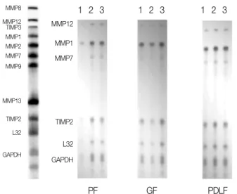
After stimulation of the cells for 24 hours in medi- um with CGRP (10-5M) or TNF-α(2 ng/mL), RNase Protection Assay demostrated that the expression of MMP-12 in PF, GF, PDLF after stimulation with TNF-αwere upregulated, compared with Mock espe- cially in PDLF. The expression of MMP-1 in PF, GF, PDLF after stimulation with TNF-αwere upreg- ulated. The expression of MMP-13 in GF after stim- ulation with TNF-αwere upregulated compared with Mock. The expression of TIMP-3, that is TNF-αcon- verting enzyme inhibitor abundant in PF, GF, PDLF were downregulated in Mock compared with TNF-α (2 ng/mL) as shown in Figure 3.
After stimulation of PF, GF, PDLF for 24 hours in
Figure 1.RNase protection assay 4 hours after stimulation with Mock and Substance P.
1, Mock stimulation only with medium; 2, Stimulation with Substance P (10-8M); 3, Stimulation with Substance P (10-5 M); PF, Human dental Pulp Fibroblasts; GF, Human Gingival Fibroblasts; PDLF, Human Periodontal Ligament Fibroblasts.
1 2 3 1 2 3 1 2 3
PF GF PDLF
MMP8 MMP12 TIMP3 MMP1 MMP2 MMP7 MMP9
MMP13
TIMP2 L32
GAPDH
MMP12 MMP1 MMP7
TIMP2
L32 GAPDH
Figure 2. RNase Protection Assay 24 hours after stimulation with Mock and Substance P.
1, Mock stimulation only with medium; 2, Stimulation with Substance P (10-8M); 3, Stimulation with Substance P (10-5 M); PF, Human dental Pulp Fibroblasts; GF, Human Gingival Fibroblasts; PDLF, Human Periodontal Ligament Fibroblasts.
1 2 3 1 2 3 1 2 3
PF GF PDLF
MMP8 MMP12 TIMP3 MMP1 MMP2 MMP7 MMP9
MMP13 TIMP2 L32 GAPDH
MMP12 TIMP3 MMP1 MMP7 MMP9
TIMP2
L32 GAPDH
Figure 3.RNase Protection Assay 24 after stimulation with TNF-α, Mock, and CGRP
1, Stimulation with TNF-α(2 ng/mL); 2, Mock stimulation only with medium; 3, Stimulation with CGRP (10-5 M);
PF, Human dental Pulp Fibroblasts; GF, Human Gingival Fibroblasts; PDLF, Human Periodontal Ligament Fibroblasts.
1 2 3 1 2 3 1 2 3
PF GF PDLF
MMP8 MMP12 TIMP3 MMP1 MMP2 MMP7 MMP9
MMP13 TIMP2 L32 GAPDH
MMP8 MMP12 TIMP3 MMP1 MMP7 MMP9
MMP13
TIMP2
L32
GAPDH
Figure 4. RNase Protection Assay 24 hrs after stimulation with Mock, and various concentration of TNF-α.
1, Mock stimulation only with medium; 2, Stimulation with TNF-α(2 ng/mL); 3, Stimulation with TNF-α(10 ng/mL); 4, Stimulation with TNF-α(100 ng/mL); PF, Human dental Pulp Fibroblasts; GF, Human Gingival Fibroblasts; PDLF, Human Periodontal Ligament Fibroblasts.
1 2 3 4 1 2 3 4 1 2 3 4
PF GF PDLF
MMP8 MMP12 TIMP3 MMP1MMP2 MMP7 MMP9
MMP13 TIMP2 L32 GAPDH
MMP8 MMP12 TIMP3 MMP1 MMP7 MMP9
MMP13
TIMP2
L32
GAPDH
medium with different dose of TNF-α(2 ng/mL, 10 ng/mL, 100 ng/mL), RNase Protection Assay demostrated that the expression of MMP-1 and MMP-12 in PF after stimulation with TNF-α(2 ng/mL, 10 ng/mL, 100 ng/mL) compared to Mock were upregulated. The effects of TNF-α(2 ng/mL, 10 ng/mL, 100 ng/mL) concentration on expression of MMP-1 and MMP-12 showed dose dependency when PF were incubated in the presence of TNF-α(2 ng/mL, 10 ng/mL, 100 ng/mL) for 24 hours as shown in Figure 4.
Enzyme linked immunosorbent assay
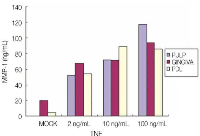
A signigicant induction of MMP-1 in the all kinds of cells studied was observed 24 hours and 48 hours after TNF-αstimulation as shown in Figures 5 and 6. Induction of MMP-1 by TNF-αwas increased dose dependently in all kinds of cells studied. MMP-1 secretion from the PF after stimulation with 2 ng/mL and 10 ng/mL of TNF-αshowed similar level, where- as 100 ng/mL of TNF-αshowed significant higher level of MMP-1 at 24 hours. MMP-1 secretion from
the GF was as same as that of PF for 24 hrs. Both 2 ng/mL and 10 ng/mL of TNF-αinduced similar level of MMP-1, whereas 100 ng/mL of TNF-αshowed higher level of MMP-1 than 2 ng/mL stimulation for 24 hours. An 11-fold increase of MMP-1 from PDLF after TNF-α(2 ng/mL) was when compared with mock stimulation for 24 hours, whereas 10 ng/mL and 100 ng/mL of TNF-αshowed similar level of MMP-1. MMP-1 secretion from all kinds of cells studied with TNF-α(10 ng/mL) was significantly higher than that of mock stimulation (Figure 6).
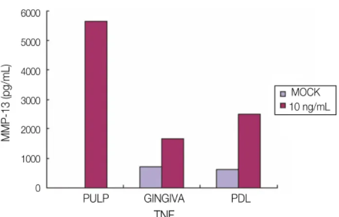
The secretion of MMP-13 was detected from PF after stimulation with TNF-α(2 ng/mL, 10 ng/mL, and 100 ng/mL) and Mock stimulation for 24 hours, nevertheless the secretion level was not dose-depen- dent shown as shown in Figure 7. The secretion from GF was detected only after 2 ng/mL of TNF-αstimu- lation for 24 hours and that from PDLF was after 10 ng/mL and 100 ng/mL of TNF-αstimulation for 24 hours (Figure 7). MMP-13 secretion from all kinds of cell studied 48 hours after stimulation with TNF-α (10 ng/mL) was significantly increased compared with Mock stimulation (Figure 8).
140 120 100 80 60 40 20 0
MMP-1 (ng/mL)
MOCK 2 ng/mL 10 ng/mL 100 ng/mL TNF
PULP GINGIVA PDL
Figure 5. Secretion of MMP-1 after stimulation with mock and various concentration of TNF-αfor 24 hrs.
PF, Human dental Pulp Fibroblasts; GF, Human Gingival Fibroblasts; PDLF, Human Periodontal Ligament Fibroblasts.
120 100 80 60 40 20 0
MMP-1 (ng/mL)
PULP GINGIVA PDL TNF
MOCK 10 ng/mL
Figure 6.Secretion of MMP-1 after stimulation with mock and TNF-α(10 ng/mL) for 48 hrs.
PF, Human dental Pulp Fibroblasts; GF, Human Gingival Fibroblasts; PDLF, Human Periodontal Ligament Fibroblasts.
Discussion
Abnormal soft tissue degradation in tooth is one of the characteristic features of dental pulp and tissues surrounding tooth (periodontal ligaments and gingi- vae). Collagens type I and III are the primary com- ponents of the connective tissue of the dental pulp.
These fibrillar collagens are resistant to most prote- olytic enzymes. Among the members of an extensive family of MMPs, only a limited numbers of these can cleave the highly structured fibrillar collagens. These include MMP-1 (collagenase-1), MMP-8 (collage- nase-2), MMP-13 (collagenase-3), and MMP-2, 9 (gelatinase). Therefore, this study demonstrated that cells cultured from pulps, periodontal ligaments, have the ability to degrade type Icollagen, which sug- gests that these cells play a major role in remodeling the extracellular matrix in pulp, gingiva, and peri- odontal ligament.
In dental pulp, the extracellular matrix (ECM) is composed mainly of type I and III collagens. MMP-1 is the only enzyme that can degrade the triple helix of type I and III collagens and render the sensitive to further digestion by other proteinases.5,6
Most of the MMPs characterized proved to be more
abundant in the odontoblasts, MMP-1, -2, -9, -10, - 11, -13, -14, -15, -16, -17, -19, -20, and -23 were expressed by mature human odontoblasts and pulp tissue.6,19On the contrary, MMP-1, -7, -12, TIMP-1, and -2 were expressed in all of cells (PF, GF, and PDLF) studied in this experiment without any stim- ulation.
Bone Morphogenic Protein-2 (BMP-2) significantly up-regulated MMP-9 mRNA in human odontoblast,20 suppressed MMP-1 in bone cells.21 However, no expression of MMP-13 could be detected in osteo- clasts, but expression of MMP-13 mRNA was detect- ed in osteoblasts and fibroblasts primarily on the inner side of calvarial bone of skull.22MMP-9 mRNA was rarely expressed from all kinds of cells studied, and the expression of MMP-9 mRNA was not increased after the stimulation with neuropeptide and proinflammatory cytokine.
IL-1 and TNF-αregulated the expression of MMP- 1 and TIMP-1 mRNA synthesis from pulp derived cells.23-25 MMP-1 mRNA expression was detected in all of cells studied, and secretion of MMP-1 was increased from all kinds of cell studied in dose depen- dent manner 24 hours after stimulation with TNF-α.
The constitutive MMP-1 serves as remodeling ele-
1800 1600 1400 1200 1000 800 600 400 200 0
MMP-13 (pg/mL)
MOCK 2 ng/mL 10 ng/mL 100 ng/mL TNF
PULP GINGIVA PDL
Figure 7.Secretion of MMP-13 after stimulation with mock and different dose of TNF-α(2 ng/mL, 10 ng/mL, 100 ng/mL) for 24 hrs
PF, Human dental Pulp Fibroblasts; GF, Human Gingival Fibroblasts; PDLF, Human Periodontal Ligament Fibroblasts.
6000
5000
4000
3000
2000
1000
0
MMP-13 (pg/mL)
PULP GINGIVA PDL TNF
MOCK 10 ng/mL
Figure 8.Secretion of MMP-13 after stimulation with mock and TNF-α(10 ng/mL) for 48 hrs.
ment of connective tissue under physiological condi- tion. Collagenase expression can be stimulated with cytokines. Providing further evidence for the sugges- tion that MMPs may participate to the regulation of defensive responses against external irrigation in the human dentin-pulp complex.
The expression of mMMP-8 was down-regulated by Transforming growth factor (TGF)-β1 in human odontoblasts and dental pulp cells,3and MMP-8 had a role in pulpal and periapical inflammation.26MMP- 8 was expressed after TNF-αstimulation from all kinds of cells studied, neither Mock and neuropeptide stimulation. Nevertheless, the expression of MMP-12 (Metalloelastase) from all of cells studied were down- regulated after TNF-αstimulation.
TIMPs may have the ability to antagonize either the activity or zymogen form of different MMPs.27 TIMPs seem to be the major local inhibitor of MMPs.17The concominant production of MMP-1 and TIMP-1 mRNA after TNF-αstimulation suggested a natural protective mechanism against dental pulp ECM degradation. Eventhough TIMP-1 mRNA intensity induced by TNF-αwas lower than MMP-1.
MMP-1 and TIMP-1 from pulp cells was increased compared with Mock stimulation.28The expression of TIMP-3 from all of cells studied (especially in dose dependent manner from PF) was incresed 24 hours after TNF-αstimulation.
The expression of MMP-1 and MMP-13 has also been documented in mineralized tissue-producing cells other than odontoblasts, for example, human odontoblasts express both MMP-1 and MMP-13.21,22 MMP-1 was equally expressed by both odontoblasts and pulp tissues, whereas MMP-13 expression was five times more abundant in pulp tissue compared with odontoblasts. The MMP-1 expression level in healthy dentin-pulp complex cells was very low but inducible.20 The constitutive MMP-1 serves as remodeling element of connective tissue under physi- ological condition. Collagenase expression can be stimulated with cytokines. Providing further evidence for the suggestion that MMPs may participate to the regulation of defensive responses against external irrigation in the human dentin-pulp complex.
TIMPs may have the ability to antagonize either the activity or zymogen form of different MMPs.
TIMPs seem to be the major local inhibitor of MMPs.
The concomitant production of MMP-1 and TIMP-1 mRNA after TNF-αstimulation suggested a natural protective mechanism against dental pulp ECM degradation. However, the amount of MMP-1 gene synthesized was considerably higher than TIMP-1 after proinflammatory cytokine stimulation.23 Resident dental pulp fibroblasts might contribute to the pathogenesis of pulpitis in that they could pro- duce a significant amount of mediators related to inflammatory tissue destruction.
The current study demonstrated that 3 kinds of cells in tooth (dental pulp, gingiva, and periodontal ligament) mediated type I collagen degradation can be enhanced by treatment with TNF-α, however, they were not regulated to induce MMPs by Substance P.
These results further demonstrate that cytokines can influence the secretion of MMPs from pulp, gingiva, and periodontal ligament, which may lead to dental pulp and periodontal tissue degradation.
Conclusions
According to this study, the results were as follows:
1. The expression of MMPs in PF, GF, PDLF after stimulation with SP and CGRP were not changed compared with Mock stimulation for 4 hours and 24 hours.
2. The expression of MMP-1 in PF, GF, PDLF 24 hours after stimulation with TNF-αwere upregu- lated.
3. The expression of MMP-12 in PF, GF, PDLF after stimulation with TNF-αwere upregulated, especially in PDLF.
4. The expression of MMP-13 in all kinds of cells studied 24 hours after stimulation with TNF-α were upregulated.
5. The expression of TIMP-3 in PF, GF, PDLF after stimulation with TNF-αwere downregulated.
6. TNF-α(2 ng/mL, 10 ng/mL, 100 ng/mL) increased MMP-1 and MMP-12 expression in PF dose dependently for 24 hours.
MMP-1 secretion in PF, GF, and PDLF can poten- tially be up regulated by TNF-αleading to type I/III collagens degradation. MMP-12 (metalloelastase) is induced in PDLF, although the tissue contains
elastin-free fibers. MMP-13 (against type I/II colla- gen) was produced only in GF responding to TNF-α.
Reduced TIMPs may enhance the action of MMPs.
Taken together, our data suggest that TNF-αin the area of inflammation may play an important role in regulating the remodeling of dentin, cementum, and alveolar bone.
References
1. Park SH, Hsiao GY, Huang GT. Role of substance P and calcitonin gene-related peptide in the regulation of interleukin-8 and monocyte chemotactic protein-1 expression in human dental pulp. Int Endod J 2004;37:185-192.
2. Patel T, Park SH, Lin LM, Chiappelli F, Huang GT.
Substance P induces interleukin-8 sectretion from human dental pulp cells. Oral Surg Oral Med, Oral Path, Oral Rad, and Endodontics 2003;96:478-485.
3. Palosaari H, Wahlgren J, Larmas M, Ro¨nkã H, Sorsa T, Salo T, Tjãderhane L. The Expression of MMPs-8 in human odontoblasts and dental pulp cells is down-reg- ulated by TGF-β1. J Dent Res 2000;79:77-84.
4. Wisithphrom K and Windsor LJ. The effects of Tumor Necrosis Factor-α, Interleukin-1β, Interleukin-6, and Transforming Growth Factor-β1 on Pulp Fibroblast Mediated Collagen Degradation. J Endod 2006;32:
853-861.
5. Krane SM. Clinical importance of metalloproteinases and their inhibitors. Ann Acad Sci 1994;732:1-10.
6. Birkedal -Hansen H, Moore WG, Bodden MK, Windser LJ, Birkedal -Hansen B, Decarlo A, Engler JA. Matrix metalloproteinases; a review. Crit Rev Oral Biol Med 1993;4:197-250.
7. Sternlight MD, Werb Z. How matrix metalloproteinases regulate cell behavior. Annu Rev Cell Dev Biol 2001;
17:463-516.
8. Douglas DA, Shi YE, Sang QA. Computational sequence analysis of the tissue inhibitor metallopro- teinases family. J Protein Chem 1997;16:237-55.
9. Dean DD, Martel-Pelletier J, Pelletier JP, Howell OS, Woessner JF Jr. Evidence for metalloproteinases and metalloproteinases inhibitor imbalance in human osteoarthritic cartilage. J Clin Invest 1989;84:678-85.
10. Stamenkovic I. Matrix metalloproteinases in tumor invasion and metastasis. Cancer Biology 2000;10:415- 433.
11. Tjãderhane L, Palosaari H, Sulkala M, Wahlgren J, Salo T. The Expression of Matrix metalloproteinase (MMPs) in Human odontoblasts. Dentin/Pulp complex 2000. I 45-51.
12. Tani-Ishii N, Wang CY, Stashenko P. Immonolocal- ization of bone resorptive cytokines in rat pulp and periodontal lesions following surgical pulp exposure.
Oral Microbiol Immunol 1995;10:213-9.
13. Mauviel A, Halcin C, Vasiloudes P, Parks WC, Kurkinen M, Uitto J. Uncoordinate regulation of colla- genase, stromelysin, and tissue inhibitor of metallopro- teinases genes by prostaglandin E2: selective enhance- ment of collagenase gene expression in human dermal fibroblasts in culture. J cell Biochem 1994;54:465-72.
14. Daphna-Iken D, Morrison AR. Interleukin-1βinduces interstitial collagenase gene expression and protein secretion in renal mesangial cells. Am J Physiol 1995;
269:F381-7.
15. Wakisaka S. Neuropeptides in the dental pulp: distrib- ution, origins, and correlation. J Endod 1998;16:67- 69.
16. Fistad I, Kvinnsland IH, Jonsson R, Heyeraas RJ.
Effect of intermittent long lasting electrical tooth stim- ulation and pulpal blood flow and immunocompetent cell: a hemodynamic and immunohistochemical study in young rat molars. Exp Nerol 1997;146:230-239.
17. Killough SA, Lundy FT, Irwin CR. Substance P expres- sion by human dental pulp fibroblasts: a potential role in neurogenic inflammation. J Endod 2009;35:73-77.
18. Wang FM, Hu T, Cheng R, Tan H, Zhou XD.
Substance P influenced gelatinolytic activity via reac- tive oxygen species in human pulp cells. Int Endod J 2008;41:856-862.
19. Coil J, Tam E, Waterfield D. Proinflammatory cytokine profile in pulp fibroblasts stimulated with Lipopoly- saccharide and Methyl Mercaptan. J Endod 2004;30:
88-91.
20. Palosaari H, Pennington CJ, Larmas M, Edwards DR, Tjãderhane L, Salo T. Expression profile of matrix metalloproteinase (MMPs) and tissue inhibitor of MMPs in human odontoblasts and pulp tissue. Eur J Oral Sci 2003;111:117-127.
21. Takiguchi T, Kobayashi M, Suzuki R, Yamaguchi R, Isatsu K, Nishihara T, Nagumo M, Hasegawa K.
Recombinant human bone morphogenetic protein-2 stimulates osteoblast differentiation and suppresses matrix metalloproteinases-1 production in human bone cells isolated from mandibule. J Period Res 1998;33:
476-485.
22. Johansson N, Saarialho-Kere U, Airola K, Herva R, Nissinen L, Westermarck J, Vuorio E, Heino J, Kãhãri VM. Collagenase-3 (MMP-3) is expressed by hyper- trophic chodrocytes, periosteal cells, and osteoblasts during human fetal bone development. Dev Dyn 1997;
208:387-397.
23. Lin SK, Wang CC, Huang S, Lee JJ, Chiang CP, Lan WH, Hong CY. Induction of dental pulp fibroblast matrix metalloproteinase-1 and tissue inhibitor of met- alloproteinase-1 gene expression by interleukin-1αand tumor necrosis factor-αthrough a prostaglandin - dependent pathway. J Endod 2001;27:185-188.
24. O’Boskey FJ, Panagakos FS. Cytokines stimulate matrix metalloproteinase production by human pulp cells during long-term culture. J Endod 1998;24:7-10.
25. Ueda I, Matsushima K. Stimulation of plasminogen activator activity and matrix metalloproteinase of human dental pulp derived cells by tumor necrosis fac- tor-α. J Endod 2001;27:175-179.
26. Wahlgren J, Salo T, Teronen O, Sorsa T, Tjaderhane L. Matrix metalloproteinase-8 (MMP-8) in pulpal and periapical inflammation and periapical root-canal exu- dates.Int Endo J. 2002;35:897-904.
27. Bond M, Murry G, Bennett MR, Amour A, Knauper V, Newby AC, Baker AH. Localization of the death domain of tissue inhibitor of metalloproteinases-3 to the N-terminus metalloproteinase inhibition is associ- ated with proapoptotic activity. J Biol Chem 2000;275:
458-468.
28. Yu WH, Yu CS-S, Meng Q, Brew K, Woessner JF.
TIMP-3 binds to sulfated glycosaminoglycans of the extracellular matrix. J Biol Chem 2000;275:31226- 31232.
29. Hummel V, Kallmann BA, Wagner S, Fu¨ller T, Bayas A, Tonn JC, Benveniste EN, Toyka KV, Rieckmann P.
Production of MMPS in Cerebral Endothelial Cells and Their Role in Shedding Adhesion Molecules. J Neuropathol EXP Neurol 2001;60:320-328.
30. Sulkala M, Larmas M, Sorsa T, Salo T, Tjãderhane L.
The localization of matrix metalloproteinase-20 (MMP- 20, Enamelysin) in mature human teeth. J Dent Res 2002;81:603-607.
31. Leeman MF, Curran S, Murray GI. The structure, reg- ulation, and function of human matrix metallopro- teinase-13. Critical Review in Biochemistry and Molecular Biology 2002;37:149-166.
32. Sawa Y, Horie Y, Yamaoka Y, Ebata N, Kim T, Yoshida S. Production of colony-stimulating factor in
human dental pulp fibroblasts. J Dent Res 2003;82:
96-100.
33. Langton KP, Barker MD, Mckie N. Localization of the functional domains of human tissue inhibitor of metal- loproteinases-3 and the effects of a Sorsby’s fundus dystrophy mutation. J Biol Chem 1998;273:16778- 16781.
34. Graves DT, Cochran D. The Contribution of inter- leukin-1 and Tumor Necrosis Factor to Periodontal Tissue Destruction. J Periodontol 2003;74:391-401.
35. Takashiba S, Naruishi K, Murayama Y. Perspective of cytokine regulation for periodontal treatment: fibrob- last biology. J Periodontol 2003;74:103-110.
36. Thomson BM, Mundy GR, Chambers TJ. Tumor necro- sis factor-αand βinduce osteoblastic cells to stimulate osteoclastic bone resorption. J Immunol 1987;138:775- 9.
국문초록
사람의 치수, 치은, 치주인대 세포에 tumor necrosis factor (TNF)-α로 자극 시 matrix metalloproteinase (MMPs)의 분비에 관한 연구
임은미1∙박상혁2,3∙김덕수2∙김선영2,3∙최경규2,3∙최기운2,3*
1가톨릭대학교 성모병원 치과보존과, 2경희대학교 구강생물학 연구소, 3경희대학교 대학원 치의학과 치과보존학교실
연구목적: 본 연구는 in vitro 상에서 치수, 치은, 치주인대 세포를 neuropeptide (substance P, Calcitonin gene related peptides (CGRP))및 inflammatory cytokine (TNF-α) 으로 자극 시 matrix metalloproteinase (MMPs)의 생성 및 발 현을 관찰한 것으로 치아와 치아 주변 조직에 염증이 존재할 경우 neuropeptide 및 inflammatory cytokine과 치아 경조직 혹은 치조골의 remodeling에 중요한 역할을 하는 matrix metalloproteinase (MMPs)와의 관계를 규명하고자 하였다.
연구 재료 및 방법: 시편으로는 우식이 없는 건전한 제3대구치(n = 10)를 사용하였으며, 발거 후 즉시 Phosphate buffered saline (PBS)에 보관하고 치아에서 치은과 치주인대 조직을 채취하였다. 치아를 종축으로 절단하고 치수 조직을 채취하여 조각으로 분리한 후 시편을 PBS에서 세 번 세척하였다. Plate에 치수, 치은, 치주인대 시편 조각을 위치시켜 Dulbecco’s Modified Eagle Medium (DMEM)을 첨가하여 세포를 배양하였다. 치수, 치은, 치주인대 세포를 culture dish에서 con- fluence에 도달할 때 까지 배양하여 Fetal Bovine Serum (FBS)가 포함되지 않은 배지로 교환하여, 37℃에서 24시간 동안 배양한 후 Phosphate buffered saline (PBS)로 1회 세척하고 Substance P (10-8M, 10-5 M)가 포함된 배양액과 Mock (배양액만 포함됨)으로 4시간, 24시간동안 자극하였다. CGRP (10-6M)을 함유한 배양액 및 TNF-α(2 ng/mL)를 포함한 배양액으로 각각 24시간동안 세포를 자극하였다. 각각 다른 농도의 TNF-α(2 ng/mL, 10 ng/mL, 100 ng/mL)를 포함한 배양액으로 24시간 동안 세포를 자극 한 후 RNase protection assay 및 Enzyme linked immunosorbent assay를 시행하 였다.
결과: SP와 CGRP는 치수, 치은, 치주인대 세포의 MMPs발현에 관여 하지 않았다. TNF-α로 24시간 자극 시 치수, 치은, 치주인대 세포에서 MMP-1,-12, -13의 발현을 증가시켰다. 반면, TNF-α는 치수, 치은, 치주인대 세포들에서 TIMP-3의 발현을 감소시켰다. 서로 다른 농도의 TNF-α(2 ng/mL, 10 ng/mL, 100 ng/mL)로 24시간 자극 시 MMP-1과 MMP- 13의 발현이 증가하였다.
결론: 치아와 치아 주변 조직에 염증이 존재 시 TNF-α가 증가함에 따라 치수, 치은, 치주인대로부터의 치아의 경조직 혹은 치조골의 remodeling에 관여하는 matrix metalloproteinase (MMPs)가 중요한 역할을 하는 것으로 사료된다.
주요단어: 치수세포; 치은세포; 치주인대세포; Matrix metalloproteinases (MMPs); Substance P; Tumor necrosis factor-alpha