Introduction
Periodontitis is an inflammatory disease of the supportive tissues surrounding the teeth and is caused by infection of mi- croorganisms such as Porphyromonas gingivalis and Fusobac- terium nucleatum resulting in the loss of teeth [1]. Periodontitis affects 40–90% of the global population, making it one of the most common diseases in the world [2]. The representa- tive symptom of periodontitis is irreversible destruction of the periodontium, which is composed of the gingiva, cementum, periodontal ligament, and the alveolar bone [3]. Periodontal pathogens such as lipopolysaccharides (LPS) are released from P.gingivalis and elicit an inflammatory response that is
characterized by the production of pro-inflammatory factors in gingival fibroblasts (GFs) and periodontal ligament (PDL) cells, the resident cells of the periodontium. These factors include tumor necrosis factor- α (TNF-α), interleukin (IL)-1β, IL-6, IL- 8, and chemokines [4]. Matrix metalloproteinase (MMP) fam- ily, matrix-degrading enzymes are also released and activated from inflamed GFs and PDL cells resulting in the degradation of the gingiva and PDL tissues [5-7]. LPS-induced inflamma- tory response also induces infiltration of various immune cells into the periodontium [8]. Alveolar bone destruction and teeth loss are also induced by accelerating osteoclastogenesis which is induced by pro-inflammatory cytokines and receptor activa- tor of nuclear factor kappa- Β ligand expressed from GFs, PDL Int J Oral Biol 44:182-190, 2019
pISSN: 1226-7155 • eISSN: 2287-6618 https://doi.org/10.11620/IJOB.2019.44.4.182
Apelin-APJ axis inhibits TNF-alpha-mediated expression of genes involved in the inflammatory response in
periodontal ligament cells
Gyuseok Lee, Won-Hyun Song, Su-Jin Kim, Young-Gwon Kim, and Je-Hwang Ryu*
Department of Pharmacology and Dental Therapeutics, School of Dentistry, Chonnam National University, Gwangju 61186, Republic of Korea
Periodontitis is an inflammatory disease of the supportive tissues surrounding the teeth, and is characterized by irreversible destruction of the gingiva, periodontal ligament (PDL), and alveolar bone, which results in the loss of teeth. In the present study, we elucidated the correlation between periodontitis and apelin (APLN), an adipokine and a regulatory peptide, respectively, which are involved in inflammation and bone remodeling. The expression of APLN is negatively correlated with periodontitis progression in gingival tissue. In addition, treatment with TNF-α downregulated the expression of APLN in PDL cells and gingival fibroblasts, indicating the protective role played by APLN against periodontitis progression. The overexpression of APLN or treatment with exogenous APLN suppressed the TNF-α- mediated catabolic gene expression of MMP1, IL6, and PTGS2 in PDL cells. Moreover, the inhibition of the APLN- APJ axis by ML221, an APJ inhibitor, induced catabolic gene expression in PDL cells. Thus, the results of this study provided evidence to support APLN as a regulatory factor of the inflammatory response during periodontitis.
Keywords: Apelin, APLN, Periodontal ligament, Periodontitis, Inflammation
Received November 11, 2019; Revised December 17, 2019; Accepted December 18, 2019
*Correspondence to: Je-Hwang Ryu, E-mail: [email protected] https://orcid.org/0000-0002-8708-8943 Copyright © The Korean Academy of Oral Biology
Original Article IJOB
cells, and infiltrated immune cells [9,10]. Though decades of research, periodontitis is recognized as a systemic disease that affects other diseases such as diabetes [11], hypertension [12], cardiovascular disease [13], chronic kidney disease [14-16], and alzheimer’s disease [17,18]. Recent studies have shown that metabolic syndromes including obesity and diabetes have been significantly related to periodontitis pathogenesis, indicating that adipokines could be a key factor in regulating pathogenesis [19]. Researchers are currently working to iden- tify the role of adipokines in GF and PDL cells that act as es- sential catabolic regulators for the pathogenesis of periodonti- tis. There are several studies that report the regulatory role of adipokines in periodontitis pathogenesis [20-23].
Among the adipokines, apelin (APLN) is a small regulatory peptide, whose activity is mediated by a receptor named APJ, a G-protein-coupled receptor [24]. APLN-APJ axis is ex- pressed in diverse tissues, including the gastrointestinal tract, brain, kidney, liver, lung, cardiovascular system, and adipose tissues [25]. Especially, several studies have reported the regulatory role of the APLN-APJ axis in bone homeostasis.
According to these previous studies, APLN could regulate the apoptosis, proliferation, and differentiation of osteoblasts and skeletal phenotypes in mice [26-30]. In addition, many stud- ies have indicated that the APLN-APJ axis has certain roles in inflammation-related diseases such as atherosclerosis, diabetic nephropathy, and ischemia reperfusion injury [31].
These previous reports have revealed the relevance of APLN in bone homeostasis and inflammation, the major events during periodontitis progression, allowing us to investigate the role of APLN in periodontitis. In this study, we demonstrate the decreased expression of APLN in the progression of the peri- odontitis, and its protective role in periodontitis by modulating the expression of catabolic factors in human PDL cells and partially in GFs.
Materials and Methods
1. Human gingival tissues
Human gingival tissues containing both epithelial and con- nective tissues were obtained from 16 patients (20–73 years;
40.80 ± 18.80) during tooth extraction comprising eight healthy patients for non-inflamed gingiva and eight chronic periodontitis patients for inflamed gingiva. The Institutional Review Board at the Chonnam National University Dental Hospital (Gwangju, Republic of Korea) approved this study
(CNUDH-2018-003). After all procedures had been fully ex- plained, written informed consent was obtained from each study subject. Gingival tissues were promptly maintained in liquid nitrogen and stored at –80℃ until further use.
2. Cell culture and stimulation
Human PDL cells and GFs were isolated from gingival papil- lary explants, obtained from clinically healthy donors with no systemic and/or periodontal disease who were informed of the purpose of this study. Briefly, after the dissection of gin- gival biopsies using dispase (Gibco BRL, Gaithersburg, MD, USA), the epithelial cell layer was microscopically dissected from the underlying connective tissue and GF was extracted from the subepithelial tissue as described in a previous study [32]. Pieces of PDL tissue were harvested from the middle of the tooth root and digested with 3 mg/mL of collagenase type 1 and 4 mg/mL of dispase [32]. Isolated primary human PDL cells and GFs were treated with the indicated amount of recombinant human APLN protein (MyBioSource, San Diego, CA, USA), human IL-1 β (GenScript, Piscataway, NJ, USA), and human TNF- α (Merck-Millipore, Billerica, MA, USA). Primary cultured human PDL cells and GFs were infected with empty vector (Ad-C) and APLN-expressing adenovirus (Ad-APLN) for 2 hours at the indicated multiplicity of infection (MOI) and incubated for an additional 24 hours alone or in the presence of IL-1 β or TNF-α. The APJ inhibitors, ML221 (Abmole Bio- Science, Houston, TX, USA) was added at the indicated con- centration to the PDL cells. Dimethyl sulfoxide or phosphate- buffered saline was used as a vehicle.
3. RNA isolation, reverse transcription-polymerase chain reaction, and quantitative real time-PCR
Total RNA was isolated from human gingival tissues and
primary cultures of human GFs and PDL cells using TRIzol
reagent (Ambion, Carlsbad, CA, USA). Normal or periodontitis
human gingival tissues were homogenized in TRIzol reagent
using a glass tissue grinder. RNA was reverse-transcribed,
and the complementary DNA was amplified by polymerase
chain reaction (PCR) using Taq polymerase (GeneAll, Seoul,
Korea). Quantitative real time (qRT)-PCR was performed using
an iCycler (Bio-Rad Laboratories, Hercules, CA, USA) and the
SYBR premix Ex Taq (Takara Bio, Shiga, Japan). All qRT-PCR
was performed in duplicates, and the target gene amplification
signal was normalized to that of glyceraldehyde-3-phosphate
dehydrogenase (GAPDH) in the same reaction. The relative levels of APLN (sense:5’-ATG AAT CTG CGG CTC TGC GTG- 3’, antisense:5’-GGA ATT TCC TCC GAC CTC CCT G-3’), MMP1 (sense:5’-GGA GGG GAT GCT CAT TTT GAT G-3’, antisense:5’-TAG GGA AGC CAA AGG AGC TGT-3’), MMP3 (sense:5’-GAT GCG CAA GCC CAG GTG TG-3’, antisense:5’
-GCC AAT TTC ATG AGC AGC AAC GA-3’), IL6 (sense:5’-GTA CAT CCT CGA CGG CAT CTC AG-3’, antisense:5’-TGG CAT TTG TGG TTG GGT CAG G-3’), and PTGS2 (sense:5’-AAT CCT AGC TGT TCC CAC CCA TG-3’, antisense:5’-AAG GGA GTC
GGG CCA TCA TCA GG-3’) gene expression were analyzed using the comparative C
t(cycle threshold) method, as de- scribed in a previous study [22].
4. Statistical analysis
The data obtained with qRT-PCR were initially tested for confirmation of normal distribution using the Shapiro-Wilk test and subsequently analyzed with Student’s t-test (pair-wise comparisons). The threshold for significance was set at p <
Nomal PD
1.2
1.0
0.8
0.6
0.4
0.2
APLN mRNA levels (fold change )
0.0 APLN
MMP3
GAPDH
Nomal PD
n = 3
< 0.01 p
a
APLN
MMP1
IL6
PTGS2
GAPDH
0 1 2.5 25 50
IL-1 (ng/mL)
0
TNF-1 (ng/mL)
0 25 50
1.2
1.0
0.8
0.6
0.4
0.2
APLN mRNA levels (fold change )
0.0
a a
TNF-1 (ng/mL) APLN
MMP1
IL6
PTGS2
GAPDH
0 1 2.5 0 25 50 0 25 50
1.2
1.0
0.8
0.6
0.4
0.2
APLN mRNA levels (fold change )
0.0
a a
A
B
C
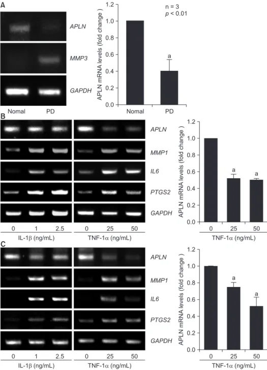
Fig. 1. Downregulated expression of APLN in human gingiva during periodontal disease progression. (A) Total RNA was extracted from whole gingival tissues of normal or periodontitis patients (PD). mRNA levels of indicated genes were determined by real time-polymerase chain reaction (RT-PCR) and relative APLN mRNA levels (compared to those of normal tissues) were quantified by quantitative real time (qRT)-PCR (n = 3). Pri- mary cultures of human periodontal ligament (PDL) cells (B) and GFs (C) were treated with the indicated amounts of cytokines interleukin (IL)-1β and tumor necrosis factor-α (TNF-α) for 24 hours in serum-free condition. The mRNA levels of the indicated genes were detected by RT-PCR analysis. Relative APLN mRNA levels (compared to those of untreated cells) were quantified by qRT-PCR (n = 3).
Values are presented as a mean ± standard
error of the mean.
0.05.
Results
1. Expression of APLN was downregulated in human gingiva affected with periodontitis
To investigate the correlation between APLN and periodonti- tis, we examined the expression levels of APLN in the gingiva of patients with periodontitis. In contrast to the upregulation of MMP3 expression in inflamed gingival tissues, APLN ex- pression was significantly downregulated (Fig. 1A). To confirm the downregulation of APLN expression in the periodontium during periodontitis, we detected the expression levels of APLN in human PDL cells and GFs that were treated with pro- inflammatory cytokines. Treatment with TNF- α dramatically suppressed the expression of APLN in both types of cells.
However, IL-1 β treatment could not regulate the expression of APLN in PDL cells (Fig. 1B) and GFs (Fig. 1C). The upregulation of MMP1, IL6, and PTGS2 represents the severity of inflam-
mation induced by treatment with pro-inflammatory cytokines.
These data suggest a negative correlation between APLN and periodontal inflammation.
2. Adenoviral overexpression of APLN suppressed the expression of genes involved in inflammation and tissue degradation in human periodontal ligament cells
To explore the function of APLN in PDL tissue, we induced overexpression of APLN by adenovirus infection in PDL cells.
Overexpression of APLN by infection of Ad-APLN in human PDL cells led to a marked reduction in the gene expression of MMP1, IL6, and PTGS2 (Fig. 2A). Moreover, overexpression of APLN significantly suppressed the upregulation of MMP- 1, IL-6, and Cox2 mRNA levels in inflamed PDL cells treated with TNF- α (Fig. 2C). However, APLN overexpression only suppressed the IL6 expression among the IL-1 β-induced upregulated genes in inflamed PDL cells (Fig. 2B). These data suggest that APLN could suppress inflammation in periodontal
Con Ad-C 200 400 800
Ad-APLN 1.0
1.0
0.8
0.6
0.4
0.2
Relative mRNA level s
0.0
A B
C
MOI
a MMP1
IL6 PTGS2
Con Ad-C 200 400 800
Ad-APLN 100
80
60
40
Relative mRNA level s 20
0 MOI
a MMP1 IL6 PTGS2
None
IL-1 (2 ng/mL)
Con Ad-C 200 400 800
Ad-APLN 40
30
20
Relative mRNA level s 10
0 MOI
a MMP1 IL6 PTGS2
None
TNF- (50 ng/mL) a
a
a
b b
Fig. 2. Protective effect of adenoviral APLN overexpression against peri- odontitis in periodontal ligament (PDL) cells. Total RNA was extracted from a primary culture of human PDL cells that were infected with the indicated multiplicity of infection (MOI) of Ad-APLN without (A) or with 2 ng/mL of interleukin (IL)-1β (B) and 50 ng/mL of tumor necrosis factor-α (TNF-α) (C) for 24 hours in serum-free condition. The relative mRNA levels of the indi- cated genes were quantified by quantitative real time real time-polymerase chain reaction (n = 3). Values are presented as mean ± standard error of the mean.
Con, concentration.
a
p < 0.05 and
bp < 0.001 compared with Ad-C.
ligament tissues during periodontitis progression.
3. Adenoviral overexpression of APLN partially suppressed the expression of genes involved in inflammation in human gingival fibroblasts
To explore the function of APLN in gingiva tissue, we induced overexpression of APLN by adenovirus infection in human GFs.
Contrary to the results in PDL cells, adenoviral overexpression of APLN in GFs reduced only the mRNA levels of PTGS2 but not the mRNA levels of MMP1 and IL6 (Fig. 3A). Moreover, overexpression of APLN also suppressed the upregulation of PTGS2 expression levels in inflamed GFs treated with IL-1 β and TNF- α. However, the expression of MMP1 and IL6 in- duced by pro-inflammatory cytokines in GFs was not regulated by overexpression of APLN (Fig. 2B and 2C). These data sug- gest that APLN partially suppresses the inflammation of gum tissues during periodontitis progression.
4. APLN-APJ axis could regulate the expression of genes involved in inflammation and tissue degradation in human periodontal ligament cells
Based on the aforementioned results, we further validated the role of APLN-APJ axis in PDL tissues. We treated exog- enous APLN or an APJ blocker, ML221, to human PDL cells.
Similar to the results observed on using Ad-APLN, treatment with recombinant APLN led to a marked reduction in the gene expression of MMP1, IL6, and PTGS2 in PDL cells (Fig. 4A). In addition, recombinant APLN inhibited the expression of MMP1, IL6, and PTGS2 induced by TNF- α in inflamed PDL cells (Fig.
4C). However, IL-1 β-induced expression of MMP1, IL6, and PTGS2 was not regulated by the treatment with recombinant APLN in PDL cells (Fig. 4B). At the highest dose of recombi- nant APLN with TNF- α co-treatment, the expression of MMP1 is rather increased (Fig. 4C). Although not statistically signifi- cant, this reversed expression pattern also appeared at the highest dose of recombinant APLN with IL-1 β co-treatment (Fig. 4B). These data indicated the cytotoxicity or unexpected
Con Ad-C 200 400 800
Ad-APLN 2.0
1.5
1.0
Relative mRNA level s 0.5
0.0
A B
C
MOI MMP1 IL6 PTGS2
Con Ad-C 200 400 800
Ad-APLN 6.0
4.0
2.0
Relative mRNA level s
0.0 MOI
a MMP1 IL6 PTGS2
None
IL-1 (2 ng/mL)
Con Ad-C 200 400 800
Ad-APLN 80.0
60.0
40.0
Relative mRNA level s 20.0
0.0 MOI
a MMP1 IL6 PTGS2
None
a b
Fig. 3. Effect of adenoviral APLN overexpression on periodontitis in gin-
gival fibroblasts (GFs). Total RNA was extracted from the primary culture
of human GFs that were infected with the indicated multiplicity of infection
(MOI) of Ad-APLN without (A) or with 2 ng/mL of interleukin (IL)-1β (B) and
50 ng/mL of tumor necrosis factor-α (TNF-α) (C) for 24 hours in serum-free
condition. The relative mRNA levels of indicated genes were quantified by
quantitative real time real time-polymerase chain reaction (n = 3). Values
are presented as mean ± standard error of the mean.
collateral effect of recombinant APLN at the high dose. In con- trast to the result using recombinant APLN, APJ inhibition by ML221 rather upregulated the mRNA levels of MMP1, IL6, and Cox2 in PDL cells (Fig. 4D). Although the APLN-APJ axis could not block the IL-1β-induced inflammation and tissue degra- dation, these data suggest that the APLN-APJ axis partially suppresses the inflammation and tissue degradation and con- sequently serves as a new therapeutic target for periodontitis.
Discussion
In this study, our results demonstrated that APLN expres- sion in gingival tissues is inversely correlated to periodontitis.
In addition, APLN expression was dramatically downregulated in PDL cells and GFs that were stimulated with TNF-α. This result was unexpected because it was known that TNF-α upregulates APLN expression in human and mouse adipose tissue via phosphoinositide 3-kinase (PI3K), c-Jun N-terminal kinase (JNK), and mitogen-activated protein kinase (MAPK) pathways [33]. Although IL-1β could not significantly decrease the expression of APLN in PDL cells and GFs, we speculated that the expression of APLN was sufficiently inhibited by TNF-α produced by PDL cells, GFs, and other infiltrated im- mune cells. Based on these results, we postulated that APLN could have a protective role in the inflammatory response dur- ing periodontitis progression.
Fig. 4. Regulation of periodontitis by treatment with recombinant APLN or APLN receptor inhibitor (ML221) in periodontal ligament (PDL) cells. Total RNA was extracted from the primary culture of human PDL cells that were treated with the indicated amount of recombinant APLN without (A) or with 2 ng/ml of interleukin IL-1β (B) and 50 ng/mL of tumor necrosis factor-α (TNF-α) (C) for 24 hours in serum-free condition. (D) Total RNA was extracted from the primary culture of human PDL cells treated with the indicated amount of ML221 for 24 hours in serum-free condition. The relative mRNA levels of the indicated genes were quantified by quantitative real time real time-polymerase chain reaction (n = 3). Values are presented as mean ± standard error of the mean.
a