43(1) : 39∼ 45 (2012)
39
엉겅퀴 70% 에탄올 추출물의 RAW264.7 세포에서 Heme oxygenase-1 발현을 통한 항염증 효과
이동성#·김경수#·리 빈·최현규·케오썸엘·전기용·박준형·김윤철* 원광대학교 약학대학
Anti-inflammatory Effect of the Cirsium japonicum var. ussuriense 70%
Ethanolic Extract in RAW264.7 Cells by Heme oxygenase-1 Expression
Dong-Sung Lee#, Kyoung-Su Kim#, Bin Li, Hyun-Gyu Choi, Samell Keo, Ki-Young Jun, Jun-Hyeong Park and Youn-Chul Kim*
Standardized Material Bank for New Botanical Drugs, College of Pharmacy, Wonkwang University, Iksan 570-749, Republic of Korea
Abstract − Cirsium japonicum var. ussuriense has long been used in herbal medicine for the treatment of arthritis, dyspepsia, and bleeding in Korea. In the present study, we investigated anti-inflammatory effects of C. japonicum var. ussuriense against lipopolysaccharide(LPS)-induced activation in RAW264.7 cells by the expression of heme oxygenase (HO)-1. The 70% EtOH extract of the aerial parts of C. japonicum var. ussuriense (CJE), showed the potent anti-inflammatory effects on LPS-induced inflammation in RAW264.7 cells. The anti-inflammatory effect of CJE was demonstrated by the suppression of pro-inflam- matory mediators, including pro-inflammatory enzymes (inducible nitric oxide synthase and cyclooxygenase-2). Furthermore CJE induced HO-1 expression through nuclear translocation of nuclear factor E2-related factor 2 (Nrf2) and increased HO activity in RAW264.7 macrophages. The effects of CJE on LPS-induced NO and PGE2 productions were partially reversed by an HO-1 inhibitor, tin protoporphyrin (SnPP). Therefore, it is suggested that CJE-induced HO-1 expression plays a role of the resulting anti-inflammatory effects in macrophages. These results suggest that CJE may be a promising candidate for the treat- ment of inflammatory diseases.
Key words − Cirsium japonicum var. ussuriense, Heme oxygenase-1, RAW264.7, iNOS, COX-2, Inflammation
엉겅퀴(Cirsium japonicum var. ussuriense)는 국화과(Com- positae)에 속하는 다년생 초본으로 한국 및 일본, 중국에 자 생한다. 지상부 전체에 흰털 및 거미줄과 같은 털이 밀생한 다. 잎은 우상으로 갈라지며 가장자리에는 결각상의 거친 톱니와 가시가 발달되어 끝이 가시와 같다. 꽃은 가지 끝에 자색으로 6~8월에 핀다. 과실은 수과로 백색의 긴 관모가 있다. 한방에서는 전초를 건조한 것을 대계(Cirsii Herba)라 고 하여, 건위, 소염, 지혈, 진통약으로 사용한다.1,2) 지금까 지 보고된 약리활성은 엉겅퀴 분말의 고지혈증 흰쥐의 지 질대사 개선기능3,4)과 면역증가활성,5) 추출물의 항산화,6) 지 질과산화 억제7)및 암 억제작용8)이 보고되고 있다. 엉겅퀴
는 flavonoid, polyacetylene, phenylpropanoid 계열의 화합 물을 함유하고 있으며1), flavonoid 성분인 pectolinarin은 지 혈작용9)이 보고되었고, apigenin은 혈관이완작용,10,11) 항암
효과12-17)가 보고 되어 있다.
염증반응은 생체나 조직에 물리적 작용이나 화학적 물질, 세균감염 등에 의한 손상이 일어 날 때, 그 손상부위를 수 복 재생하려는 기전이다. 일단 자극이 가해지면 국소적으로 histamine, serotonine, bradykinin, prostaglandins 과 같은 혈 관 확장 물질이 유리되어 혈관 투과성이 증대되면서 염증 을 유발한다. 지속적인 염증반응은 점막손상을 촉진하고, 암 발생 등의 질환을 유도하기도 한다.18) 염증과정 중에 많은 양의 염증유도 사이토카인(proinflammatory cytokines), nitric oxide (NO), prostaglandin E2 (PGE2)가 생성된다.19) Nitric oxide는 nitric oxide synthases (NOS)에 의해 L-arginine으로
#These authors contributed equally to this work.
*교신저자(E-mail):[email protected] (Tel):+82-63-850-6823
부터 생성된 것으로 염증이나 자가면역과 같은 면역학적 반 응에서 중요한 역할을 하는 염증매개조절 인자이다.20) iNOS (inducible nitric oxide synthase)는 염증 반응을 보이는 대식 세포에서 NO를 생성하여 염증 반응을 촉진하는 역할을 한 다21). 외부 자극 또는 자가면역으로 인한 NO의 과도한 분 비는 염증 반응을 촉진시켜 다양한 염증성 질환을 야기한 다. 그러므로 iNOS의 발현 및 그에 따른 NO생성의 조절은 염증 조절에 중요하다.21) Cyclooxygenase (COX)-2 또한 염 증반응 과정에 발현되는 유도성 효소이며, 통증과 발열을 유발하는 prostaglandins (PGs)과 같은 매개물질들을 생성한 다.22, 23)
Heme oxygenase (HO) 유도체 중 하나인 HO-1은 세포내 의 heme을 분해하여 일산화탄소, 철, billiverdin을 만든다.24) 이렇게 분해된 세가지 형태의 생성물들과 HO-1 그 자체는 세포손상 및 사멸의 억제, 항염증 및 항산화 작용을 하는 것 으로 알려져 있으며, 특히 최근에는 HO-1의 발현이 항염증 작용을 통하여 세포 손상을 억제한다는 연구 결과가 보고 되었다.25)본 연구에서는 LPS로 활성화된 RAW264.7 대식 세포에서 엉겅퀴 지상부의 70% 에탄올 추출물인 CJE의 HO-1 발현을 통한 항염증 효과 및 기전을 밝혔다.
재료 및 방법
실험재료 − 본 실험에 사용한 엉겅퀴 지상부는 2011년 6 월 전북 진안군 성수면 좌포리에서 채집 하였으며, 형태학 적 평가를 통하여 동정하였고 표본시료는 천연물 신약 표 준화 소재은행에 보관하였다. 엉겅퀴 지상부 70% 에탄올 추출물 (CJE) 은 천연물 신약 표준화 소재 은행(소재번호 : NNMBS000283)에서 분양 받아 사용하였다.
시약 및 기기 − DMEM 배지와 trypsin-ethylenediamine- tetraacetic acid (EDTA), fetal bovine serum (FBS)는 Gibco Laboratories사에서 구입하였다. Lipopolysaccharide (LPS)와 3'-(4,5-dimethylthiazol-2-yl)-2,5-diphenyltetrazolium bromide (MTT)는 Sigma사에서 구입하였다. 96-Well tissue culture plates와 기타 tissue culture dishes는 Falcon사 제품을 이용 하였다. 흡광도는 BioRad사의 Microplate Reader를 이용하 여 측정하였다. TLC plate는 Merck사의 Silica gel 60 F254 plate를 이용하였다.
시료 제조 – 엉겅퀴 지상부 30 g을 70% 에탄올 수용액 600 ml 로 2시간 동안 가열 환류 추출하고 여과한 다음 여 액을 감압 농축하여 엉겅퀴지상부의 70% 에탄올 추출물 (CJE) 7.35 g을 얻었다. 시료는 실험에 사용한 배지에 녹여 서 사용하였다. 제조된 시료(CJE)는 박층크로마토그래프 (TLC)법으로 함유 성분 패턴 분석을 하였다. 시료를 2.5 mg/
ml의 농도로 메탄올에 용해시켜 검액을 만들고, 10 µl 씩 TLC판에 점적하여 CHCl3 : MeOH (20 : 1)의 용매 조건으
로 전개시키고 이를 10% 황산에 발색하였으며, Rf값이 각 각 0.3, 0.45, 0.75, 0.91인 네 개의 반점들이 확인 되었다.
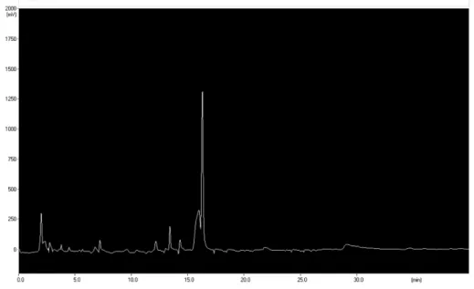
HPLC 분석 − 본 연구에서 사용한 엉겅퀴 지상부 70%
에탄올 추출물(CJE)은 HPLC를 사용하여 함유 성분의 패턴 을 분석하였다. 사용한 HPLC 장치는 Sykam S2100 system (Sykam, 독일)이었으며, 컬럼은 Inertsil ODS-3 (GL Sciences Inc., 4.6 × 150 m, 5µm, 일본)을 사용하였다. 이동상은 acetonitrile, water (JT Baker, 미국)을 사용하였으며, 온도는 실온이었으며, 유속은 분당 0.7 ml이었다. 크로마토그램은 Sykam S3200 UV/VIS Detector (Sykam, 독일)를 이용하여 210 nm에서 검출하였다. 이동상 용매로는 acetonitrile:water (1:4)에서 acetonitrile:water (4:1)의 조성으로 40분 동안 변 화시켰다.
세포배양 − RAW264.7 대식세포는 ATCC에서 동결상태로 구입하였다. RAW264.7 대식세포는 FBS (10%)과 penicillin (100 U/ml)와 streptomycin sulfate (100 µg/ml)가 함유된 DMEM배지에서 습한 조건의 37oC, 5% CO2 Incubator에서 배양하였다.
세포독성 − 본 실험에서 RAW264.7 대식세포에 대한 세 포독성 및 실험 시 처리 농도를 결정하고, 엉겅퀴 지상부 추 출물의 세포독성을 측정하기 위해 MTT assay를 사용하였 다.26) 간단히 기술하면 먼저 96 well plate에 1×10⁴ cells/
well로 동일하게 분주하고 24시간 동안 배양하였다. 기존의 배지를 제거하고 새로운 배지를 넣어준 후 DMSO에 녹인 시료를 다양한 농도(10, 20, 40, 80 µg/ml)로 DMEM 배지 에 희석하여 첨가하였다. DMSO의 처리 농도는 배지 대비 0.1% 이하가 되도록 하였다. 이를 다시 24시간 배양한 후에 배지를 제거하고 MTT시약(5 mg/ml)을 넣고, 4시간 동안 방 치한 후 상등액을 제거하였다. 형성된 formazan의 각 well 에 DMSO 20 µl를 첨가한 후 orbital shaker를 이용하여 녹 이고, 30분 후 595 nm에서 흡광도를 측정하였다. 실험은3회 반복 실시하여 평균값을 구하였으며, Control의 흡광도 값 을 기준으로 세포 생존율을 비교하였다.
Western Blot Analysis − RAW264.7 대식세포를 60 mm dish에 3×10⁵ cells/well 밀도로 24시간 배양한 후 각각의 시료를 농도별로 처리하였다. RAW264.7 대식세포에 RIPA buffer를 첨가한 다음, 4oC, 14,000×g에서 25분간 원심분 리하고 상등액을 튜브에 옮겼다. 단백질 정량은 BSA 단 백질 실험 키트를 이용하였고 각각의 시료를 12% SDS- polyacrylamide gel에서 영동하고 nitrocellulose membrane (NC membrane)으로 전사하였다. 전사된 NC membrane을 5% 무지방유가 포함된 신선한 blocking buffer (0.1% Tween 20 in Tris-buggered saline)에서 blocking한 후 iNOS, COX- 2 antibody를 1:1000으로 희석하여 넣고 1시간 동안 반응시 켰다. 다시 2차 antiboby (Anti-mouse IgG)를 1:1000으로 희 석하여 넣고 1시간 동안 반응한 다음, ECL 용액을 1:1로 잘
섞어서 NC membrane 위에 가하여 발광시키고 암실에서 X 선 필름에 감광한 후 현상하였다. 같은 방법으로 actin antibody를 이용하여 actin을 측정한다.
Heme Oxygenase Activity − HO 효소 활성을 Tenhunen 등의 방법27)에 의해 다음과 같이 측정하였다. 세포로부터 얻어진 microsome에 nicotinamide adenine dinucleotide phosphate (NADPH)와 랫의 간 cytosol에서 얻어진 biliverdin reductase를 포함하는 반응용액인 100 mM PBS, 2 mM MgCl2, 3 mg의 랫의 간 cytosol, 0.8 mM NADPH, 2 mM glucose-6-phosphate, 0.2 U의 glucose-6-phosphate dehydro- genase 등을 첨가하고 기질로서 hemin을 20 µM 처리한 후 37oC에서 1시간 동안 암실에서 반응한 뒤 1 ml의 chloroform 으로 반응을 종결하고 450 nm에서 흡광도를 측정하였다.
핵과 세포질 분획의 분리 − RAW264.7 대식세포에 protease inhibitor cocktail I 과 1 mM phenylmethylsulfonyl fluoride (PMSF)를 첨가한 PER-mammalian protein extraction buffer 를 첨가하여 균질화하고 4oC에서 15,000×g로 원심분리 하 였다. 10분간 원심분리 후 상등액을 세포질 분획으로 사용 하기 위하여 -80oC에서 저장하였다. 나머지 침전물은 PBS 로 세척한 후 RIPA buffer [150 mM NaCl, 1% NP-40, 0.5% sodium deoxycholate, 0.1% SDS, 50 mM Tris-HCl (pH 7.4), 50 mM glycerophosphate, 20 mM NaF, 20 mM ethylene glycol tetraacetic acid (EGTA), 1 mM dithiothreitol (DTT), 1 mM Na3VO4, protease inhibitors]를 첨가하고 4oC 에서 15분간 혼합한 후 4oC, 16,000×g에서 15분간 원심분 리 하였다. 이후의 과정은 앞에서 설명한 western blotting 방법을 이용하였다.
Nitrite Assay − 배양된 세포를 5×10⁵ cells/well 수준으로 96 well plate에 100 µl씩 배양한 다음 24시간 동안 배양하
고, 24시간 후 medium을 제거한 후 DMEM으로 희석된 각 농도 별 시료 처리 후 LPS (1 µg/ml)를 처리하여 24시간 후 세포에서 medium으로 분비되어 나온 NO의 양을 Griess 시 약 (0.1% (w/v) N-(1-naphathyl)-ethylenediamine and 1%
(w/v) sulfanilamide in 5% (v/v) phosphoric acid)을 사용하 여 반응하였다. 반응 후 ELISA micro plate reader (Bio Rad Laboratories Inc., California, USA, Model 550)를 사용하여 540 nm에서 측정 하였다.
EIA에 의한 PGE2 측정 − PGE2의 측정은 항체(lyophilized prostaglandin E2 conjugate to horseradish peroxidase)를 사 용하여 prostglandin E2 enzymeimmunoassay system (EIA, Amersham Pharmacia Biotech, Inc., Piscataway, NJ)을 이 용하였다.
통계처리 − 본 실험의 통계처리는 Student’s test 방법을 사 용하였으며, 값은 평균과 표준오차로 나타내었다.
결과 및 고찰
NO는 IL-1β, TNF-α, LPS 등과 같은 염증유발인자 및 암 발생인자에 의해 발현되는 inducible nitric oxide synthase (iNOS)에 의해 과량 발생하며,28) 생성된 NO의 지속적 과발 현은 점막손상을 촉진하고, 그 결과 일부에서는 암 발생 등 의 질환을 유도한다.29) 본 연구에서는 엉겅퀴 지상부의 70%
에탄올 추출물(CJE; Fig. 1)이 갖는 항염증 효과를 측정하 고자 LPS로 iNOS, COX-2 단백질 발현과 그에 따른 NO, PGE2 생성을 유발시킨 RAW264.7 대식세포를 이용하여 항 염증 효과와 그 기전을 연구하였다.
CJE를 농도별로 RAW264.7 대식세포에 처리 하였을 때 세포 독성을 나타내지 않은 10, 20, 40, 80 µg/ml을 실험 가
Fig. 1. HPLC chromatogram of CJE
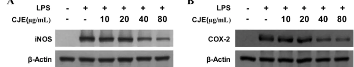
능한 농도로 정하고 실험을 진행 하였다(Fig. 2). RAW264.7 대식세포에 CJE를 10, 20, 40, 80 µg/ml을 처리한 후 3시간 배양하고 LPS 1 µg/ml을 처리하였다. LPS 처리 후 24시간 후에 대표적인 염증 mediator에 영향을 주는지 확인하기 위 해 iNOS, COX-2 발현을 Western blot으로 관찰하였다. LPS 에 의해 증가하던 iNOS, COX-2 단백질의 발현은 CJE (10~
80µg/ml)에 의해 농도 의존 적으로 억제 되었다(Fig. 3).
iNOS와 COX-2에 의해 각각 생성되는 NO와 PGE2 역시 CJE (10~80 µg/ml)에 의해 농도 의존적으로 생성이 억제 되
었다(Fig. 4). 또한, 산화적 스트레스로부터의 뇌세포 보호 기전에 관여하는 중요한 단백질인 heme oxygenase (HO)-1 의 발현 정도를 알아보기 위하여 시료를 12 시간 동안 RAW264.7 대식세포에 처리한 결과, CJE (10~80 µg/ml)을 처리하였을 때 HO-1의 발현이 농도 의존적으로 뚜렷하게 증가 하였고 18시간 후에 극대 효과를 나타냈으며, HO activity 역시 증가함을 나타냈다(Fig. 5). HO-1의 발현 실험 에는 HO-1의 유도물질로 알려진 CoPP (20 µM)를 양성 대 조약물로 사용하였다.
한편, HO-1 단백질 발현에 관여하는 메커니즘 중 nuclear factor-E2-related factor 2 (Nrf2)의 핵 내로의 전사가 매우 중요한 인자로 작용한다고 알려져 있다. 전사 인자로서의 Nrf2는 HO-1과 같은 항산화 단백질 유전자에 존재하는 antioxidant response element (ARE)에 결합하여 이들 유전 자의 발현과 단백질 생성을 항진시킴으로써 산화적 스트레 스에 대한 생체방어기전의 핵심적 역할을 담당한다.30,31) CJE 에 의한 Nrf2의 전사 여부를 알아보기 위하여 80 µg/ml 농 도의 CJE 를 시간별로 처리하고 Western blot을 이용하여 분석하였다. 시간이 경과함에 따라 세포질의 Nrf2는 점점 감소하는 반면, 핵 내부의 Nrf2는 증가하는 양상을 보이는 것으로 보아 Nrf2의 핵 내로의 전사가 이루어졌음을 확인 할 수 있었다(Fig. 6). CJE 의 항염증 효과와 HO-1의 발현 Fig. 2. Effects of CJE on cell viability. RAW264.7 macrophages
were incubated for 24 h with various concentrations of CJE (5- 160µg/mL). Cell viability was determined as described under Materials and method. Data represent the mean values of three experiments ± SD.
Fig. 3. Effects of CJE on protein iNOS (A) and COX-2 (B) expression in RAW264.7 macrophages stimulated with LPS. Cells were pre-treated for 12 h with indicated concentrations of CJE, and 18 h with LPS (1µg/mL). Western blot analysis (A, B) were performed as described in Materials and method, and representative blots of three independent experiments are shown.
Fig. 4. Effects of CJE on nitrite (A), PGE2 (B) production in RAW264.7 macrophages stimulated with LPS. Cells were pre-treated for 12 h with indicated concentrations of CJE, and stimulated 18 h with LPS (1µg/mL). The concentration of nitrite (A), PGE2 (B) were determined as described under Materials and method. Data represent the mean values of three experiments ± SD. *p<0.05 compared to the group treated with LPS.
간의 직접적인 관계를 알아보기 위하여, HO-1 발현 억제제 인 SnPP를 이용하여 실험하였다. HO-1 발현 억제제인 SnPP 를 CJE (80 µg/ml)와 함께 12시간 동안 처리한 실험에서 CJE 에 의해 억제 되었던 NO와 PGE2가 SnPP를 병용투여 함으로써 부분적으로 증가되었다(Fig. 7). 이와 같은 결과로 부터 CJE 의 항염증 효과는 HO-1 단백질 발현과 관련 있 음을 확인 하였다.
따라서, 본 연구를 통해 엉겅퀴 지상부 70% 에탄올 추출 물(CJE) 은 RAW264.7 대식세포에서 Nrf2를 핵 내로 전사 하고 이를 통하여 HO-1 단백질 발현을 함으로써 LPS에 의 한 염증성 사이토카인으로부터 보호활성을 나타낸다는 사 실을 확인하였다.
결 론
엉겅퀴 지상부 추출물의 항염증 효과와 그 보호 기전을 탐 색하였으며, 그 결과 엉겅퀴의 70% 에탄올 추출물(CJE)이 LPS로 유발한 RAW264.7 대식세포의 NO 및 PGE2생성 억 제와 iNOS 및 COX-2 발현 억제를 통한 유의한 항염증 효 과를 나타내었다. 이는 Nrf2의 핵 내 전사 유도를 통한 HO- 1 단백질 발현 기전과 관여 되어 있다고 판단된다. 이러한 결과를 바탕으로 향후 CJE 의 추가적인 다른 항염증 기전 연구와 활성물질의 분리 동정 연구가 필요하다고 판단된다.
Fig. 5. Effects of CJE on HO-1 expression and HO activity in RAW264.7 macrophages. Cells were incubated for 12 h with indicated concentrations of CJE (A, C) and periods with 80 µg/mL of CJE (B). Western blot analysis for HO-1 expression (A, B) was performed as described in Materials and method and representative blots of three independent experiments are shown. HO activity (C) was determined as described in Materials and method. Data represent the mean values of three experiments ± SD.
*p<0.05 compared to the control group; **p<0.05 compared to the group treated with CJE (80 ìg/mL) alone.
Fig. 6. Effects of CJE on nuclear translocation of Nrf2. Cells were treated with 80µg/mL of CJE for 0.5, 1.0 and 1.5 h. The nuclei were fractionated from the cytosol using PER- Mammalian Protein Extraction buffer as described in Experimental part. Nrf2 protein was detected by Western blot analysis, and representative blots of three independent experiments are shown.
Fig. 7. Effects of SnPP on the inhibition of nitrite (A), PGE2 (B) production by CJE pre-treatment in LPS-stimulated macrophages. Cells were pre-treated for 12 h with CJE (80 µg/
mL) in the presence or absence of SnPP (50µM), and stimulated for 18 h with LPS (1 µg/mL). The concentrations of nitrite (A), PGE2 (B) were determined as described in Materials and method. Data represent mean values of three experiments ± SD. *p<0.05 compared to the control group;
**p<0.05 compared to the group treated with LPS alone;
#p<0.05 compared to the group treated with CJE plus LPS.
인용문헌
1.생약학교재편찬위원회 (2007) 생약학, 439-440. 동명사, 서 울
2.한국 약용식물학 연구회 (2001) 종합약용식물학, 297. 학 창사, 서울.
3. Lim, S. S. and Lee, J. H. (1997) Effect of Artemisia princeps var. orientalis and Circium japonicum var. ussuriense on car- diovascular system of hyperlipidemic rat. Korean J. Nutr. 30:
12-18.
4. Lim, S. S., Kim, M. H. and Lee, J. H. (1997) Effect of Arte- misia princeps var. orientalis and Circium japonicum var.
ussuriense on liver function, body lipid, and bile acid of hyperlipidemic rat. Korean J. Nutr. 30: 797-802.
5. Lee, M. K., Moon, H. C., Lee, J. H., Kim, J. D., Yu, C. Y. and Lee, H. Y. (2002) Screening of immune enhancing activities in medicinal herbs, Compositae. Korean J. Med. Crop Sci.
10: 51-57.
6. Lee, H. K., Kim, J. S., Kim, N. Y., Kim, M. J., Park, S. U. and Yu, C. Y. (2003) Antioxidant, antimutagenicity, and anti- cancer activities of extracts from Cirsium japonicum var.
ussuriense Kitamura. Korean J. Med. Crop Sci. 11: 53-61.
7. Park, J. C., Hur, J. M., Park, J. G., Kim, S. C., Park, J. R., Choi, S. H. and Choi, J. W. (2004) Effects of methanol extract of Cirsium japonicum var. ussuriense and its prin- ciple, hispidulin-7-O-neohesperidoside on hepatic alcohol- metabolizing enzymes and lipid peroxidation in ethanol- treated rats. Phytother. Res. 18: 19-24.
8. Liu, S., Luo, X., Li, D., Zhang, J., Qiu, D., Liu, W., She, L.
and Yang, Z. (2006) Tumor inhibition and improved immu- nity in mice treated with flavone from Cirsium japonicum DC. Int. Immunopharmacol. 6: 1387-1393.
9. Shida, H., Umino, T., Tsuji, K. and Kosuge, T. (1987) Studies on antihemorrhagic substances in herbs classified as hemo- statics in Chinese medicine. VII. On the antihemorrhagic principle in Cirsium japonicum DC. Chem. Pharm. Bull. 35:
861-864.
10. Chan, E. C., Pannangpetch, P. and Woodman, O. L. (2000) Relaxation to flavones and flavonols in rat isolated thoracic aorta: mechanism of action and structure–activity relation- ships. J. Cardiovasc. Pharmacol. 35: 326-333.
11. Xu, Y .C., Yeung, D. K. Y., Man, R. Y. K. and Leung, S. W.
S. (2006) Kaempferol enhances endothelium-independent and dependent relaxation in the porcine coronary artery. Mol.
Cell. Biochem. 287: 61-67.
12. McVean, M., Weinberg, W. C. and Pelling, J. C. (2002) A p21(waf1)-independent pathway for inhibitory phosphory- lation of cyclin-dependent kinase p34(cdc2) and concomitant G(2)/M arrest by the chemopreventive flavonoid apigenin.
Mol Carcinog. 33: 36-43.
13. Wang, I. K., Lin-Shiau, S. Y. and Lin, J. K. (1999) Induction of apoptosis by apigenin and related flavonoids through cyto-
chrome c release and activation of caspase-9 and caspase-3 in leukaemia HL-60 cells. Eur. J. Cancer. 35: 1517-1525.
14. Vargo, M. A., Voss, O. H., Poustka, F., Cardounel, A. J., Grotewold, E. and Doseff, A. I. (2006) Apigenin-induced- apoptosis is mediated by the activation of PKC and caspases in leukemia cells. Biochem. Pharmacol. 72: 681-692.
15. Fotsis, T., Pepper, M. S., Aktas, E., Breit, S., Rasku, S., Adler- creutz, H., Wähälä, K., Montesano, R. and Schweigerer, L.
(1997) Flavonoids, dietary-derived inhibitors of cell prolif- eration and in vitro angiogenesis. Cancer Res. 57: 2916-2921.
16. Yin, F., Giuliano, A. E., Law, R. E. and Van Herle, A. J.
(2001) Apigenin inhibits growth and induces G2/M arrest by modulating cyclin-CDK regulators and ERK MAP kinase activation in breast carcinoma cells. Anticancer Res. 21: 413- 420.
17. Chen, D., Daniel, K. G., Chen, M. S., Kuhn, D. J., Landis- Piwowar, K. R. and Dou, Q. P. (2005) Dietary flavonoids as proteasome inhibitors and apoptosis inducers in human leu- kemia cells. Biochem. Pharmacol. 69: 1421-1432.
18. Willoughby, D. A. (1975) Human arthritis applied to animal models. Towards a better therapy. Ann. Rheum. Dis. 34: 471- 478.
19. Posadas, I., Terencio, M. C., Guillén, I., Ferrándiz, M. L., Coloma, J., Payá, M. and Alcaraz, M. J. (2000) Co-regulation between cyclo-oxygenase-2 and inducible nitric oxide syn- thase expression in the time-course of murine inflammation.
Naunyn Schmiedebergs Arch. Pharmacol. 361: 98-106.
20. Berlet, B. S. and Stadtman, E. R. (1997) Protein oxidation in aging, disease, and oxidative stress. J. Biol. Chem. 272:
20313-20316.
21. Palmer, R. M., Ashton, D. S. and Moncada, S. (1988) Vas- cular endothelial cells synthesize nitric oxide from L-arginine.
Nature. 333: 664-666.
22. Liao, C. H., Sang, S., Liang, Y. C., Ho, C. T. and Lin, J. K.
(2004) Suppression of inducible nitric oxide synthase and cyclooxygenase-2 in downregulating nuclear factor-kappa B pathway by Garcinol. Mol. Carcinog. 41: 140-149.
23. Salvemini, D., Manning, P. T., Zweifel, B. S., Seibert, K., Connor, J., Currie, M. G., Needleman, P. and Masferrer, J. L.
(1995) Dual inhibition of nitric oxide and prostaglandin pro- duction contributes to the antiinflammatory properties of nitric oxide synthase inhibitors. J. Clin. Invest. 96: 301-308.
24. Lee, M. S., Lee, J., Kwon, D. Y. and Kim, M. S. (2006) Ondamtanggamibang protects neurons from oxidative stress with induction of heme oxygenase-1. J. Ethnopharmacol.
108: 294-298.
25. Choi, B. M., Kim, H. J., Oh, G. S., Pae, H. O., Oh, H., Jeong, S., Kwon, T. O., Kim, Y. M. and Chung, H. T. (2002) 1,2,3,4,6-Penta-O-galloyl-beta-D-glucose protects rat neu- ronal cells (Neuro 2A) from hydrogen peroxide-mediated cell death via the induction of heme oxygenase-1. Neurosci. Lett.
328: 185-189.
26. Mosmann, T. (1983) Rapid colorimetric assay for cellular growth and survival: application to proliferation and cyto- toxicity assay. J. Immunol. Methods. 65: 55-63.
27. Tenhunen, R., Marver, H. S. and Schmid, R. (1970) The enzymatic catabolism of hemoglobin: stimulation of microso- mal heme oxygenase by hemin. J. Lab. Clin. Med. 75: 410- 421.
28. Knowles, R. G. and Moncada, S. (1994) Nitric oxide syn- thases in mammals. Biochem. J. 298: 249-258.
29. Balkwill, F. and Mantovani, A. (2001) Inflammation and can- cer: back to Virchow? Lancet. 357: 537-545.
30. Balogun, E., Hoque, M., Gong, P., Killeen, E., Green, C. J.,
Foresti, R., Alam, J. and Motterlini, R. (2003) Curcumin acti- vates the heme oxygenase-1 gene via regulation of Nrf2 and the antioxidant-responsive element. Biochem. J. 371: 887- 895.
31. Itoh, K., Chiba, T., Takahashi, S., Ishii, T., Igarashi, K., Katoh, Y., Oyake, T., Hayashi, N., Satoh, K., Hatayama, I., Yama- moto, M. and Nabeshima, Y. (1997) An Nrf2/small Maf het- erodimer mediates the induction of phase II detoxifying enzyme genes through antioxidant response elements. Bio- chem. Biophys. Res. Commun. 236: 313-322.
(2012. 1. 27 접수; 2012. 3. 8 심사; 2012. 3. 9 게재확정)