• 검색 결과가 없습니다.

Calcifying Aponeurotic Fibroma : A Case Report

N/A
N/A
Protected

Academic year: 2021

Share "Calcifying Aponeurotic Fibroma : A Case Report"

Copied!
6
0
0

로드 중.... (전체 텍스트 보기)

전체 글

(1)

Vol.20 No.2. 223-228. Dec., 2003.

INTRODUCTION

1)

Calcifying aponeurotic fibroma, also known as juvenile aponeurotic fibroma, is a very uncommon soft tissue neoplasm which was first described by Keasy in 1953 (1). It occurs predominantly in the hands and feet of children and adolescents. The tumor is characterized by a diffuse or nodular proliferation of fibroblastic cells with scattered foci of peculiar calcification and chondroid area (2). It tends to recur locally after surgical excision. The nature and origin of

책임저자:최준혁, 대구시 남구 대명동 317-1. 영남대학교 의과대학 병리학교실 TEL.(053) 620-3335 FAX.(053) 656-1429 E-mail: [email protected]

the tumor are controversy. To our knowledge, only three substantial series have been published in the past 50 years in English literature (3-5). Two cases have been reported in Korean literature (6,7).

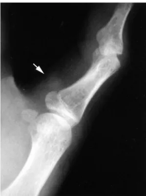
We report a case of calcifying aponeurotic fibroma that occurred in the right thumb of a 14 year-old boy, and review the literature.

CASE REPORT

A 14 year old boy presented with a mass on ulnar aspect of right thumb. The mass

Calcifying Aponeurotic Fibroma : A Case Report

Joon Hyuk Choi, Jae Sung Seo*, Kil Ho Cho

Department of Pathology, Orthopedic Surgery*, and Diagnostic Radiology

College of Medicine, Yeungnam University, Daegu, Korea

-Abstract-

Calcifying aponeurotic fibroma is a rare benign soft tissue tumor that usually involves distal extremities in children and adolescents, especially the hands and feet. We report a case of calcifying aponeurotic fibroma arising in a 14-year-old boy who complained of right thumb mass. Surgical excision was performed. The resected specimen showed a 2.0×1.5 cm grayish white, fibrotic tissue. Histologic examination showed proliferation of fibroblastic cells with infiltrative growth pattern. Foci of calcification and chondroid differentiation were present.

Key Words: Calcifying aponeurotic fibroma, Soft tissue, Immunohistochemistry

(2)

occurred in childhood. There were no pain and tenderness. Physical examination revealed a firm, fixed mass on ulnar aspect of the right thumb at the level of the metacarpo- phalangeal joint. The motion of the right thumb was normal. He had a history of myopathy at 4 years of age. Radiographs of the right hand demonstrated a soft tissue mass on the right thumb (Fig. 1). Surgical excision of the mass was performed. The resected specimen showed a 2.0x1.5 cm grayish white, fibrotic tissue. Microscopic examination showed uniform, fibroblastic

cells separated by a densely collagenous stroma with infiltrative growth pattern (Fig.

2). Multiple foci of calcification were present (Fig. 3). The amorphous calcification was surrounded by rounded cells (Fig. 4). These rounded cells were arranged in parallel rows.

The chondroid differentiation were focally found (Fig. 5). The osteoclastic giant cells Fig. 1. Radiograph of the right hand shows

a soft tissue mass (arrow) on the metacarpophalangeal joint area of the right thumb.

Fig. 2. Photomicrograph shows an infiltrative growth pattern of uniform fibroblastic cells into adipose tissue (Hematoxylin- eosin stain, x100).

Fig. 3. Photomicrograph shows multiple foci

of calcification (Hematoxylin-eosin

stain, x100).

(3)

were occasionally present. The necrosis was not present. On the immunohistochemical stain, the cells showed diffuse positivity for vimentin and CD99, and focal positivity for S-100 protein and α-smooth muscle actin.

A pathologic diagnosis of calcifying aponeurotic fibroma was made. The patient has remained free from recurrence over a period of 23 months.

DISCUSSION

Calcifying aponeurotic fibroma is a very rare soft tissue tumor. It was previously designated as juvenile aponeurotic fibroma or aponeurotic fibroma (2). This tumor occurs almost exclusively in children and young adults, nearly half in the first decade with the median age of 11 years (2). Occasional cases are seen in older individuals. There is a 2:1 male predominance. There is no evidence of familial or racial prevalence (3).

Eighty percent of the tumors occurs in distal extremities, especially in the palmar side of hand and fingers, and 20% occurs in the proximal parts of extremities and trunk (8).

It arises near tendons, fascia, and aponeuroses.

Arm (5), neck (9), leg (10) and lumbosacral region (11) are rarely affected. The patho- genesis of calcifying aponeurotic fibroma is unresolved. Fetsch and Miettinen (5) favored an origin from fibroblastic cells attempting to differentiate into dense regular connective tissue and also fibrocartilage and hyaline cartilage. Clinically, it presents as a solitary, small, slowly growing, poorly circumscribed non-tender mass (2). It rarely presents multiple discrete masses (12). Radiographs show a soft tissue mass, possibly with stippled calcification (2).

Macroscopically, calcifying aponeurotic fibroma forms a firm, pale, infiltrative mass, It has a rubbery consistency and may have visibly calcified foci. It is usually small with Fig. 4. Photomicrograph shows an amorphous

calcification surrounded by rounded cells (Hematoxylin-eosin stain, x200).

Fig. 5. Photomicrograph shows chondroid

differentiation (Hematoxylin-eosin

stain, x100).

(4)

median size of 2 to 3 cm (8). Histologically, these tumors have a spindle cell fibroblastic component that often extends to subcutaneous fat. But mitotic activity is low (<2/10 HPF).

Most cases also contain distinctive round or oval fibrocartilaginous foci, often with central calcification. Nodular foci of epithelioid fibroblasts may precede the formation of such cartilaginous foci. Some lesions have a fibromatosis-like component. The predominance of fibrous component in children and cartilaginous component in older individuals suggest that the cartilaginous component increases during the evolution of the lesion (8). Genetic data have not been reported.

On the immunohistochemical study, the limited number of cases examined had variably expressed vimentin, α-smooth muscle actin, CD99 and S-100 protein (5). The present case showed positivity for vimentin, CD99, S-100 protein, and α-smooth muscle actin.

Cells with features of chondrocytes, fibroblastic cells and occasional myofibroblastic cells were found on the electron microscopic study (13). This results suggest the tumor have a capacity for bidirectional differentiation into cartilage and fibrous tissue. The present case was morphologically typical of calcifying aponeurotic fibroma. The radiologic findings support a soft tissue mass on the right thumb.

The differential diagnosis for calcifying aponeurotic fibroma includes fibrous hamartoma of infancy, infantile (juvenile) fibromatosis,

palmar fibromatosis, soft-tissue chondroma.

fibrosarcoma, and monophasic synovial sarcoma (14). Because fibrous hamartoma of infancy shows infiltrative growth pattern and occurs in infant and young children, it can resemble calcifying aponeurotic fibroma.

However, fibrous hamartoma of infancy does not occur in the hands and feet, and have no cartilage and calcification. Infantile (juvenile) fibromatosis does not occur in the hands and feet, and is not associated with calcification and cartilage. Although calcifying aponeurotic fibroma having prominent fibromatosis-like component may mimic palmar fibromatosis, the presence of calcification and chondroid differentiation elsewhere is helpful for diagnosis. Calcifying aponeurotic fibroma can be confused with soft tissue chondroma, which is mostly found in adults and well- marginated. Fibrosarcoma and monophasic synovial sarcoma may be mistaken calcifying aponeurotic fibroma. Monophasic synoial sarcoma is lack of chondroid differentiation and shows immunoreactivity for epithelial markers.

The maturation or regression of the tumor

may occur over time. Up to 50% of patients

experience local recurrence, usually within 3

years of diagnosis (5). In our case, the

patient showed no evidence of recurrence at

follow-up of 23 months. Rare cases of

malignant transformation was reported (15,

16). A complete but function preserving

excision is the recommended treatment. But

(5)

radical surgical therapy is required in those vary rare cases that display evidence of malignant transformation (16).

-요 약-

석회화건막섬유종은 매우 드문 양성 연부조 직 종양으로 소아와 청소년기에 팔다리, 특히 손과 발에 발생한다. 저자들은 오른쪽 엄지손 가락에 발생한 석회화건막섬유종 1예를 경험하 였기에 문헌 고찰과 함께 보고한다. 14세 남자 가 오른쪽 엄지손가락의 종괴를 주소로 내원하 였다. 방사선 소견에서 연부조직 병변이 오른 쪽 엄지손가락의 손허리손가락관절 부위에 관 찰되었다. 종괴에 대한 절제가 시행되었다. 절 제된 종괴는 2.0×1.5 cm 크기의 회백색을 띠 는 섬유성 조직이었다. 조직학적으로 종괴는 섬유모세포의 증식과 주위조직으로 침윤성 성 장을 보였다. 종괴 내부에 부분적인 석회화와 연골 분화가 관찰되었다.

REFERENCES

1. Keasbey LE: Juvenile aponeurotic fibroma (calcifying fibroma): a distinctive tumor arising in the palms and soles of young children. Cancer 6: 338-346, 1953.

2. Fletcher CDM, Unni K, Mertens F: WHO classification of tumors. Pathology and genetics of tumors of soft tissue and bone. IARCPress, Lyon, 2002, pp. 69-70.

3. Allen PW, Enzinger FM: Juvenile aponeurotic fibroma. Cancer 26: 857-867, 1970.

4. Goldman RL: The cartilage analogue of fibromatosis (aponeurotic fibroma). Further observations based on 7 new cases.

Cancer 26: 1325-1331, 1970.

5. Fetsch JF, Miettinen M: Calcifying aponeurotic fibroma: a clinicopathologic study of 22 cases arising in uncommon sites. Hum Pathol 29: 1504-1510, 1998.

6. Kang JS, Kim TH, Park KB: A case of calcifying aponeurotic fibroma. Korean J Dermatol 30: 253-257, 1992.

7. Lim TG, Park JH, Kim YK, Choi GS: A case of calcifying aponeurotic fibroma.

Korean J Dermatol 39: 930-932, 2001.

8. Miettinnen M: Diagnostic soft tissue pathology. Churchill Livingstone, New York, 2003, pp. 179.

9. Sharma R, Punia RS, Sharma A, Marwah N: Juvenile (calcifying) aponeurotic fibroma of the neck. Pediatr Surg Int 13: 295- 296, 1998.

10. Aprin H, Schwartz G, Lipper S: Juvenile nodular aponeurotic fibroma in the area of the knee joint. Clin Orthop 190: 257- 259, 1984.

11. Murphy BA, Kilpatrick SE, Panella MJ, White WL: Extra-acral calcifying aponeurotic fibroma: a distinctive case with 23-year follow-up. J Cutan Pathol 23: 369-372, 1996.

12. Hassel B: Calcifying aponeurotic fibroma.

A case of multiple primary tumours. Scand

J Plast Resconstr Hand Surg 26: 115-

116, 1992.

(6)

13. Iwasaki H, Kikuchi M, Eimoto T: Juvenile aponeurotic fibroma: an ultrastructural study. Ultrastruct Pathol 4: 75-83, 1983.

14. Kempson RL, Fletcher CDM, Evans HL, Hendrickson MR, Silbey RK: Atlas of tumor pathology. Tumor of the soft tissues.

AFIP, 3rd series, Washignton, D.C., 2001, pp. 55-57.

15. Lafferty KA, Nelson EL, Demuth RJ, Miller SH, Harrison MW: Juvenile aponeurotic fibroma with disseminated fibrosarcoma. J Hand Surg [Am] 11:

737-740, 1986.

16. Weiss SW, Goldblum JR: Soft tissue

tumors. 4th ed, Mosby, St. Louis, 2001,

pp. 388-395.

수치

Fig. 3. Photomicrograph  shows  multiple  foci  of  calcification  (Hematoxylin-eosin  stain,  x100).
Fig. 5. Photomicrograph  shows  chondroid  differentiation (Hematoxylin-eosin  stain,  x100).

참조

관련 문서

Florid reactive periostitis is a rare benign lesion and is ac- companied by aggressive periosteal reaction associated with soft tissue swelling, which usually occurs in the

Bizarre parosteal osteochondromatous proliferation (BPOP) is a rare benign lesion arising from the surface of cortical bone which usually occurs in the small bones of distal

Pilomatricoma, or calcifying epithelioma of Malherbe, is a benign, usually asymptomatic tumor arising from hair follicular matrix cells.. It is usually a solitary lesion and is

이러한 COC의 종양 성 본태를 표현하기 위하여 calcifying odongogenic ghost cell tumor(COGCT), cystic calcifying odontogenic tumor(CCOT), dentinogenic ghost cell

Histologically, the tumor consists of a cementum-like or bony mass. Ossifying fibroma shares many histopathologic features with fibrous dysplasia. Their

However, oral and maxillofacial surgeons should be aware of the rare possibility that cemento-ossifying fibroma can trans- form into osteosarcoma and that careful diagnosis and

Angioleiomyomas account for approximately 4.4% of all benign soft tissue tumors. 1) Most angioleiomyomas occur in the extremities, especially the lower

Herein, we report a rare and interesting case of a soft fibroma aris- ing from the nipple, showing an unusual