─ 300 ─ elSSN 2287-1683
plSSN 1738-8767
Journal of Trauma and Injury Vol. 26, No. 4, December, 2013
� Case Report � [ J Trauma Inj 2013;26:300-303 ]
� Address for Correspondence : Seong Yup Kim, M.D.
Department of Trauma Surgery, National Medical Center, Euljiro 6-Ga, Jung-Gu, Seoul, 100-799, Korea
Tel : 82-2-2260-7540, Fax : 82-2-2269-0750, E-mail : [email protected]
Submitted : October 29, 2013 Revised : December 19, 2013 Accepted : December 19, 2013 This case report was presented at the poster session, 1
stPPTC, 2013.
복강내 종양으로 오인된 외상성 혈종: 증례보고
국립중앙의료원 외상외과
박종민, 김성엽, 정일용, 김우식, 신용철, 김영철, 박세혁
- Abstract -
Traumatic Organized Hematoma Mimicking Intra-peritoneal Tumor : A Case Report
Jong-Min Park, M.D., Seong Yup Kim, M.D., Il Yong Chung, M.D., Woo-Shik Kim, M.D., Yong-Chul Shin, M.D., Yeong Cheol Kim, M.D., Sei Hyeog Park, M.D.
Department of Trauma Surgery, National Medical Center, Seoul, Korea
Blunt abdominal trauma is commonly encountered in the emergency department. The lack of historical data and the presence of distracting injuries or altered mental status, from head injury or intoxication, can make these injuries difficult to diagnose and manage. We experienced a case of traumatic organized hematoma misdi- agnosed as intra-peritoneal tumor with intestinal obstruction. A 52-year-old homeless male patient who have chronic alcoholism was admitted via emergency room with infra-umbilical abdominal pain. At admission, he was drunken status and so we could not be aware of blows to the abdomen. He had a unknown large operation scar on mid abdomen. A computed tomography (CT) scan showed the intestinal obstruction of the ileum level with 5.5cm sized mesenteric tumor. We performed adhesiolysis and widely segmental resection of small bowel including tumor with side-to-side anastomosis due to great discrepancy in size. He stated later that he was a vic- tim of the violence before 3 weeks. A final pathologic report revealed well encapsulated, traumatic mesenteric hematoma with organizing thrombi, ischemia and abscess formation with multiple adhesion bands. Finally, the patient was discharged without complications on postoperative day 14.
Key Words: Abdominal injuries, Mesentery, Hematoma, Neoplasms
─ 301 ─
Jong-Min Park, et al.: Traumatic Organized Hematoma Mimicking Intra-peritoneal Tumor: A Case Report
I.
Introduction
Blunt abdominal trauma is commonly encountered in the emergency department. The lack of historical data and the presence of distracting injuries or altered mental status, from head injury or intoxica- tion, can make these injuries difficult to diagnose and manage. As a result, delay in the diagnosis and surgical management of bowel injury increases the incidence of intra-abdominal complications, such as, abscess and sepsis, and mortality.(1) In order of fre- quency, gastrointestinal injury occurred more com- monly in the small bowel followed by colon/rectum, duodenum, stomach, and appendix. Mesenteric injuries are about three times more frequent than bowel perforation, with extra-gastrointestinal injuries in at least half of all cases.(2,3)
We report a case of traumatic organized hematoma misdiagnosed as intra-peritoneal tumor with intestinal obstruction.
II.
Case
A 52-year-old homeless male patient who has chronic alcoholism was admitted via emergency room with infra-umbilical abdominal pain. At admission, he was drunken and so we could not be aware of blows to the abdomen. He has stable vital
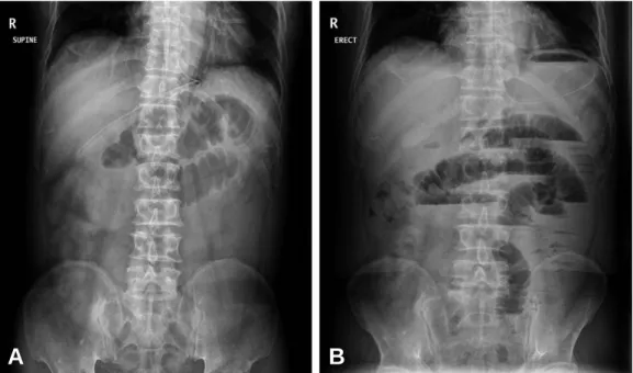
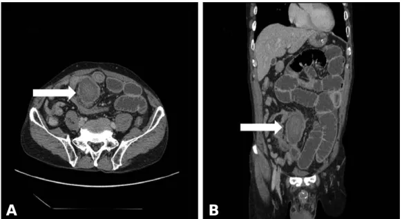
signs but mild direct tenderness on infra-umbilical area. A unknown large operation scar was noted on mid abdomen. Initial hemoglobin level was 12.5 g/dL and there was no leukocytosis. There were no remarkable changes in these laboratory findings within the first 24 hrs. Brain computed tomography (CT) scan showed no evidence of abnormal parenchymal density in brain or abnormal fracture line in skull. Simple X-ray (Fig. 1) showed dilated small bowel loops and abdominal CT scan (Fig. 2) revealed a well-defined homogenous mesenteric soft tissue mass measuring 7×5.5 cm with intestinal obstruction at the ileum level. Diagnosis of a mesenteric tumor was considered.
At first, we conservatively treated him by hydra- tion and nasogastric tube drainage for one day.
However, his physical examination and X-ray find- ings were not improved. The next day, as a mesen- teric tumor with intestinal obstruction of uncertain etiology was suspected, a explorative laparotomy was performed. Small bowel was hardly fixed and twisted to this tumor-like lesion with obstruction.
We performed adhesiolysis and widely segmental resection of small bowel (about 1m in length) includ- ing mesenteric tumor with side-to-side anastomosis due to great discrepancy in size. He stated later that he was a victim of the violence before 3 weeks. A final pathologic report a revealed well encapsulated,
Fig. 1. Supine (A) and erect (B) simple X-ray shows dilated small bowel loops with step ladder sign.
A B
traumatic mesenteric hematoma with organizing thrombi, ischemia and abscess formation with multiple adhesion bands. Finally, the patient was discharged without complications on postoperative day 14.
III. Discussion
A history of any previous abdominal trauma or surgical intervention is an important clue in differ- entiating mesenteric hematomas from true neoplas- tic process.(3) The radiological appearance of a well- defined mass is an additional characteristic that help in arriving at the right diagnosis.(4,5) CT has proved to be an excellent imaging modality for diag- nosis and managing hemodynamically stable patients with abdominal injuries, contributing towards a significant reduction of morbidity and mortality in trauma victims.(6) In this case, CT scans of the mesenteric tumor were retrospectively reviewed, its highest density of 82 Hounsfield units most strongly suggesting a hematoma. However, CT cannot be used as the sole indicator in the evalua- tion of the most common encountered blunt trauma.
A combination of clinical and laboratory reassess- ment and review of CT scans of the whole abdomen and pelvis using lung, bone and soft tissue window settings is recommended.(7)
In conclusion, the diagnosis of bowel and mesen-
teric injuries remains difficult and requires and high index of suspicious. We need to remind the impor- tance of the judicious history taking and the appro- priate clinical context to identify the cause of unex- plained acute abdomen.
REFERENCES