185
<원례보저>
Trimethyltin에 의한 랫드 해마의 신경세포 사멸과 iNOS의 연관성
장석원
1·최성영
1·박창남
1·안미정
2·신태균
1,*·김승준
3,*
1제주대학교 수의과대학, 2제주대학교 의학전문대학원, 3경북대학교 수의과대학 (접수: 2011년 6월 3일, 수정: 2011년 8월 22일, 게재승인: 2011년 8월 22일)
Inducible nitric oxide synthase is involved in neuronal death induced by trimethyltin in the rat hippocampus
Sukwon Jang
1, Sungyoung Choi
1, Changnam Park
1, Meejung Ahn
2, Taekyun Shin
1,*, Seungjoon Kim
3,*
1
College of Veterinary Medicine and Veterinary Medical Research Institute, Jeju National University, Jeju 690-756, Korea
2
Department of Anatomy, School of Medicine, Jeju National University, Jeju 690-756, Korea
3
College of Veterinary Medicine, Kyungpook National University, Daegu 702-701, Korea (Received: June 03, 2011; Revised: August 22, 2011; Accepted: August 22, 2011) Abstract : Trimethyltin chloride (TMT) has been used as a neurotoxin for inducing brain dysfunction and neuronal death. Neuronal death in the hippocampus by TMT may generate excessive nitric oxide, but there are few studies about nitric oxide synthase enzyme involved in the synthesis of nitric oxide. The purpose of present study is to analyze the TMT toxicity in each region of rat hippocampus. To evaluate the involvement of nitric oxide, we analyzed the effects of aminoguanidine known as a selective inhibitor for inducible nitric oxide synthase on behavioral changes and the hippocampus of rat by TMT toxicity. 6-week- old male Sprague-Dawley rats were administered with a single dose of TMT (8 mg/kg b.w., i.p.) and the control group was similarly administered with distilled water. TMT + aminoguanidine-treated groups were administered with aminoguanidine (10 mg/kg or 100 mg/kg b.w., i.p.) for 3 days prior to TMT injection.
The rats were sacrificed 2 days after TMT administration. In the TMT-treated group, a number of cell losses were seen in CA1, CA3 and the dentate gyrus. In the TMT + aminoguanidine-treated group, neuronal death was seen in CA1 and CA3, but reduced in the dentate gyrus compared to the TMT-treated group.
Western blot analysis showed that cleaved caspase-3 expression was increased in the TMT-treated group compared to the control group. However, the expression significantly declined in the TMT + aminoguanidine- treated group. The present findings suggest that inducible nitric oxide synthase is involved in neuronal death induced by TMT.
Keywords : aminoguanidine, hippocampus, iNOS, trimethyltin
서 론
Trimethyltin chloride(TMT)
는신경세포의사멸과신경 계의기능손상을유도할수있는신경독성제로알려져있다
[13]. TMT
을실험적으로랫드에복강내주입하면꼬리를쫓는행동
,
자학행동,
발성,
경련,
학습장애와같 은여러가지증상들을나타내며이러한증상들을총괄 하여“TMT
증후군”
이라고보고되어있다[5]. TMT
가투여된동물의경우해마의신경세포사멸이특징적으 로관찰된다고알려져있다
[14].
이와같은신경세포의 사멸은hydrogen peroxide
또는nitric oxide
와같은산화적
stress
와 밀접한 관련이있는 것으로 알려져 있고,
catalase
와같은항산화제에의해세포사멸이억제될수있음이알려져있다
[8].
그러나아직까지nitric oxide
가TMT
에의한행동변화및세포사멸에어떤영향을미칠 수있는지에관한실험적연구는많지않다.
*Corresponding authors
Tel: +82-64-754-3363, Fax: +82-64-702-9920 (Taekyun Shin) E-mail: [email protected]
Tel: +82-53-950-5971, Fax: +82-53-950-5955 (Seungjoon Kim)
E-mail: [email protected]
Nitric oxide
는뇌에서신경전달과같은중요한역할을 하며[3, 6], L-arginine
으로부터nitric oxide synthase
(NOS)
에의해생성되며NOS
는초기발견된세포형에따라신경세포유래의
neuronal NOS,
혈관내피세포유 래의endothelial NOS,
감작된macrophage
에서관찰되는inducible NOS
로나눌수있다[15].
정상적인동물에서 생성된nitric oxide
는항상성유지에필수불가결한물 질이지만[2],
염증시과다하게생성된nitric oxide
는생체에해로울수있다고한다
[7]. TMT
에의한해마신경세포의사멸도과다하게생성된
nitric oxide
가관련되 어있을개연성이높으나nitric oxide
생성과관련이있 는NOS
효소에관한연구는많지않다.
본연구의목적은
TMT
독성에의한rat
의해마손상을부위별로분석하고
,
다음으로nitric oxide
의관여여부를확인하기위하여
inducible NOS
의억제제로알려진
aminoguanidine [1]
이TMT
독성에의한rat
의행동 변화및해마조직에어떤영향을미치는지분석하고자 하였다.
재료 및 방법
실험동물과실험약물
실험동물은
6
주령수컷Sprague-Dawley
랫드(
오리엔 트바이오,
한국)
를 구입하여사육상자에넣어사육하였 다.
실험기간동안의사육환경은온도22
±2
oC,
상대습 도50
±10%,
환기횟수10~15
회/h,
인공조명(12
시간점 등, 12
시간소등),
조도150~300 lux
의조건을유지하였 다.
사육관리는 실험동물전용고형 사료(pellet chew:
5L79 diets; PMI Nutrition, USA)
를급여하였고,
음수는 제한없이공급하였다.
본연구의모든동물실험은제주 대학교동물실험윤리위원회에의해승인을받아수행되 었다. Trimethyltin chloride
와aminoguanidine
은Sigma Chemical(USA)
에서구입하였다.
TMT
적정용량측정해마에적절한손상을줄수있는
TMT
용량을확인하기
6
주령수컷Sprague-Dawley
랫드를그룹(TMT 6 mg/kg
투여군, 8 mg/kg
투여군, 10 mg/kg
투여군, 12 mg/
kg
투여군)
당3
마리씩나누었고, 48
시간후에희생시켰다.
TMT
투여후적정희생시간측정실험에 적합한 적정 희생 시간을 측정하기 위해
TMT(8 mg/kg)
를복강투여한후24
시간, 48
시간및72
시간희생군으로그룹당
3
마리씩나누고,
각실험군을 희생시킨후해마의조직학적변화를관찰하였다.
행동학적평가
TMT
를복강투여하고48
시간후동물을희생시키기 전1
분간앞발을드는횟수를측정하였다.
랫드가앞발 을드는행동은먹이를찾거나,
몸단장을하고,
호기심에 의한행동으로신경증상이나행동학적이상을확인할수 있으며본연구에서앞발을드는횟수측정을신경학적 활동평가로사용하였다.
해마의크기가정서불안과같 은행동이상을보이고[9],
이러한증상은해마의구성세 포들의세포사에의한결과와도일치한다고생각된다.
실험군의구성
Aminoguanidine
이해마에미치는영향을평가하기위 해실험동물은PBS
를단독으로투여한대조군(control), TMT
를단독으로투여한실험군(vehicle), TMT
투여전aminoguanidine
을 주입한 실험군은10 mg/kg
투여군(TMT + AG10), 100 mg/kg
투여군(TMT + AG100)
으로 나누었다(n = 3, per group).
TMT
투여와조직표본준비및조직검사TMT
는실험동물의복강에8 mg/kg
의용량으로투여 하였고, aminoguanidine
은10 mg/kg
및100 mg/kg
의용 량으로TMT
투여전3
일간복강내로투여하였다.
실 험동물은에테르로마취하여방혈하였고,
대뇌의해마 부분을적출하여,
각조직의일부분은단백질발현을보 기위한웨스턴샘플로급속냉동하여사용까지–70
oC
의
deep freezer
에 보관하였다.
조직염색용조직은4%
paraformaldehyde
용액에고정하고, ethanol
과xylene
으로탈수와투명화과정을거쳐파라핀에포매한후
5 µm
의두께로 조직절편을 만들어
hematoxylin-eosin
염색 및terminal deoxynucleotidyl transferase-mediated dUTP nick end-labeling(TUNEL)
을실시하였다. Cresyl violet
염색용 조직절편은10 µm
의두께를사용하였다.
Terminal deoxynucleotidyl transferase-mediated dUTP nick end-labeling(TUNEL)
적출한장기조직은
TMT
투여후48
시간후에적출되었다
.
조직은PBS
에녹인4% paraformaldehyde
용액 에 고정한후 파라핀에포매하고,
슬라이드글라스에5
µm
두께로조직절편을만들었다. DNA
분열은in situ nick end-labeling
을통해검출되었고,
염색방법은제조 사(Calbiochem, Germany)
의 지시사항에따라시행되었 다.
파라핀절편은 파라핀을제거,
수세후Proteinase K(20
µg/mL)
로15
분간상온에서반응시킨후, 0.15 U/
µL
의
TdT
와0.0004 nmol/
µL
의digoxigenin-dUTP
을포함한TdT
반응완충용액(140 mM sodium cacodylate, 1 mM
CoCl
2, 30 mM Tris-HCl, pH 7.2)
에60
분간37
oC
에서반응시켰고
, 10
분간TBS
용액(300 mM NaCl, 30 mM sodium citrate)
에 담가두었다.
그 후30
분간 상온에서peroxidase-labeled anti-digoxigenin antibody
로반응시킨 후, diaminobenzidine
으로발색을하고광학현미경을이 용하여관찰하였다.
현미경시야에서dentate gyrus
의세 포자멸사를확인하였고, dentate gyrus
에서의TUNEL-
양 성세포들의숫자는각그룹별평균 ± 평균의표준오차(mean
±SEM)
로 표시하였다.
각 실험군의데이터분석 은Student-Newman-Keuls test
를 이용하여통계처리를 한후통계학적유의성( p < 0.05)
을검정하였다.
Western blot analysis
적출한 장기 조직은
leupeptin(0.5 ug/mL), PMSF(1 mM), aprotinin(5
µg/mL)
등의protein inhibitor
가포함된40 mM Tris-HCl, pH 7.4, 120 mM NaCl, 0.1% Nonidiet P-40(polyoxyethylene [9] p-t-octyl phenol)
의buffer
에넣 어완전히파쇄한후, 14,000 rpm
으로30
분간원심분리 하여상층액을회수하였다.
이들을단백질정량하여변 성시킨후동량(20
µg/20
µL)
으로sodium dodecyl sulfate- polyacrylamide gel
에서전기영동하고, gel
상의단백질 밴드를다시nitrocellulos membrane
에100 V
에서2
시간 동안이동시켰다.
옮겨진membrane
을5% skim milk
로blocking
한후, 1
차항체로rabbit anti-cleaved caspase-3(1 :
1,000; Cell Signaling Technology, USA)
을실온에서1
시 간 반응시킨 후2
차 항체는horseradish peroxidase- conjugated horse anti-rabbit IgG(Vector Laboratories, USA)
를실온에서60
분간반응시켰다.
면역반응이끝난membrane
은Amersham ECL reagents(Amersham Life Science, UK)
로반응시켜, X-ray
필름에노출시키고,
그 결과를densitometer(M GS-700 Imaging Densitometer, Bio-Rad laboratories, USA)
를사용하여측정하였다.
결 과
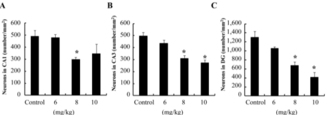
TMT
적정용량측정TMT
의용량에따른해마신경세포의손상을연구한결과
, 6 mg/kg
용량의실험군은신경세포의손상이나타나지않았으며
, 8 mg/kg
용량의실험군은CA1
부위, CA3
부위및
dentate gyrus
에서유의성있는신경세포사멸이 관찰되었다. 10 mg/kg
용량의실험군은해마각부위세포층의현저한뉴런의손실이나타났고
, 12 mg/kg
용량의실험군은투여후
24
시간내에실험동물이폐사하였 다.
이러한실험결과에따라본실험에서는TMT
에의 한 신경세포의사멸에inducible nitric oxide synthase
가 연관됨을연구하기위해적정TMT
의용량을뉴런사멸 이유의성있게증가하는8 mg/kg
으로정하였다(Fig. 1).
Fig. 1. Dose dependent changes in rat hippocampus after Trimethyltin chloride (TMT) injection.
*p< 0.01
vs.normal control.
Fig. 2. Time course of changes in rat hippocampus after TMT injection.
*p< 0.01
vs.normal control.
TMT
투여후적정희생시간측정TMT
투여후24
시간희생실험군은해마신경세포의 뚜렷한손실이관찰되지않았다
. 48
시간희생실험군은해마신경세포유의성있는손상이
CA1
부위, CA3
부위및
dentate gyrus
에서관찰되었다. 72
시간실험군은 해마세포층전체에서현저한신경세포손상을나타내 었다.
따라서본실험에서는각부위별유의성있는해마신경세포손상이관찰되는
TMT
투여후48
시간후가적절하다고판단하였다
(Fig. 2).
앞발을드는횟수의변화
1
분 동안랫드가앞발을드는횟수를측정하여비교한결과
, TMT
를투여하지않은정상군에비하여TMT
를복강투여한실험군은앞발을드는횟수가유의하게 증가되었고
, TMT
와aminoguanidine
을병용 투여한실험군은
TMT
대조군에비하여횟수가유의하게감소되었다
(Fig. 3).
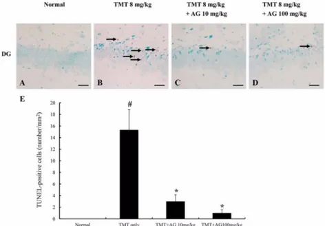
Dentate gyrus
에서의TUNEL
염색형태학적소견상
CA1
부위, CA3
부위및dentate gyrus
에서관찰된
pyknotic
세포가세포자멸사에의한것인지확인하기위해
TUNEL
염색을 실시한결과,
정상군은양성세포가관찰되지않았고
, dentate gyrus
에서TMT
를투여한실험군은유의성있는양성세포의증가를나 타내었다
.
그러나TMT
와10 mg/kg
용량의aminoguani- dine
을병용투여한실험군에서는TMT
실험군에비해 양성세포가확연히감소되었다. TMT
와100 mg/kg
용량 의aminoguanidine
을병용투여한실험군에서도10 mg/
kg
용량을투여한실험군과같은양상을나타내었다(Fig.
4).
그러나CA1
부위와CA3
부위에서는TMT
투여군 과aminoguanidine
병용군에서큰차이를보이지않았다.
Fig. 3. Frequency of raising forelimbs for a min.
#p< 0.05
vs.
normal control and
*p< 0.05
vs.TMT-treated group.
Fig. 4. TUNEL staining of rat hippocampus with normal control (A), TMT-treated group (B), TMT + aminoguanidine (10 mg/kg) group (C), TMT + aminoguanidine (100 mg/kg) group (D). In normal DG (A), no apoptotic cell was found. Many apoptotic cells (arrows) were seen in the TMT-treated rat hippocampus (B). Some apoptotic cells (arrows) were seen in TMT + aminoguanidine (10 mg/kg) group (C) and TMT + aminoguanidine (100 mg/kg) group (D). The number of apoptotic cells was reduced in the dentate gyrus of the TMT + aminoguanidine group compared to the TMT-treated group (E). The number of apoptotic cells (mean SEM) is indicated for four groups: the control rats (N-3), TMT-treated rats (N-3), TMT + aminoguanidine (10 mg/kg)-treated rats (N-3) and TMT + aminoguanidine (100 mg/kg)-treated rats (N-3).
#p< 0.05
vs.normal control and
*p< 0.05
vs.TMT-treated group. AG: aminoguanidine, DG: Dentate gyrus. Scale bars = 30
µm.
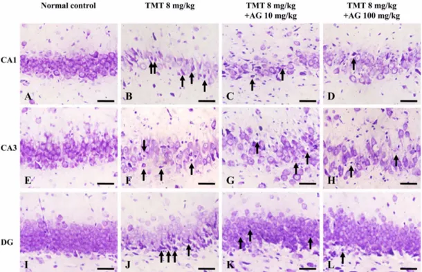
해마의조직학적변화및
CA1
부위와CA 3
부위에서 의세포수측정해마의조직학적관찰결과는최종적으로신경세포에 특징적으로염색이잘되는
cresyl violet
염색을실시하였으며
,
정상군은해마의CA1
부위, CA3
부위및dentate
gyrus
에서신경세포손실이없는정상적인조직소견을나타내었고
, TMT
를투여한실험군은CA1
부위, CA3
부위및
dentate gyrus
에서다수의뉴런손실이관찰되Fig. 5. Cresyl violet staining of rat hippocampus with normal control (A, E, I), TMT (8 mg/kg, 48 h)-treated group (B, F, J), TMT + aminoguanidine (10 mg/kg)-treated group (C, G, K), TMT + aminoguanidine (100 mg/kg)-treated group (D, H, L). The loss of neurons in CA1 and CA3 regions was seen in the TMT-treated rat hippocampus (B and F). Some pyknotic cells (arrows) were seen in the TMT-treated rat dentate gyrus (J). The loss of neurons in CA1 and CA3 regions was seen in the TMT + aminoguanidine (10 mg/kg)-treated group (C and G) and TMT + aminoguanidine (100 mg/kg)-treated group (D and H). AG: aminoguanidine, DG: Dentate gyrus, CA: Cornu ammonis. Scale bars = 30
µm.
Fig. 6. Number of surviving neurons in CA1 (A) and CA3 (B) regions of normal control and experimental group. The number of surviving cells (mean
±SEM) is indicated for four groups: the control rats (N-3), TMT-treated rats (N-3), TMT + aminoguanidine (10 mg/kg)-treated rats (N-3) and TMT + aminoguanidine (100 mg/kg)-treated rats (N-3).
*p< 0.05
vs.normal control. AG: aminoguanidine, CA: Cornu ammonis.
었다
. TMT
와10 mg/kg
용량의aminoguanidine
을투여한실험군은
CA1
부위와CA3
부위에서뉴런손실이관찰되었으나
,
형태학적소견은aminoguanidine
병용효과 가 뚜렷하게 관찰되었다. TMT
와100 mg/kg
용량의aminoguanidine
을투여한실험군에서도10 mg/kg
용량을 투여한실험군과같은조직양상을나타내었다(Fig. 5).
CA1
부위와CA3
부위에서양성세포의숫자를 측정한 결과
,
정상군에 비해TMT
실험군과TMT
와aminoguanidine
을병용투여한실험군에서세포의유의성있는감소를나타내었다
(Fig. 6). TMT
와aminoguani-
dine
병용투여한실험군에서는TMT
단독투여군에비해유의성있는차이는없었지만신경세포손실이좀더 미미한수준으로관찰되었다
.
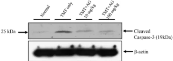
Western blot analysis
Caspase-3
단백질은TMT
독성에의한산화적stress
에관여한다고보고되어있다
[11].
본실험에서cleaved caspase-3
단백질의발현양상을Western blot
을이용하여관찰한결과
,
정상군에비해TMT
실험군은발현이유의하게증가하였고
, TMT
와aminoguanidine
을병용투여한실험군모두에서
TMT
실험군에비해유의성있는감소를나타내었다
(Fig. 7).
고 찰
본 연구에서는
TMT
독성에 의한 랫드의 해마손상 을 부위별로 분석하였고, nitric oxide
의 관여여부를확인하기 위하여
inducible NOS
의 억제제로 알려진aminoguanidine [4]
이TMT
독성에의한rat
의행동변화 및해마조직에어떤영향을미칠수있는지분석하였다.
랫드에서
TMT
독성에의한해마손상연구는많이이 루어져있으나, CA1
부위, CA3
부위및dentate gyrus
를각부위별로차이점을분석한연구는아직없다
.
이 번연구는8 mg/kg
의 용량으로48
시간후랫드해마의 각부위별차이점을분석한첫번째논문이다.
TMT
독성에의한랫드의해마손상은CA1
부위와CA3
부위에서일어난다고보고되어있다
[10].
본연구에서 조직학적으로확인한TMT
독성에의한랫드의CA1
부위와
CA3
부위의해마손상도 이와 일치한다.
그리고TMT
독성에의한랫드의dentate gyrus
에서의손상에대 한연구의결과는[16],
본연구에서확인한TMT
독성에 의한dentate gyrus
의조직학적변화소견과유사하다.
TMT
독성에의한신경세포의사멸은hydrogen peroxide
또는
nitric oxide
와같은산화적stress
와밀접한관련이 있는 것으로 알려져있다[8].
그러나 아직까지nitric
oxide
가TMT
에의한행동변화및세포사멸에어떤영향을미칠수있는지에관한실험적연구는많지않다
.
본연구의결과
, TMT
실험군에서행동학적으로꼬리를쫓는행동
,
발성,
경련과같은증상이발생되었으며,
앞발을드는횟수를측정한결과
aminoguanidine
을 투 여함에따라TMT
에의한증상이완화되는효과를확인 할수있었으며이러한행동학적결과는해마의전체적인양적크기에의한행동학적이상으로 생각된다
[9].
아울러 조직병리학적으로도
TMT
실험군과TMT
와aminoguanidine
을병용투여한실험군간에dentate gyrus
에서차이점이큰것으로보아
TMT
에의한dentate gyrus
에서의신경세포사멸에
inducible nitric oxide synthase
의 선택적
inhibitor
인aminoguanidine
이 관여하는것으 로사료된다.
또한본연구의cleaved caspase-3 western blotting
결과는inducible nitric oxide synthase
의inhibitor
인
aminoguanidine
이세포자멸사에관여하는caspase-3
의작용을줄여준다는
[12]
보고와같은결과이다.
결 론
6
주령의Sprague-Dawley
랫드에서8 mg/kg
용량의TMT
에의한48
시간후해마의신경세포의사멸은CA1
부위
, CA3
부위와dentate gyrus
에일어나며,
특히세포 자멸사는dentate gyrus
에서크게나타나는것으로사료 된다.
본실험의결과로보아이러한신경손상은산화적stress
에의해 생성되는nitric oxide
와관련된inducible nitric oxide synthase
가 높은연관성을갖는다고 생각된다
. TMT
에의한신경세포의사멸이각부위에따라신호전달경로의정도가다를것으로여겨지나
,
구체적인 신호전달경로의차이를규명하기위해서추가적인연 구가이루어져야할것이다.
감사의 글
이연구는한국연구재단의일부지원에의해이루어 진것임