Cilostazol Protects Rat Chondrocytes Against
Nitric Oxide–Induced Apoptosis In Vitro and Prevents
Cartilage Destruction in a Rat Model of Osteoarthritis
Sung Won Lee,
1Yeon Suk Song,
1Sang Hwa Shin,
1Kyung Taek Kim,
1Young Chul Park,
2Bong Soo Park,
2Il Yun,
2Kunhong Kim,
3Sang Yeob Lee,
1Won Tae Chung,
1Hye Jeong Lee,
1and Young Hyun Yoo
1Objective. To examine whether cilostazol, a
selec-tive phosphodiesterase type III inhibitor, protects rat
articular chondrocytes against nitric oxide (NO)–
induced apoptosis and prevents cartilage destruction in
mono-iodoacetate–induced osteoarthritis (OA) in a rat
model in which inducible nitric oxide synthase (iNOS) is
expressed.
Methods. The NO donor sodium nitroprusside
was administered to rat articular chondrocytes that had
been pretreated with cilostazol. Induction of apoptosis
was evaluated by DNA electrophoresis and pulsed-field
gel electrophoresis. The expression level and the
subcel-lular location of apoptosis-associated factors were
ex-amined by Western blot analysis and confocal
micros-copy, respectively. Protein kinase CK2 (PKCK2) activity
was also assayed. To examine whether orally
adminis-tered cilostazol prevents cartilage destruction in vivo,
cartilage samples obtained from rats with
experimen-tally induced OA were subjected to hematoxylin and
eosin, Safranin O, and TUNEL staining and
immuno-histochemical analysis of iNOS expression.
Results. Cilostazol prevented NO-induced
reduc-tion in viability, in a dose-dependent manner. It also
prevented the up-regulation of phosphorylated p53 and
p38, the down-regulation of heme oxygenase 1, the
subcellular translocation of apoptosis-inducing factor
and cytochrome c, and the activation of caspases 3, 7,
and 8 induced by NO treatment, indicating that
cilosta-zol prevented NO-induced cell death by blocking
apo-ptosis. In addition, cilostazol prevented NO-induced
translocation of cleaved Bid onto mitochondria, and
caused phosphorylated Bid to accumulate in the nucleus
and cytosol. Cilostazol prevented the down-regulation of
PKCK2 and the reduction in PKCK2 activity induced by
NO, indicating that its apoptosis-preventing activity
was mediated via PKCK2. It also prevented chondrocyte
apoptosis and cartilage destruction in a rat model of
experimentally induced OA.
Conclusion. Our findings indicate that cilostazol
prevents NO-induced apoptosis of chondrocytes via
PKCK2 in vitro and prevents cartilage destruction in a
rat model of OA.
Osteoarthritis (OA) is characterized by
degrada-tion of matrix and destrucdegrada-tion of articular cartilage (1).
Articular chondrocytes are solely responsible for the
production and maintenance of the extracellular matrix.
Therefore, chondrocyte apoptosis is implicated in
carti-lage degeneration (2,3). Several stimuli, such as nitric
oxide (NO) (4–7), prostaglandin E
2(8), Fas ligand
(9,10), tumor necrosis factor
␣ (TNF␣) (11), and TRAIL
(12) have been reported to induce apoptosis in
chondro-cytes. Since those apoptosis-inducing substances might
play a part in the pathogenesis of OA, manipulation of
the mechanism mediated by those stimuli has the
poten-tial for substanpoten-tial therapeutic impact.
NO is believed to be an important mediator of
Supported by the Korea Science and Engineering Foundation (grants R01-2004-000-10002-0 and R01-2007-000-20100-0).
1Sung Won Lee, MD, PhD, Yeon Suk Song, MS, Sang Hwa
Shin, MD, Kyung Taek Kim, MD, Sang Yeob Lee, MD, Won Tae Chung, MD, Hye Jeong Lee, MD, Young Hyun Yoo, MD, PhD: Dong-A University, Seo-Gu, Busan, South Korea;2Young Chul Park,
PhD, Bong Soo Park, PhD, Il Yun, PhD: Pusan National University, Seo-Gu, Busan, South Korea;3Kunhong Kim, MD: Yonsei University,
Seodaemun-Gu, Seoul, South Korea.
Address correspondence and reprint requests to Young Hyun Yoo, MD, PhD, Department of Anatomy and Cell Biology, Dong-A University College of Medicine, Medical Science Research Center, 3-1 Dongdaesin-Dong, Seo-Gu, Busan 602-7143, South Korea. E-mail: [email protected].
Submitted for publication May 16, 2007; accepted in revised form November 14, 2007.
the dedifferentiation and apoptosis of articular
cytes in arthritic cartilage (6). It is produced in
chondro-cytes by the action of proinflammatory cytokines such as
interleukin-1
. NO production in chondrocytes causes
activation of matrix metalloproteinases (MMPs),
de-creased production of interleukin-1
 receptor
antago-nist, inhibition of proteoglycan synthesis and type II
collagen expression, and apoptosis of chondrocytes
(2,13,14).
Cilostazol, 6-[4-(1-cyclohexyl-1H-tetrazol-5-yl)
butoxy]-3,4-dihydro-2-(1H)-quinolinone, is a potent
se-lective phosphodiesterase type III (PDE III) inhibitor. It
elevates intercellular cyclic cAMP by blocking its
hydro-lysis by PDE III (15). Cilostazol functions as a platelet
aggregation inhibitor (15) and vasodilator (16), and is
mainly used for treating patients with peripheral arterial
disease (17) and intermittent claudication (18). In
addi-tion, it seems to reduce inflammation and inhibit MMP
expression by increasing AMP and inhibiting the p38/
JNK pathway (19). Previous studies have demonstrated
that cilostazol inhibits apoptosis under several
circum-stances (12,20–22). In those studies, cilostazol was
shown to prevent lipoprotein particle–induced apoptosis
in human umbilical vein endothelial cells (HUVECs)
(22), lipopolysaccharide-induced apoptosis in HUVECs
(20), and ischemic injury–induced apoptosis in neural
cells (12,21). Notably, protein kinase CK2 (PKCK2) was
reported to be involved in the prevention of apoptosis by
cilostazol (12,23).
PKCK2 has typically been classified as a
messenger-independent protein serine/threonine kinase
(24,25). It is distributed ubiquitously in the cell
cyto-plasm and nucleus of eukaryotic organisms. Although
the role of PKCK2 is unknown, it may potentially
participate in a complex series of cellular functions,
including cell growth and proliferation, by catalyzing the
phosphorylation of a large number of proteins (26).
PKCK2 also participates in the regulation of apoptosis
by phosphorylating some apoptosis-related factors (26–
31). Despite the many previous studies of PKCK2, little
progress has been made in understanding its molecular
role in chondrocytes.
This study was undertaken to determine whether
cilostazol prevents NO-induced chondrocyte apoptosis
and to examine the role of PKCK2 in this effect. Our
findings indicated that cilostazol protects rat
chondro-cytes against NO-induced apoptosis via PKCK2 in vitro,
and that oral administration of cilostazol prevents
carti-lage destruction in rats with experimentally induced OA.
MATERIALS AND METHODS
Reagents. Rabbit polyclonal anti-human caspase 3,
caspase 7, and cytochrome c, heme oxygenase 1 (HO-1), apoptosis-inducing factor (AIF), iNOS, phospho-p53 (pSer15),
Bid, GAPDH, and goat anti-human PKCK2 antibodies were obtained from Santa Cruz Biotechnology (Santa Cruz, CA). Mouse polyclonal anti-human poly(ADP-ribose) polymerase (PARP) antibody was obtained from Oncogene (Cambridge, MA), and mouse monoclonal anti-human caspase 8 and phospho-p38 antibodies were from Cell Signaling Technology (Beverly, MA). Rabbit polyclonal anti-human Bid antibody (cleaved product), pSer antibody, and TUNEL reaction mix-ture were from Chemicon (Temecula, CA). Fluorescein iso-thiocyanate (FITC)–conjugated goat anti-rabbit IgG antibody and avidin–biotin–peroxidase (ABC) complex were obtained from Vector (Burlingame, CA), and Dulbecco’s modified Eagle’s medium (DMEM) and fetal bovine serum (FBS) were from Gibco (Gaithersburg, MD). Cilostazol (OPC-13013) was generously donated by Otsuka Pharmaceutical (Tokushima, Japan). Sodium nitroprusside (SNP) was obtained from Fluka (Buchs, Switzerland). DMSO, Hoechst 33342, RNase A, proteinase K, protease inhibitor cocktail, propidium iodide, 3,3⬘-diaminobenzidine (DAB), emodin, type II collagenase, mono-iodoacetate (MIA), and Safranin O were all obtained from Sigma (St. Louis, MO). The enhanced chemiluminescent Western blotting detection reagent (SuperSignal West Pico chemiluminescent substrate) was obtained from Pierce (Rock-ford, IL).
Cell culture of articular chondrocytes. Rat articular
chondrocytes for primary culture were isolated from slices of knee joint cartilage from 5-week-old Sprague-Dawley rats. Chondrocytes were isolated by enzymatic digestion for 2 hours with 0.2% solid type II collagenase (381 units/mg) in DMEM. After collection of individual cells by brief centrifugation, cells were resuspended in DMEM supplemented with 10% (volume/ volume) FBS, 50 g/ml of streptomycin, and 50 units/ml of penicillin. Cells were plated on culture dishes at a density of 5⫻ 104cells/cm2. Medium was replaced every 2 days, and cells
reached confluence at ⬃5 days of culture. Cells from day-4 cultures were treated with the NO donor SNP. After incuba-tion in the presence or absence of indicated pharmacologic reagents, such as cilostazol or emodin, for 24 hours, the cells were further exposed to 1.5 mM SNP for 24 hours.
Cell viability assay. Cell viability was determined with
the Vi-Cell cell counter (Beckman Coulter, Fullerton, CA), which performs an automated trypan blue exclusion assay.
Nuclear morphology analysis of apoptosis.
Twenty-four hours after SNP treatment, the cell suspension was cytospun onto a clean fat-free glass slide with a cytocentrifuge. Cytocentrifuged samples were fixed for 10 minutes in 4% paraformaldehyde and stained in 4g/ml Hoechst 33342 for 30 minutes at 4°C. The total cell number (ⱖ300 cells from each experiment) was counted using differential interference con-trast (DIC) optics, and the number of cells showing condensed or fragmented nuclei on Hoechst staining was calculated using epifluorescence optics, by an observer who was blinded with regard to the experimental group.
DNA electrophoresis. Cells (2 ⫻ 106) were
resus-pended in 1.5 ml of lysis buffer (10 mM Tris [pH 7.5], 10 mM EDTA [pH 8.0], 10 mM NaCl, and 0.5% sodium dodecyl
sulfate [SDS]) to which proteinase K (200g/ml) was added. After samples were incubated overnight at 48°C, 200 l of ice-cold 5M NaCl was added, and the supernatant containing fragmented DNA was collected after centrifugation. The DNA was then precipitated overnight at⫺20°C in 50% isopropanol and treated with RNase A for 1 hour at 37°C. A loading buffer containing 100 mM EDTA, 0.5% SDS, 40% sucrose, and 0.05% bromphenol blue was added at 1:5 (v/v). Separation was achieved in 2% agarose gels in Tris–acetic acid/EDTA buffer (containing 0.5g/ml ethidium bromide) at 50 mA for 1.5 hours.
Pulsed-field gel electrophoresis (PFGE). Cells (2 ⫻
106) were suspended in 50 l of phosphate buffered saline
(PBS) containing 1% agarose with a low melting temperature. The cell suspension was poured into a template (5⫻ 2 ⫻ 10 mm), plugged, and cooled on ice. The hardened agarose gel blocks were incubated with 250l of a mixture of proteinase K (1 mg/ml), N-lauroylsarcosine sodium (1% [weight/volume]), and 0.5M EDTA (pH 9.2) at 50°C for 48 hours. After incubation, half the volume of the digested agarose gel block was loaded into a sample well of a 1% (w/v) agarose gel (type II; 150⫻ 150 ⫻ 4.4 mm) (Sigma) in 0.5⫻ Tris–borate–EDTA (TBE) buffer (89 mM Tris–boric acid, 2 mM EDTA [pH 8.0]). PFGE was carried out in 0.5⫻ TBE maintained at 14°C by circulating cool water for 16 hours (constant 6V; switch times initial 60 seconds and final 90 seconds), using the CHEF Mapper XA System (Bio-Rad, Hercules, CA). DNA in the gel was stained with ethidium bromide and detected with LAS-3000PLUS (Fuji Photo Film Company, Kanagawa, Japan). Chromosomal DNA from Saccharomyces cerevisiae (Bio-Rad, Hercules, CA) and a mixture of DNA, its concatemers, and
Hind III–digested DNA (Sigma) were used as DNA size
markers.
Subcellular fractionation. Cells (5⫻ 107) were washed
in Tris-based, Mg2⫹/Ca2⫹-deficient buffer (135 mM NaCl, 5 mM KCl, and 25 mM Tris Cl [pH 7.6]) and allowed to swell for 10 minutes in ice-cold hypotonic CaRSB buffer (10 mM NaCl, 1.5 mM CaCl2, 10 mM Tris HCl [pH 7.5], and 1⫻
protease inhibitor cocktail). Cells were dounced with 60 strokes, and mitochondria stabilization buffer (210 mM man-nitol, 70 mM sucrose, 5 mM EDTA, and 5 mM Tris [pH 7.6]) was added to stabilize mitochondria (2 ml of 2.5⫻ per 3 ml of homogenate). After collecting the nucleus (by centrifuging twice at 3,000 revolutions per minute for 15 minutes), the supernatant was spun at 14,000 rpm for 20 minutes at 4°C. The pellet and the supernatant included mitochondria and cyto-plasm, respectively.
Western blot analysis. Cells (2 ⫻ 106) were washed
twice in ice-cold PBS, resuspended in 200l ice-cold solubi-lizing buffer (300 mM NaCl, 50 mM Tris Cl [pH 7.6], 0.5% Triton X-100, 2 mM phenylmethylsulfonyl fluoride [PMSF], 2l/ml aprotinin, and 2 l/ml leupeptin) and incubated at 4°C for 30 minutes. The lysates were centrifuged at 14,000 rpm for 20 minutes at 4°C. Protein concentrations of cell lysates were determined with Bradford protein assay reagent (Bio-Rad, Richmond, CA) and 40 g of proteins was loaded onto 7.5–15% SDS–polyacrylamide gels. The gels were transferred to nitrocellulose membranes (Amersham Pharmacia Biotech, Piscataway, NJ) and reacted with each antibody. Immunostain-ing with antibodies was performed usImmunostain-ing SuperSignal West
Figure 1. Prevention of sodium nitroprusside (SNP)–induced cell
death in rat articular chondrocytes by cilostazol (C). A, Viability of cells pretreated with 1–100M cilostazol 24 hours prior to treatment with 1.5 mM of the nitric donor SNP. Cells were harvested 24 hours after treatment with SNP, and viability was determined by automated trypan blue exclusion assay with a cell counter. Cilostazol pretreatment significantly prevented SNP-induced cell death in a dose-dependent manner, and with 50 M cilostazol, prevention of SNP-induced apoptosis was substantial. Bars show the mean and SD percent cell viability, as compared with untreated controls (Ctrl).ⴱ ⫽ P ⬍ 0.05 versus controls. B, Results of DNA electrophoresis (left) and pulsed-field gel electrophoresis (PFGE) (right) of cells left untreated, treated with 50M cilostazol, treated with 1.5 mM SNP, or treated with both cilostazol and SNP. Conventional DNA electrophoresis of SNP-treated cells did not demonstrate ladder-like DNA fragments. PFGE of SNP-treated cells revealed the disintegration of nuclear DNA into giant fragments of 1–2 Mbp and high molecular weight fragments of 100–800 kbp, which was completely prevented by cilostazol treat-ment. C, Nuclear morphology of cells 24 hours after SNP treattreat-ment. Hoechst staining showed that cilostazol pretreatment significantly prevented SNP-induced nuclear condensation. C⫹S ⫽ cells treated with both cilostazol and SNP. Values are the mean⫾ SD percent-age of apoptotic cells with condensed nuclei.ⴱ ⫽ P ⬍ 0.05 versus controls.
Pico enhanced chemiluminescent substrate and detected with LAS-3000PLUS.
Coimmunoprecipitation. Cells were collected and
lysed in 1 ml of immunoprecipitation lysis buffer (300 mM NaCl, 500 mM Tris Cl [pH 7.6], 0.5% Triton X-100, protease inhibitors, 10 mM Na4P2O7, 1 mM Na3VO4, 25 mM NaF, and
1mM-glycerophosphate). Protein concentrations of cell ly-sates were determined using the Bradford method, and 500g of protein was precleared and then incubated with anti-phosphorylated serine antibody in extraction buffer at 4°C overnight. The immune complexes were precipitated with protein A/G–agarose beads (Santa Cruz Biotechnology) for 2 hours, and washed 5 times with extraction buffer prior to boiling in SDS sample buffer. Immunoprecipitated proteins were separated by SDS–polyacrylamide gel electrophoresis, and Western blot analysis was performed as described above.
Immunofluorescence staining and confocal micros-copy. A cell suspension was cytospun onto a clean fat-free glass
slide with a cytocentrifuge. Cells were incubated with anti– cytochrome c, AIF, and Bid antibodies for 1 hour, washed 3 times for 5 minutes each, and then incubated with FITC-conjugated secondary antibody for 1 hour at room tempera-ture. Fluorescent images were observed and analyzed under a Zeiss LSM 510 confocal laser scanning microscope (Zeiss, Gottingen, Germany).
PKCK2 activity assay. PKCK2 activity was measured
using a casein kinase 2 assay kit (Upstate Biotechnology, Lake Placid, NY). A recombinant full-length human PKCK2 protein that contains an ␣-subunit N-terminal 6⫻ His-Tag and a -subunit N-terminal genome signature tag (Upstate Biotech-nology) was used as a positive control.
Rat model of OA. Animals. Fifteen Sprague-Dawley
rats weighing 175–200 gm were used. The study was approved by the ethics committee of Dong-A University and fulfilled the guidelines for animal experiments established by The Korean Academy of Medical Sciences. OA was induced in 5 rats by intraarticular injection of MIA, as described previously (12). Briefly, 1 mg of MIA in 0.1 ml of physiologic saline solution was injected once a week for 4 weeks into the right knees. Cilostazol (30 mg/kg/day) was administered orally once daily beginning the day after the first injection of MIA. Five additional rats received MIA alone, and 5 rats received no MIA or cilostazol and were used as controls.
Tissue preparation and histologic examination. At the
end of the 4 weeks, animals were killed by ether inhalation. Both knee joints from each animal were dissected, and femoral condyles were fixed in PBS (pH 7.4) containing 4% parafor-maldehyde, decalcified in 12.5% EDTA, dehydrated, and embedded in paraffin blocks. Five-micrometer microsections were prepared and stained with hematoxylin and eosin and with Safranin O.
TUNEL staining. Sections were incubated with
protein-ase K (20g/ml) for 15 minutes at room temperature, in 2% H2O2 solution for 5 minutes at room temperature, and then
with terminal deoxynucleotidyl transferase (TdT) enzyme for 1 hour at 37°C. Next, antidigoxigenin–peroxidase was applied to the sections for 30 minutes at room temperature and developed with 0.05% DAB. Staining for the negative control was undertaken by skipping the TdT application. Sections were observed and photographed under DIC optics without any counterstaining.
Immunohistochemistry. Sections were incubated in goat
serum solution (diluted 1:70) for 30 minutes at room temper-ature, and then with primary antibody (diluted 1:100) for 2 hours at room temperature. Next, sections were incubated with secondary antibody for 1 hour at 37°C and developed using ABC complex, and peroxidase was revealed by DAB.
Statistical analysis. Four independent experiments
were carried out in vitro. Results are expressed as the mean⫾ SD from 4 experiments performed in triplicate.
RESULTS
Prevention of NO-induced apoptosis in rat
artic-ular chondrocytes by cilostazol. Cilostazol prevented
NO-induced cell death in a dose-dependent manner
(Figure 1A). Since cilostazol substantially prevented
NO-induced cell death at 50
M, this concentration was
used for further exploration of the mechanism of
pre-venting cell death. Various assays were performed in
order to confirm that cilostazol prevented NO-induced
cell death by blocking apoptosis. Although cells treated
with the NO donor SNP failed to show ladder-like DNA
fragments from their genomic DNA on agarose gel,
PFGE revealed the disintegration of nuclear DNA into
giant fragments of 1–2 Mbp and high molecular weight
fragments of 100–800 kbp. Cilostazol appeared to
com-pletely prevent these giant and high molecular weight
fragmentations (Figure 1B). We also observed that
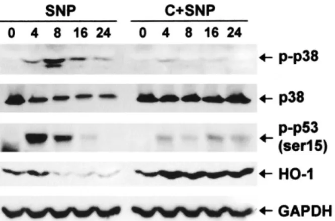
Figure 2. Prevention of SNP-induced down-regulation of heme
oxy-genase 1 (HO-1) and up-regulation of phosphorylated p38 and phos-phorylated p53 by 50M cilostazol. Western blots of antibodies to anti–phosphorylated p38, anti–phosphorylated p53 (Ser15), and anti–
HO-1, before pretreatment (0) and 4, 8, 16, and 24 hours after treatment with SNP alone or with SNP and cilostazol, are shown; p38 was used as the loading control for phosphorylated p38. Cilostazol pretreatment prevented SNP-induced down-regulation of HO-1 as well as SNP-induced up-regulation of phosphorylated p38 and phos-phorylated p53. GAPDH was used as a loading control. See Figure 1 for other definitions.
cilostazol prevented NO-induced nuclear condensation
(Figure 1C). These findings support the notion that
cilostazol protects cells against NO-induced apoptosis.
Prevention of NO-induced down-regulation of
HO-1 and of up-regulation of phosphorylated p38 and
p53 by cilostazol. Activation of p38 and resultant
phos-phorylation of p53 is crucial in NO-induced cell death of
cultured articular chondrocytes (32). Thus, we examined
whether the prevention of cell death by cilostazol was
mediated via these factors. Our results showed that
cilostazol prevented NO-induced up-regulation of
phos-phorylated p38 and p53. Since HO-1, a cytoprotective
factor, is also known to be involved in NO-induced
chondrocyte injury (33), we next examined whether
cilostazol modulates the expression of HO-1 in
chondro-cytes treated with SNP. We found that cilostazol
pre-vented NO-induced down-regulation of HO-1 (Figure 2).
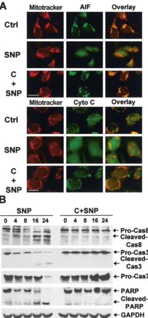
Prevention of the release of mitochondrial
apo-ptotic factors and of the activation of caspases by
cilostazol. NO-induced apoptosis has been
demon-strated to be accompanied by the opening of
mitochon-drial permeability transition pores and by the release of
AIF and cytochrome c from mitochondria (34,35).
Therefore, we examined whether cilostazol prevented
these mitochondrial events. Our results showed that NO
induced the release of AIF and cytochrome c from
mitochondria in rat chondrocytes, and that cilostazol
prevented the release of these factors from
mitochon-dria (Figure 3A). Caspases play an essential role in most
types of apoptosis, including NO-induced apoptosis. We
found that caspase 3 and caspase 7 were activated in
NO-induced apoptosis and that cilostazol prevented the
activation of these effector caspases. In addition, the
processing of initiator caspase 8 was observed in
NO-induced apoptosis, and this was also inhibited by
cilosta-zol. Cilostazol also prevented SNP-mediated cleavage of
a caspase substrate, PARP (Figure 3B).
Cilostazol causes phosphorylated Bid to
accumu-late in both the nucleus and cytosol and prevents
NO-induced down-regulation of PKCK2 expression and
reduction of PKCK2 activity. It is well known that Bid
plays an essential role in caspase 8–mediated apoptosis.
Thus, we next examined whether cilostazol prevents
NO-induced apoptosis by modulating Bid. Western
blot-ting using the antibody detecblot-ting cleaved Bid
demon-strated that SNP induced mitochondrial translocation of
cleaved Bid (Figure 4A). Confocal microscopy also
showed that SNP induced mitochondrial translocation of
Bid (Figure 4B). In addition, coimmunoprecipitation
showed that SNP downregulated phosphorylated Bid in
the nucleus and cytosol (Figure 4C). Notably, cilostazol
Figure 3. Prevention of the release of mitochondrial apoptotic
fac-tors and the activation of caspases by 50M cilostazol. A, Confocal microscopy images showing localization of Mitotracker stains, apoptosis-inducing factor (AIF), and cytochrome c (Cyto c) on the mitochondria in control cells, and release of both AIF and cytochrome
c from mitochondria by SNP treatment. Cilostazol pretreatment
prevented SNP-induced release of AIF and cytochrome c from mito-chondria. Bars⫽ 20m. B, Western blots of caspase 8 (pro-Cas8), caspase 3 (pro-Cas3), caspase 7 (pro-Cas7), and poly(ADP-ribose) polymerase (PARP), before pretreatment (0) and 4, 8, 16, and 24 hours after treatment with SNP alone or with SNP and cilostazol. Cilostazol pretreatment prevented SNP-induced degradation of caspase 8, caspase 3, and caspase 7 and SNP-induced production of caspase 8 and caspase 3 cleaved products. Cilostazol pretreatment also prevented the degradation of the caspase substrate PARP and its cleavage by SNP. GAPDH was used as a loading control. See Figure 1 for other definitions.
prevented not only the mitochondrial translocation of
Bid but also the down-regulation of phosphorylated Bid
in the nucleus and cytosol (Figures 4A–C).
We next tested whether the protection of cells
against NO-induced apoptosis by cilostazol was
medi-ated via PKCK2. Western blot analysis revealed that the
expression level of PKCK2 catalytic subunit
␣ was
reduced after SNP treatment (Figure 4D). In addition,
SNP treatment resulted in a decrease in PKCK2 activity
(Figure 4E). Importantly, cilostazol prevented not only
NO-induced down-regulation of PKCK2 but also the
NO-induced reduction of PKCK2 activity (Figures 4D
and E).
Prevention of NO-induced apoptosis by cilostazol
through PKCK2. To corroborate that
cilostazol-mediated prevention of NO-induced apoptosis occurs
via PKCK2, we performed several assays using a PKCK2
inhibitor, emodin (36). A cell viability assay showed that
50
M emodin counteracted the effect of cilostazol on
NO-induced chondrocyte death (Figure 5A). Western
blot analysis also showed that 50
M emodin abolished
the ability of cilostazol to prevent NO-induced
down-regulation of Bid (Figure 5B). Emodin also abolished
the ability of cilostazol to prevent NO-induced
mito-chondrial translocation of cleaved Bid (Figure 5C).
Furthermore, it abolished the ability of cilostazol to
prevent NO-induced down-regulation of phosphorylated
Bid in cytosol (Figure 5D). An assay for PKCK2 activity
showed that emodin abolished the ability of cilostazol to
prevent NO-induced reduction of PKCK2 activity
(Fig-ure 5E).
Prevention of cartilage destruction and of
chondro-cyte apoptosis by cilostazol in rats with MIA-induced OA.
Cartilage obtained from rats injected with MIA showed
Figure 4. Accumulation of phosphorylated Bid in both the nucleus and cytosol and prevention of SNP-induced down-regulation of
protein kinase CK2 (PKCK2) expression and decrease of PKCK2 activity by 50M cilostazol. A, Western blot showing SNP-induced mitochondrial translocation of cleaved Bid, which was prevented by cilostazol pretreatment. B, Confocal microscopy images showing localization of Bid in the nucleus or cytosol in control (CON) cells, and translocation of Bid onto mitochondria as a result of SNP treatment. Cilostazol pretreatment prevented SNP-induced mitochondrial translocation of Bid. Bar⫽ 20m. C, Coimmunoprecipi-tation assay showing that cilostazol pretreatment prevented SNP-induced down-regulation of phosphorylated Bid in the nucleus and cytosol. IP⫽ immunoprecipitation. D, Western blot of PKCK2␣, before pretreatment (0), and 4, 8, 16, and 24 hours after treatment with SNP alone or with cilostazol and SNP. Cilostazol pretreatment prevented SNP-induced down-regulation of PKCK2. GAPDH was used as a loading control. E, Results of a PKCK2 activity assay, showing that cilostazol pretreatment prevented SNP-induced reduction in PKCK2 activity. Values are the mean and SD. See Figure 1 for other definitions.
structural changes, including changes characteristic of OA,
such as irregular surfaces, the disappearance of
surface-layer cells, slightly diffuse cell growth in the transitional and
radial zones, and slightly reduced Safranin O staining.
Most chondrocytes displayed a positive TUNEL reaction.
Cilostazol prevented this MIA-induced cartilage
degener-ation. A previous study showed that iNOS was expressed in
the early stages of experimental OA (37). Consistent with
those results, our immunohistochemical analysis also
showed that iNOS was expressed in most chondrocytes.
Notably, cilostazol reduced the level of expression of iNOS
(Figure 6).
DISCUSSION
The results of this study support the hypothesis
that PKCK2 is involved in NO-induced apoptosis of rat
articular chondrocytes and that cilostazol protects rat
chondrocytes against NO-induced apoptosis via PKCK2
in vitro. Furthermore, our findings support the notion
that cilostazol prevents cartilage destruction in
MIA-induced OA in a rat model in which iNOS is expressed.
The recent observation that cilostazol inhibits
apoptosis under several circumstances (12,20–22) raises
the interesting possibility that it can be considered as a
therapy for diseases associated with excess apoptosis.
Although apoptotic chondrocyte death is not always a
widespread phenomenon in cartilage aging or OA
car-tilage degeneration (38), obviously any kind of cell death
is detrimental to the tissue. Therefore, any strategy to
prevent chondrocyte death or to manipulate the
mech-anism mediated by stimuli that cause cell death has
potential for substantial therapeutic impact (39).
Although understanding of the mechanism by
which cilostazol protects cells against apoptosis remains
Figure 5. Prevention of SNP-induced apoptosis by cilostazol via protein kinase CK2 (PKCK2). After simultaneous pretreatment with
50M emodin (E) and 50 M cilostazol for 24 hours, cells were further exposed to 1.5 mM SNP for 24 hours. A, Cell viability, determined by automated trypan blue exclusion assay with a cell counter. Emodin significantly abolished the ability of cilostazol to prevent SNP-induced chondrocyte death. Bars show the mean and SD percent cell viability, as compared with untreated controls. ⴱ ⫽ P ⬍ 0.05 versus controls. B, Western blot showing that emodin abolished the ability of cilostazol to prevent SNP-induced down-regulation of Bid expression. C, Western blot showing that emodin abolished the ability of cilostazol to prevent SNP-induced mitochondrial translocation of cleaved Bid. D, Coimmunoprecipitation showing that emodin abolished the ability of cilostazol to prevent SNP-induced down-regulation of phosphorylated Bid in cytosol. IP⫽ immunoprecipitation. E, Results of a PKCK2 activity assay, showing that emodin significantly abolished the ability of cilostazol to prevent SNP-induced reduction in PKCK2 activity. Bars show the mean and SD.ⴱ ⫽ P ⬍ 0.05 versus untreated cells. See Figure 1 for other definitions.
far from complete, recent evidence has yielded certain
insights. Previous studies (20,21) have shown that
cilostazol reverses decreases in Bcl-2 protein and
in-creases in Bax protein production and cytochrome c
release. The maxi-K channel opening–coupled
up-regulation and down-up-regulation of PTEN
phosphoryla-tion with resultant increases in Akt and CREB
phos-phorylation and increased Bcl-2 protein levels have also
been demonstrated (12). In addition, suppression of
NAD(P)H oxidase–dependent superoxide formation
and of cytokine induction has been demonstrated (22).
In a few apoptosis systems showing the prevention of
apoptosis by cilostazol, PKCK2 has also been reported
to play a crucial role (12,23).
PKCK2 is a constitutively active, growth factor–
independent serine/threonine protein kinase (40)
com-posed of 2 catalytic
␣ (and/or ␣⬘) subunits and 2
regulatory
 subunits (41). Although it is well known
that PKCK2 participates in various cellular functions, its
biologic activity on chondrocytes has not been previously
described. In the present study, we demonstrated the
presence of PKCK2 activity on rat articular
chondro-cytes.
We also demonstrated that PKCK2 is involved in
the regulation of rat articular chondrocyte apoptosis. It
has been well documented that PKCK2 is involved in the
regulation of apoptosis by phosphorylating some
apoptosis-related factors. PKCK2 activity has been
found to be consistently enhanced in various tumor cells
(42–44), and increased expression of PKCK2 has
pro-tected cells against Fas- and drug-induced apoptosis
(45,46). Furthermore, inhibitors of PKCK2 have been
reported to trigger apoptosis and increase the
suscepti-bility of cancer cells to chemotherapeutic agents or
apoptotic stimuli (47–49). Thus, PKCK2 appears to have
a general antiapoptotic function.
Through recent studies, advances have been
made in understanding the mechanism by which PKCK2
supports cell survival. Phosphorylation of Max, the
transcription partner of the c-Myc protooncogene, by
Figure 6. Prevention of cartilage destruction and chondrocyte apoptosis by cilostazol in rats with mono-iodoacetate (MIA)–induced
osteoarthritis. Hematoxylin and eosin (H&E) staining showed that cilostazol prevented MIA-induced cartilage surface irregularity and slightly diffuse cell growth in the transitional and radial zones. Cilostazol also prevented MIA-induced reduction of Safranin O staining and increased expression of TUNEL-positive chondrocytes. Immunohistochemical analysis showed that cilostazol prevented MIA-induced expression of inducible nitric oxide synthase (iNOS [NOS2]). See Figure 1 for other definitions.
PKCK2 protected it against caspase-mediated cleavage
(50). It has also been demonstrated that
phosphoryla-tion by PKCK2 is required for the apoptotic protein
ARC to exert its inhibitory activity on caspase 8 (30) and
that PKCK2-regulated procaspase 2 activation plays a
central role in both death receptor–mediated and
stress-mediated apoptosis (31). Furthermore, overexpression
of PKCK2 blocked the mitochondrial apoptosis
machin-ery engaged by TRAIL (51). Thus, further
determina-tion of the exact molecular mechanisms through which
PKCK2 modulates apoptosis of chondrocytes could lead
to better prevention and treatment of OA.
This study showed that cilostazol prevented
NO-induced apoptosis in articular chondrocytes by
modulat-ing signalmodulat-ing events, such as activation of p38,
phosphor-ylation of p53, and down-regulation of HO-1, and
mitochondrial events, such as the release of cytochrome
c and AIF. We also demonstrated that cilostazol
pre-vented the degradation of Bid and its mitochondrial
translocation, and caused phosphorylated Bid to
accu-mulate in the nucleus and cytosol.
Bid, which is the best-characterized connection
between the intrinsic and extrinsic apoptosis pathways,
translocates to mitochondria after cleavage by caspase 8,
causing proapoptotic changes. Previous studies have
shown that Bid was processed by caspases in
NO-induced apoptosis (21,52). However, the molecular role
of Bid in NO-induced apoptosis has not been fully
delineated. This is the first report to describe the role of
Bid in NO-induced apoptosis of chondrocytes. Our
findings, which showed that the apoptosis-preventing
effect of cilostazol resulted at least in part from
modu-lation of Bid, raise another important point regarding
the efficacy of cilostazol. According to the findings of
previous studies, PKCK2 regulates Bid activity, and Bid
phosphorylated by PKCKs is insensitive to caspase
cleavage (45,47,53). We therefore conclude that
cilosta-zol phosphorylates Bid by modulating PKCK2
expres-sion and activity, which confers the resistance of Bid to
caspase cleavage, resulting in the prevention of
NO-induced apoptosis.
Although findings in rats with MIA-induced
ex-perimental OA cannot be translated to human OA, as
indicated by a recent study showing that there is little
transcriptional similarity between iodoacetate-induced
arthritis in rats and human OA-derived cartilage (54),
experimental models have often been used for new
target discovery/validation and drug evaluation in OA.
MIA is known to disturb chondrocyte metabolism and to
induce iNOS, resulting in induction of chondrocyte
apoptosis and loss of cartilage matrix. The present study
showed that cilostazol prevented apoptosis of articular
chondrocytes and destruction of cartilage in
MIA-induced OA in rats. Furthermore, we observed that
cilostazol prevented the induction of iNOS. Thus, we
propose that cilostazol inhibits iNOS expression in
chon-drocytes in an experimental model of OA, leading to the
prevention of chondrocyte apoptosis and cartilage
de-struction, although we did not examine
antiinflamma-tory action and inhibiantiinflamma-tory effects of cilostazol on MMPs.
Taken together, our findings show that cilostazol
protects rat chondrocytes against NO-induced apoptosis
via PKCK2 in vitro and prevents cartilage destruction in
a rat model of OA. Further studies are needed to
determine the exact mechanism of action of cilostazol.
AUTHOR CONTRIBUTIONS
Dr. Yoo had full access to all of the data in the study and takes responsibility for the integrity of the data and the accuracy of the data analysis.
Study design. Sung Won Lee, Yoo.
Acquisition of data. Song, Shin, Kyung Taek Kim, Kunhong Kim, Sang
Yeob Lee, Chung.
Analysis and interpretation of data. Young Chul Park. Manuscript preparation. Bong Soo Park, Yun, Hye Jeong Lee. Statistical analysis. Song.
REFERENCES
1. Mankin HJ, Dorfman H, Lippiello L, Zarins A. Biochemical and metabolic abnormalities in articular cartilage from osteoarthritic human hips. II. Correlation of morphology with biochemical and metabolic data. J Bone Joint Surg Am 1971;53:523–37.
2. Hashimoto S, Ochs RL, Komiya S, Lotz M. Linkage of chondro-cyte apoptosis and cartilage degradation in human osteoarthritis. Arthritis Rheum 1998;41:1632–8.
3. Kim HA, Lee YJ, Seong SC, Choe KW, Song YW. Apoptotic chondrocyte death in human osteoarthritis. J Rheumatol 2000;27: 455–62.
4. Hashimoto S, Takahashi K, Amiel D, Coutts RD, Lotz M. Chondrocyte apoptosis and nitric oxide production during exper-imentally induced osteoarthritis. Arthritis Rheum 1998;41: 1266–74.
5. Blanco FJ, Ochs RL, Schwarz H, Lotz M. Chondrocyte apoptosis induced by nitric oxide. Am J Pathol 1995;146:75–85.
6. Amin AR, Abramson SB. The role of nitric oxide in articular cartilage breakdown in osteoarthritis. Curr Opin Rheumatol 1998; 10:263–8.
7. Kim SJ, Ju JW, Oh CD, Yoon YM, Song WK, Kim JH, et al. ERK-1/2 and p38 kinase oppositely regulate nitric oxide-induced apoptosis of chondrocytes in association with p53, caspase-3, and differentiation status. J Biol Chem 2002;277:1332–9.
8. Miwa M, Saura R, Hirata S, Hayashi Y, Mizuno K, Itoh H. Induction of apoptosis in bovine articular chondrocyte by prosta-glandin E2 through cAMP-dependent pathway. Osteoarthritis
Cartilage 2000;8:17–24.
9. Hashimoto S, Setareh M, Ochs RL, Lotz M. Fas/Fas ligand expression and induction of apoptosis in chondrocytes. Arthritis Rheum 1997;40:1749–55.
10. Kuhn K, Lotz M. Regulation of CD95 (Fas/APO-1)–induced apoptosis in human chondrocytes. Arthritis Rheum 2001;44: 1644–53.
11. Aizawa T, Kon T, Einhorn TA, Gerstenfeld LC. Induction of apoptosis in chondrocytes by tumor necrosis factor-␣. J Orthop Res 2001;19:785–96.
12. Lee SW, Lee HJ, Chung WT, Choi SM, Rhyu SH, Kim DK, et al. TRAIL induces apoptosis of chondrocytes and influences the pathogenesis of experimentally induced rat osteoarthritis. Arthritis Rheum 2004;50:534–42.
13. Cao M, Westerhausen-Larson A, Niyibizi C, Kavalkovich K, Georgescu HI, Rizzo CF, et al. Nitric oxide inhibits the synthesis of type-II collagen without altering Col2A1 mRNA abundance: prolyl hydroxylase as a possible target. Biochem J 1997;324: 305–10.
14. Notoya K, Jovanovic DV, Reboul P, Martel-Pelletier J, Mineau F, Pelletier JP. The induction of cell death in human osteoarthritis chondrocytes by nitric oxide is related to the production of prostaglandin E2via the induction of cyclooxygenase-2. J Immunol
2000;165:3402–10.
15. Kimura Y, Tani T, Kanbe T, Watanabe K. Effect of cilostazol on platelet aggregation and experimental thrombosis. Arzneimittel-forschung 1985;35:1144–9.
16. Tanaka K, Gotoh F, Fukuuchi A, Amano T, Uematsu D, Kawamura J, et al. Effects of a selective inhibitor of cyclic AMP phosphodiesterase on the pial microcirculation in feline cerebral ischemia. Stroke 1989;20:668–73.
17. Jaff MR. Pharmacotherapy for peripheral arterial disease: emerg-ing therapeutic options. Angiology 2002;53:627–33.
18. Dawson DL, Cutler BS, Meissner MH, Strandness DE Jr. Cilosta-zol has beneficial effects in treatment of intermittent claudication: result from a multicenter, randomized, prospective, double-blind trial. Circulation 1998;98:678–86.
19. Tsai CS, Lin FY, Chen YH, Yang TL, Wang HJ, Huang GS, et al. Cilostazol attenuates MCP-1 and MMP-9 expression in vivo in LPS-administrated balloon-injured rabbit aorta and in vitro in LPS-treated monocytic THP-1 cells. J Cell Biochem 2007. E-pub ahead of print.
20. Kim KY, Shin HK, Choi JM, Hong KW. Inhibition of lipopolysac-charide-induced apoptosis by cilostazol in human umbilical vein endothelial cells. J Pharmacol Exp Ther 2002;300:709–15. 21. Choi JM, Shin HK, Kim KY, Lee JH, Hong KW. Neuroprotective
effect of cilostazol against focal cerebral ischemia via antiapoptotic action in rats. J Pharmacol Exp Ther 2000;300:787–93.
22. Shin HK, Kim YK, Kim KY, Lee JH, Hong KW. Remnant lipoprotein particles induce apoptosis in endothelial cells by NAD(P)H oxidase-mediated production of superoxide and cyto-kines via lectin-like oxidized low-density lipoprotein receptor-1 activation: prevention by cilostazol. Circulation 2004;109:1022–8. 23. Kim KY, Shin HK, Lee JH, Kim CD, Lee WS, Rhim BY, et al. Cilostazol enhances casein kinase 2 phosphorylation and sup-presses tumor necrosis factor-␣-induced increased phosphatase and tensin homolog deleted from chromosome 10 phosphorylation and apoptotic cell death in SK-N-SH cells. J Pharmacol Exp Ther 2004;308:97–104.
24. Hanks SK, Hunter T. Protein kinases 6. The eukaryotic protein kinase superfamily: kinase (catalytic) domain structure and classi-fication. FASEB J 1995;9:576–96.
25. Hunter T, Plowman GD. The protein kinases of budding yeast: six score and more. Trends Biochem Sci 1997;22:18–22.
26. Allende JE, Allende CC. Protein kinases. 4. Protein kinase CK2: an enzyme with multiple substrates and a puzzling regulation. FASEB J 1995;9:313–23.
27. Luscher B, Kuenzel EA, Krebs EG, Eisenman RN. Myc oncopro-teins are phosphorylated by casein kinase II. EMBO J 1989;2: 1111–9.
28. McElhinny JA, Trushin SA, Bren GD, Chester N, Paya CV.
Casein kinase II phosphorylates IB␣ at S-283, S-289, S-293, and T-291 and is required for its degradation. Mol Cell Biol 1996;16: 899–906.
29. Keller DM, Zeng X, Wang Y, Zhang QH, Kapoor M, Shu H, et al. A DNA damage-induced p53 serine 392 kinase complex contains CK2, hSpt16, and SSRP1. Mol Cell 2001;7:283–92.
30. Li PF, Li J, Muller EC, Otto A, Dietz R, von Harsdorf R. Phosphorylation by protein kinase CK2: a signaling switch for the caspase-inhibiting protein ARC. Mol Cell 2002;10:247–58. 31. Shin S, Lee Y, Kim W, Ko H, Choi H, Kim K. Caspase-2 primes
cancer cells for TRAIL-mediated apoptosis by processing pro-caspase-8. EMBO J 2005;24:3532–42.
32. Kim SJ, Hwang SG, Shin DY, Kang SS, Chun JS. p38 kinase regulates nitric oxide-induced apoptosis of articular chondrocytes by accumulating p53 via NFB-dependent transcription and sta-bilization by serine 15 phosphorylation. J Biol Chem 2002;277: 33501–8.
33. Fernandez P, Guillen MI, Gomar F, Alcaraz MJ. Expression of heme oxygenase-1 and regulation by cytokines in human osteo-arthritic chondrocytes. Biochem Pharmacol 2003;66:2049–52. 34. Jin HO, Park IC, An S, Lee HC, Woo SH, Hong YJ, et al.
Up-regulation of Bak and Bim via JNK downstream pathway in the response to nitric oxide in human glioblastoma cells. J Cell Physiol 2006;206:477–86.
35. Moriya R, Uehara T, Nomura Y. Mechanism of nitric oxide-induced apoptosis in human neuroblastoma SH-SY5Y cells. FEBS Lett 2000;484:253–60.
36. Yim H, Lee YH, Lee CH, Lee SK. Emodin, an anthraquinone derivative isolated from the rhizomes of Rheum palmatum, selec-tively inhibits the activity of casein kinase II as a competitive inhibitor. Planta Med 1999;65:9–13.
37. Dumond H, Presle N, Pottie P, Pacquelet S, Terlain B, Netter P, et al. Site specific changes in gene expression and cartilage metabolism during early experimental osteoarthritis. Osteoarthri-tis Cartilage 2004;12:284–95.
38. Aigner T, Hemmel M, Neureiter D, Gebhard PM, Zeiler G, Kirchner T, et al. Apoptotic cell death is not a widespread phenomenon in normal aging and osteoarthritic human articular knee cartilage: a study of proliferation, programmed cell death (apoptosis), and viability of chondrocytes in normal and osteoar-thritic human knee cartilage. Arthritis Rheum 2001;44:1304–12. 39. Aigner T, Haag J, Martin J, Buckwalter J. Osteoarthritis: aging of
matrix and cells—going for a remedy. Curr Drug Targets 2007;8: 325–31.
40. Litchfield DW, Dobrowolska G, Krebs EG. Regulation of casein kinase II by growth factors: a reevaluation. Cell Mol Biol Res 1994;40:373–81.
41. Xu X, Toselli PA, Russell LD, Seldin DC. Globozoospermia in mice lacking the casein kinase II␣⬘ catalytic subunit. Nat Genet 1999;23:118–21.
42. Munstermann U, Fritz G, Seitz G, Lu YP, Schneider HR, Issinger OG. Casein kinase II is elevated in solid human tumours and rapidly proliferating non-neoplastic tissue. Eur J Biochem 1990; 189:251–7.
43. Landesman-Bollag E, Romieu-Mourez R, Song DH, Sonenshein GE, Cardiff RD, Seldin DC. Protein kinase CK2 in mammary gland tumorigenesis. Oncogene 2001;20:3247–57.
44. Faust RA, Niehans G, Gapany M, Hoistad D, Knapp D, Cherwitz D, et al. Subcellular immunolocalization of protein kinase CK2 in normal and carcinoma cells. Int J Biochem Cell Biol 1999;31: 941–9.
45. Desagher S, Osen-Sand A, Montessuit S, Magnenat E, Vilbois F, Hochmann A, et al. Phosphorylation of bid by casein kinases I and II regulates its cleavage by caspase 8. Mol Cell 2001;8;601–11. 46. Guo C, Yu S, Davis AT, Wang H, Green JE, Ahmed K. A
protection against drug-induced apoptosis in cancer cells. J Biol Chem 2001;276:5992–9.
47. Ruzzene M, Penzo D, Pinna LA. Protein kinase CK2 inhibitor 4, 5, 6, 7-tetrabromobenzotriazole (TBB) induces apoptosis and caspase-dependent degradation of haematopoietic lineage cell-specific protein 1 (HS1) in Jurkat cells. Biochem J 2002;364:41–7. 48. Ravi R, Bedi A. Sensitization of tumor cells to Apo2 ligand/ TRAIL-induced apoptosis by inhibition of casein kinase II. Cancer Res 2002;62:4180–5.
49. Faust RA, Tawfic S, Davis AT, Bubash LA, Ahmed K. Antisense oligonucleotides against protein kinase CK2-␣ inhibit growth of squamous cell carcinoma of the head and neck in vitro. Head Neck 2000;22:341–6.
50. Krippner-Heidenreich A, Talanian RV, Sekul R, Kraft R, Thole H, Ottleben H, et al. Targeting of the transcription factor Max during apoptosis: phosphorylation-regulated cleavage by caspase-5
at an unusual glutamic acid residue in position P1. Biochem J 2001;358(Pt 3):705–15.
51. Wang G, Ahmad KA, Ahmed K. Role of protein kinase CK2 in the regulation of tumor necrosis factor-related apoptosis inducing ligand-induced apoptosis in prostate cancer cells. Cancer Res 2006;66:2242–9.
52. Yabuki M, Tsutsui K, Horton AA, Yoshioka T, Utsumi K. Caspase activation and cytochrome c release during HL-60 cell apoptosis induced by a nitric oxide donor. Free Radic Res 2000;32:507–14. 53. Litchfield DW. Protein kinase CK2: structure, regulation and role in cellular decisions of life and death. Biochem J 2003;369:1–15. 54. Barve RA, Minnerly JC, Weiss DJ, Meyer DM, Aguiar DJ,
Sullivan PM, et al. Transcriptional profiling and pathway analysis of monosodium iodoacetate-induced experimental osteoarthritis in rats: relevance to human disease. Osteoarthritis Cartilage 2007;15:1190–8.